Review of Medical Physiology - William F. Ganong 2002
Functions of the Nervous System
Regulation of Posture and Movement
Corticospinal and Corticobulbar System - Structure and Function
Descending Pathways
Nerve fibers running from the motor cortex to the cranial nerve nuclei form the corticobulbar tract. The nerve fibers that cross the midline in the region of the medullary pyramids, forming the lateral corticospinal tract, account for 80% of the corticospinal pathway. The remaining 20% form the anterior, or ventral, corticospinal tract (Fig. 12-2), which does not cross the midline until it reaches the level where these fibers form synaptic contacts with motor Neurons. Additionally, this pathway contains corticospinal neurons that terminate on the same side of the body. The anterior pathway, which is phylogenetically older, initially terminates on interneurons that form synapses on neurons in the medial part of the anterior horn, from which the musculature of the Axial Skeleton and proximal limbs is innervated. Accordingly, the lateral corticospinal tract projects to neurons in the lateral part of the anterior horn, which innervate the musculature of the distal limbs and, consequently, regulate fine and precise movements. In humans, fibers of this phylogenetically newer system terminate directly on lateral motor neurons.
Cortical Motor Fields
Stimulation of the cortical fields from which the corticospinal and corticobulbar tracts originate elicits rapid and differentiated movements. The best-known such area is the motor cortex (MI) of the precentral gyrus (Fig. 12-3). However, there is also a supplementary motor area located dorsal to the cingulate sulcus on the Medial surface of the hemisphere, extending toward the premotor cortex and onto the lateral surface (see Fig. 12-3). Motor responses are also evoked by stimulation of somatosensory area I, located in the postcentral gyrus, and somatosensory area II, located on the banks of the Sylvian fissure (see Chapter 7). These observations are consistent with the fact that 30% of the fibers of the corticospinal and corticobulbar tracts arise from the motor cortex, 30% from the premotor cortex, and 40% from the parietal lobe, particularly the somatosensory region.
Class="center">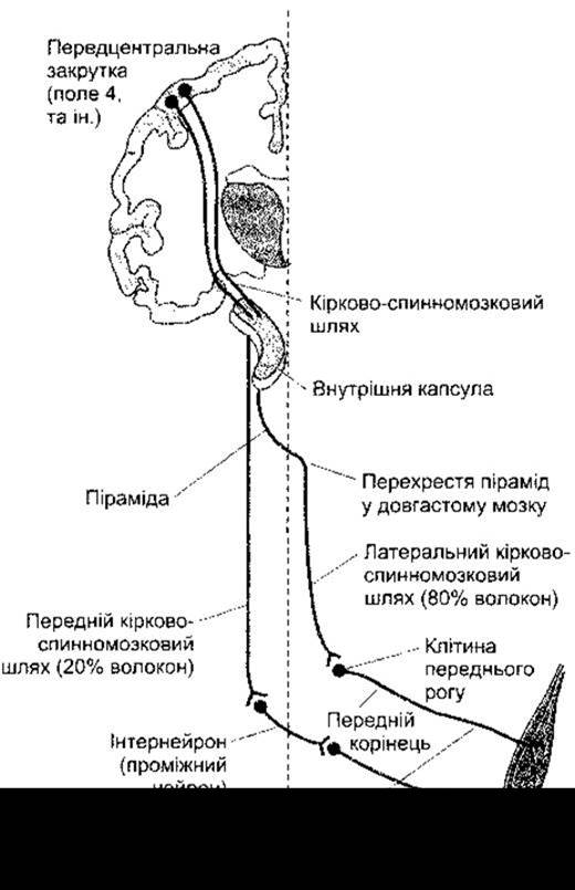
Fig. 12-2. Corticospinal pathways.

Fig. 12-3. Medial (top) and lateral (bottom) surfaces of the human cerebral hemisphere. Shown are the motor cortex (Brodmann area 4) and other areas involved in The regulation of voluntary movements, numbered according to Brodmann (reprinted with permission from Kandel ER, Schwartz JH, Jessell TM [editors]: Principles of Neural Science, 4th ed. McGraw-Hill, 2000).
Experimental stimulation of the cortex in patients undergoing Craniotomy under local anesthesia has made it possible to identify most of the motor projection areas of the cortex. These findings have been confirmed by PET and MRI Brain scans in conscious, unanesthetized humans (Fig. 12-4). The various PARTS OF THE body are represented in the precentral gyrus such that the FOOT is projected at the top of the gyrus and the face at the bottom (Fig. 12-5). The facial area is represented bilaterally (in both hemispheres), whereas the rest of the body is represented unilaterally, such that the cortical motor field regulates the musculature of the contralateral side of the body. The cortical representation of each body part is proportional in size to the skill required for its voluntary movements. For example, the cortical areas associated with speech articulation or movements performed by hand Muscles are exceptionally large; The Use of the Larynx, Lips, and Tongue to produce words, as well as the fingers, particularly the thumb, provides capabilities that reach their highest level in humans. The conditions under which the human Cerebral Cortex can be studied do not permit stimulation of the banks of sulci and other inaccessible areas. Detailed studies in monkeys have revealed that the body parts are correspondingly mapped in the cortex, with the axial musculature and proximal limb regions represented along the anterior edge of the precentral gyrus, and the distal limb regions along its posterior edge. Another feature of MI is the presence of considerable overlap in Muscle innervation zones, along with significant segregation of the innervation zones of synergistic muscles. There is an ongoing debate as to whether individual muscles or specific motor patterns are projected in MI; the latest data indicate that both are represented. Cells within the motor cortex are organized into distinct columns. Cells of each Column receive numerous sensory inputs from the peripheral region where they regulate movements, thereby providing a basis for feedback-controlled movement. Some of these inputs are direct, whereas others are relayed through somatosensory area I of the postcentral gyrus.
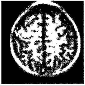
Fig. 12-4. Hand area of the motor cortex in a 7-year-old boy, visualized by MRI. Activity changes resulting from squeezing a rubber ball with the right hand are shown in white, and with the left hand, in black. In this particular case, activation of the left motor cortex by left-hand motor activity (see text) is not apparent (reprinted with permission from Waxman SG: Correlative Neuroanatomy, 24th ed. McGraw-Hill, 2000).

Fig. 12-5. Motor homunculus. Diagram of a coronal section of the precentral gyrus showing the cortical representation of various body parts. The sizes of the homunculus's body parts are proportional to the corresponding cortical fields of the gyrus (reprinted with permission from Penfield W, Rasmussen G: The Cerebral Cortex of Man. Macmillan, 1950).
Cerebral dominance, discussed in detail in Chapter 16, also applies to the human motor cortex. Movements of the left-hand fingers are predominantly accompanied by activation of the right motor cortex, and vice versa, as demonstrated by MRI studies (see Chapter 32); however, left-hand finger movements also activate the left motor cortex, particularly in right-handed individuals. This is consistent with the fact that lesions of the left motor cortex impair the function of both the left and right hands, whereas lesions of the right motor cortex cause only minor impairment to the right hand.
Plasticity
PET and functional MRI studies have revealed that in intact experimental animals as well as humans, the motor cortex exhibits the same plasticity as the sensory cortex (see Chapter 7). Thus, for example, the finger representation area in the contralateral motor cortex increases in size As a result of training in rapid finger movements of one hand; these changes are noticeable within a week and reach a maximum level by the fourth week. Cortical areas controlling the contraction of other muscles also increase in size as a result of training. Despite a small ischemic lesion in the hand representation area of the monkey motor cortex, recovery of motor function is accompanied by the restoration of the hand representation area in the adjacent, undamaged cortex. Consequently, the motor cortical map changes with experience.
Supplementary Motor Area
Most inputs to the supplementary motor area originate from the motor cortex. Accordingly, this area participates in the programming of motor sequences. Lesions to this area in monkeys cause difficulties in executing complex combined movements and impairments in bimanual coordination.
If a person counts silently, the motor cortex remains inactive, but when they begin to vocalize the numbers they are manipulating, Blood flow to the motor cortex and supplementary motor area increases. Thus, the supplementary motor area, together with the motor cortex, mediates complex voluntary movements associated with prior planning. Blood flow increases regardless of whether the planned movement is actually executed or not. This blood flow elevation is observed in either case, regardless of whether the movement is performed by the contralateral or ipsilateral hand.
Premotor Cortex
Inputs from the premotor cortex project to Brainstem regions involved in posture regulation and to the motor cortex, particularly to the areas of Water/144.html">Origin of the corticospinal and corticobulbar tracts. The function of the premotor cortex is not yet fully understood; it is believed to be associated with postural adjustment at the onset of a planned movement, specifically in preparation for its execution.
Posterior Parietal Cortex
In addition to fibers forming part of the corticospinal and corticobulbar tracts, fibers project to the premotor cortex from the somatosensory area and the adjacent posterior region of the parietal lobe. Lesions of the somatosensory area result in deficits in performing learned movement sequences, such as using a knife and fork while eating. Some neurons in area 5 (see Fig. 12-3) are involved in regulating movements such as reaching for or manipulating an object with the hand, whereas certain neurons in area 7 participate in hand-eye coordination.
Role in Movement Regulation
As noted, the corticospinal and corticobulbar tracts constitute the system responsible for fine and precise movements. However, this does not mean that movements, even fine ones, are impossible without this system. Lower vertebrates essentially lack the corticospinal and corticobulbar system, yet they move quite agilely. Cats and dogs can stand, walk, run, and even eat after the complete destruction of this system. Only in primates do such interventions produce significant alterations.
Careful pyramid sectioning, which causes highly selective destruction of the lateral corticospinal tract in laboratory primates, leads to a sudden and prolonged loss of The ability to grasp and hold small objects with two fingers and to perform isolated wrist movements. These impairments stem from the loss of control over the distal limb musculature, which normally ensures fine and precise movements. On the other hand, damage to the anterior corticospinal tract causes axial muscle dysfunction, making it difficult to maintain balance, walk, and climb.
Effects on Stretch Reflexes
Pyramid sectioning in monkeys results predominantly in prolonged hypotonia and flaccidity rather than spasticity. In humans, given the Structural Features of the Central Nervous system, pathological processes only rarely damage the corticospinal and corticonuclear tracts in isolation, i.e., without damaging the pathways involved in posture regulation. The Emergence of spasticity apparently indicates damage to these very pathways rather than to the corticospinal and corticonuclear tracts.
In the case of lateral corticospinal tract damage in humans, the Babinski sign appears: dorsiflexion of the big toe and fanning of the other toes upon stimulation of the Skin along the lateral border of the sole. Except in infants, the normal response to this stimulation is plantarflexion of all toes. The Babinski sign signifies the disappearance of this flexor reflex, which is normally used to assess lateral corticospinal tract function. It is important for determining the localization of the pathological process, although its physiological basis remains unknown.
Last update: 10/08/2026
Editorial and Educational Adaptation: This material has been compiled based on the primary/original source text. The project team performed an editorial review, corrected technical inaccuracies, structured sections, and adapted the content for an educational format.
What was processed:
- elimination of formatting defects (OCR errors, structural breaks, corrupted characters);
- editorial organization of content;
- standardization of terminology in accordance with academic sources;
- verification of factual statements against the original source text.
All mentions of the author, publication year, and origin of the primary text have been preserved in accordance with the source.