Special Histology and Embryology: Practical Course - V. K. Napkhanyuk 2001
Nervous System
Cerebral Cortex
The Brain (encephalon cerebrum) consists of the right and left hemispheres, the Brainstem comprising the Diencephalon, Midbrain, and Hindbrain (Pons, Cerebellum), as well as the Medulla Oblongata.
The cerebral cortex (cortex cerebralis) is represented by a layer of Gray matter approximately 3 mm thick. It is most developed in the precentral gyrus, where its thickness reaches 5 mm. The presence of numerous sulci and gyri significantly increases the surface area of the brain's gray matter. The cortex is formed by about 50 billion Nerve Cells. Various Regions of the cortex that differ from one another in certain features of Cell localization and Structure, fiber arrangement, and functional significance are called areas. There are no sharply defined boundaries between them. Areas are the sites of higher analysis and synthesis of nerve impulses.
The layered arrangement of cortical cells is called cytoarchitectonics. The cortex consists of six layers:
1) the molecular layer (lamina molecularis), consisting of a small number of small associative spindle-shaped cells with long horizontal dendrites and descending axons that form horizontal collaterals;
2) the external granular layer (lamina granularis externa), formed by small neurocytes up to 10 µm in size, which have a round, angular, or pyramidal shape, and stellate neurocytes; the dendrites of these cells ascend to the molecular layer of axons or go into the White matter, or, forming loops, also enter the tangential plexus of fibers of the molecular layer;
3) the pyramidal layer (lamina piramidalis), the thickest layer, formed by pyramidal-shaped cells whose size sequentially increases from 10 to 40 µm. The apices of the pyramidal neurocytes are directed toward the cortical surface. An apical dendrite extends from the apex, lateral dendrites from the lateral surface, and an axon from the base;
4) the internal granular layer (lamina granularis interna), formed by small granular neurocytes and numerous horizontal fibers. In some areas, it is very strongly developed (e.g., in the visual cortex) and may be completely absent (in the precentral gyrus);
5) the ganglionic layer (lamina ganglionaris), formed by large pyramidal cells (Betz cells). These are very large cells reaching up to 120 µm in height and 80 µm in width. The axons of these cells project to the motor nuclei of the BRAIN AND SPINAL cord, forming A large number of collaterals and exerting an inhibitory effect on the cortex;
6) the multiform layer (lamina multiformis), formed by neurocytes of various, predominantly spindle-shaped forms. The dendrites of these cells reach the molecular layer, and the axons pass into the white matter as part of the efferent tracts of the brain.
The Structural and functional unit of the cortex is the module. It is a vertical Column with a diameter of about 300 µm. The module is organized around a corticocortical fiber originating either from pyramidal Cells of the same hemisphere (association fiber) or from the opposite hemisphere (commissural fiber). The module contains two thalamocortical fibers. These are specific afferent fibers terminating in layer 4 of the cortex.
The layered arrangement of tangential bundles of nerve fibers within the cortex is called myeloarchitectonics. The cortex contains: association fibers, which connect individual cortical areas of one hemisphere; commissural fibers, which connect the cortex of different hemispheres; and projection fibers, connecting both afferent and efferent fibers with the nuclei of lower Divisions of the Central Nervous system. In the cerebral cortex, these fibers form radial rays that terminate in the pyramidal layer.
Slides for Study
Slide 3. Section of the cerebral cortex (Fig. 3).
Low magnification. Examine the layered STRUCTURE OF THE cortex. Locate the sulcus between two gyri containing the pia mater and Blood Vessels within it. The cortical surface is visible on both sides of the sulcus. The outermost cortical layer is the molecular layer, which is easily distinguished due to its low cell density. The next layer, the external granular layer, contains small neurocytes of round, angular, or pyramidal shape up to 10 µm in size, followed by the widest layer, the external pyramidal layer. Below lies the internal granular layer with small stellate cells. This layer is easier to identify by first observing the layer of large pyramidal neurocytes, which are up to 120 µm in height and immediately catch the eye. Next is the layer of spindle-shaped neurocytes. Beneath the cortex, the white matter is visible, formed predominantly by myelin fibers. Both gray and white matter also contain gliocytes, which are identified by their nuclei.
Class="center">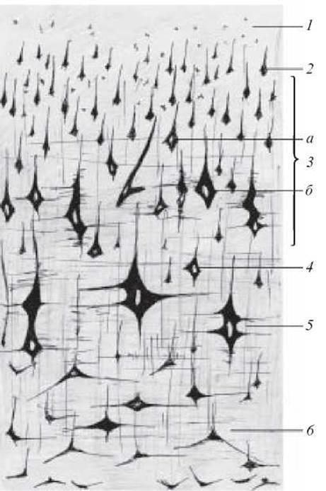
Fig. 3. Section of the cerebral cortex. Silver impregnation. × 120:
1 — molecular layer; 2 — external granular layer; 3 — pyramidal layer (a — small and medium pyramidal cells; b — large pyramidal cells); 4 — internal granular layer; 5 — ganglionic layer; 6 — layer of multiform cells
Label on the figure: 1) molecular layer; 2) external granular layer; 3) pyramidal layer; a) small and medium pyramidal cells; b) large pyramidal cells; 4) internal granular layer; 5) ganglionic layer; 6) layer of multiform cells; 7) blood vessels.
Last update: 10/08/2026
Editorial and Educational Adaptation: This material has been compiled based on the primary/original source text. The project team performed an editorial review, corrected technical inaccuracies, structured sections, and adapted the content for an educational format.
What was processed:
- elimination of formatting defects (OCR errors, structural breaks, corrupted characters);
- editorial organization of content;
- standardization of terminology in accordance with academic sources;
- verification of factual statements against the original source text.
All mentions of the author, publication year, and origin of the primary text have been preserved in accordance with the source.