Review of Medical Physiology - William F. Ganong 2002
Physiology of Nerve and Muscle Cells
Excitable Tissue: Nervous
Nerve Cells
Neurons in the Central Nervous system of mammals vary greatly in shape and size (Fig. 2-1). However, most of them consist of the same basic parts as a typical spinal motor neuron (Fig. 2-2). These Cells have five to seven branching processes extending from The Cell body, known as dendrites. Specifically, in the Cerebral Cortex AND cerebellar cortex, dendrites feature small knob-like protrusions called dendritic spines. A typical neuron also possesses a long fibrous process, the axon, which originates from a slightly thickened region of the cell body known as the axon hillock. The initial segment of the axon is called the initial segment. Near its termination, the axon branches into axon terminals, each ending in numerous synaptic swellings known as terminal boutons or axon telodendria. These contain granules or vesicles that store synaptic Transmitters synthesized within the neurons (see Chapter 4).
The axons of many neurons are myelinated, meaning they are covered by a myelin sheath—a protein-lipid complex formed by multiple layers of cell membranes belonging to Schwann cells (neurolemmocytes) (Fig. 2-3). Schwann cells are glial-like cells distributed along peripheral nerve fibers. The myelin sheath is formed by the repeated wrapping (up to 100 times) of the Schwann cell membrane around the axon. Myelin becomes compacted when the extracellular portions of a membrane protein known as protein zero (P0) adhere to the extracellular PARTS OF THE adjacent membrane. Various Mutations in the P0 Gene lead to peripheral neuropathies; 29 different mutations have been described that cause both moderate and severe disorders. The myelin sheath is absent at the terminal branching of the axon and at periodic constrictions (roughly every 1 mm, spanning about 1 μm) known as the nodes of Ranvier. The insulating function of myelin is described below. However, not all neurons in the mammalian body are myelinated; some are unmyelinated, meaning they are simply surrounded by Schwann cells without the repeated membrane wrapping and myelin formation characteristic of myelinated fibers. Most invertebrate neurons are unmyelinated.
In mammals, neurons are predominantly myelinated, but the myelin is formed mostly by oligodendrocytes rather than Schwann cells (see Fig. 2-3). Unlike Schwann cells, which form a myelin sheath between two adjacent nodes of Ranvier on a single neuron, oligodendrocytes extend their myelin-forming processes to multiple neighboring axons. In multiple sclerosis, a severe autoimmune disease, multifocal destruction of myelin occurs within the CNS. The loss of myelin is accompanied by the slowing or cessation of impulse conduction along demyelinated axons.
The dimensions of individual neurons are truly striking. For instance, assuming the cell body of a spinal motor neuron innervating the Muscles of the FOOT were the size of a tennis ball, its dendrites could fill a medium-sized living room, and its axon would stretch for 1.6 km while being 13 mm in diameter. While the conventional names for the parts of a neuron are clear when applied to spinal motor neurons and interneurons, certain inconsistencies arise when using the terms "dendrite" and "axon" for Other types of neurons. From a functional perspective (see below and Chapters 4 and 5), neurons generally consist of four main zones: the receptor or dendritic zone, which integrates multiple local potential changes generated by synaptic connections (Fig. 2-4); the Action Potential-generating zone (the initial segment in spinal motor neurons, the initial node of Ranvier in sensory cutaneous neurons); the axonal process, which conducts propagated impulses toward the nerve endings; and the nerve terminals, where action potentials trigger the release of synaptic Neurotransmitters.
Class="center">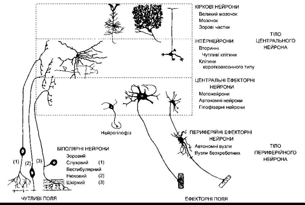
Fig. 2-1. Selected types of neurons in the mammalian nervous system (reprinted with permission, from Bodian D. Introductory survey of neurons. Cold Spring Harbor Symp Quant Biol. 1952; 17:1).
In most cases, the cell body is located in the dendritic zone at the proximal end of the axon, but it can also be situated within the axon itself (e.g., in auditory neurons) or off to the side (e.g., in sensory cutaneous neurons; see Fig. 2-1). THE POSITION OF the neuronal cell body does not affect the receptor function of the dendritic zone or the impulse-conduction function of the axon.
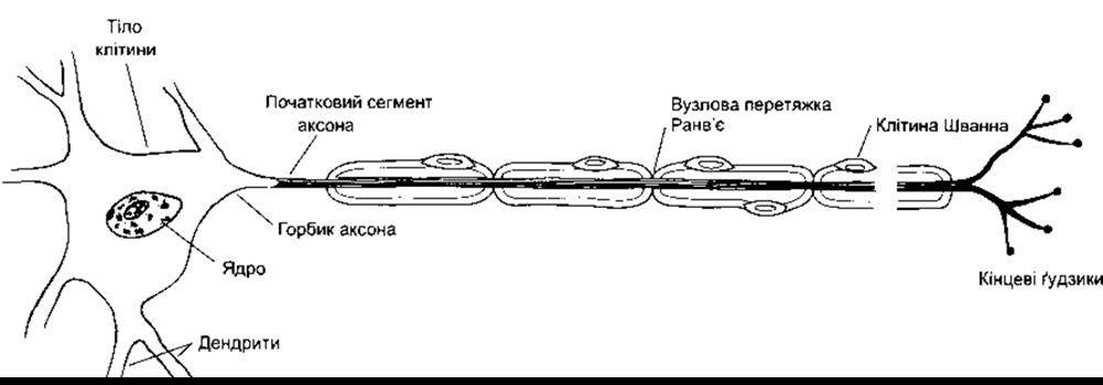
Fig. 2-2. A motor neuron with a myelinated axon.
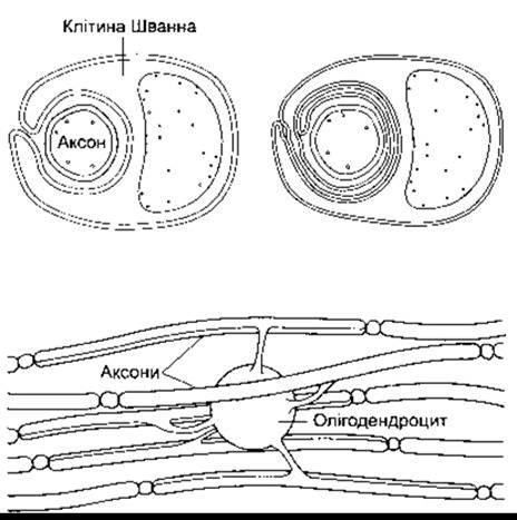
Fig. 2-3. Top: Relationship between Schwann cells and peripheral nerve axons. The cross-section of an unmyelinated axon is shown on the left, and a myelinated axon on the right. As illustrated, the Schwann cell membrane rolls up and envelops the axon. Bottom: Myelination of CNS axons by oligodendrocytes. A single oligodendrocyte extends processes to over 40 axons.
Notably, the dendritic arborizations of neurons vary remarkably in size and structural complexity (Fig. 2-1; see also Figs. 11-1 and 12-17). In addition to integrating passive electrical activity, dendrites in certain instances can also generate and propagate action potentials.
Protein Synthesis AND Axonal Transport
Nerve cells are secretory in nature, yet they differ from other secretory cell types because their secretory zone is located primarily at the axon terminal, far removed from the cell body. Ribosomes are scarce or entirely absent in the axon and its terminals; thus, the synthesis of all necessary Proteins occurs within the Endoplasmic reticulum and Golgi apparatus, after which the axon transports them to synaptic boutons via axoplasmic transport. Consequently, the neuronal cell body maintains the Functional and Structural integrity of the axon: if an axon is severed, its distal segment undergoes degeneration (Wallerian degeneration). Anterograde transport is mediated by microtubules, with molecular mechanisms detailed in Chapter 1. Fast anterograde transport operates at a rate of approximately 400 mm per day, whereas slow transport moves at about 0.5–10.0 mm per day. Retrograde transport, which involves the movement of substances in the opposite direction, proceeds at roughly 200 mm per day.
Synaptic vesicles are recycled locally at the membrane, but some used vesicles are transported back to the cell body and accumulate in Lysosomes. Certain substances, such as nerve growth factor, as well as various Viruses that enter nerve endings via endocytosis, also reach the cell body through retrograde axonal transport.
Last update: 10/08/2026
Editorial and Educational Adaptation: This material has been compiled based on the primary/original source text. The project team performed an editorial review, corrected technical inaccuracies, structured sections, and adapted the content for an educational format.
What was processed:
- elimination of formatting defects (OCR errors, structural breaks, corrupted characters);
- editorial organization of content;
- standardization of terminology in accordance with academic sources;
- verification of factual statements against the original source text.
All mentions of the author, publication year, and origin of the primary text have been preserved in accordance with the source.