General Microbiology - Schlegel, H. G. 1987
Viruses: Distribution and Structure
Viruses
Structure of viruses. A viral particle, also known as a virion, consists of a nucleic acid (DNA or RNA) surrounded by a protein coat called a capsid. Such a unit (capsid + nucleic acid = nucleocapsid) may be "naked" or, in other cases, enveloped (Figs. 4.2 and 4.3). Naked nucleocapsids include, for example, particles of tobacco mosaic virus, wart virus, and adenovirus. An additional envelope surrounds Influenza and herpes viruses.
The capsid, in turn, consists of subunits known as capsomeres and typically exhibits a symmetrical structure. Two Types of Symmetry are distinguished: helical and cubic. Table 4.1 groups various viruses according to their structure. Below, we examine four well-known disease-causing viruses: two with helical symmetry—one with naked particles (tobacco mosaic virus) and one enveloped (influenza virus)—and two with cubic symmetry—one with naked particles (poliovirus and other polyhedral viruses) and one enveloped (herpes virus).
Class="center">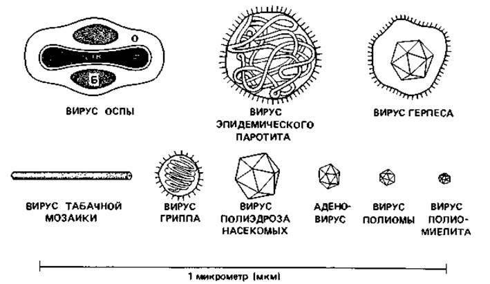
Fig. 4.2. Shape and size of particles (virions) of certain viruses. B — elliptical protein body; O — envelope.

Fig. 4.3. Structural types of Viral Particles. Four forms are depicted: two with helical symmetry and two with cubic symmetry (in both cases, one virion is "naked" and one is enveloped).
Table 4.1. Morphological classes of viruses
|
Helical structure |
||
|
Naked capsids |
Enveloped capsids |
|
|
RNA |
Tobacco mosaic RNA virus and many other plant viruses |
Myxoviruses: Influenza Viruses, parainfluenza viruses (mumps, measles) |
|
DNA |
Coliphage fd |
|
|
Polyhedral structure (icosahedra) |
||
|
Naked capsids |
Enveloped capsids |
|
|
RNA |
Picornaviruses: FOOT-and-Mouth disease RNA viruses, ECHO, poliovirus, reoviruses |
Retroviruses (oncogenic): Sarcoma, leukemia, and carcinoma viruses |
|
Circular DNA |
Papovaviruses (oncogenic RNA-associated): papilloma, polyoma, and SV40 viruses |
Togaviruses: Semliki Forest virus, encephalitis viruses, yellow fever virus |
|
Single-stranded circular DNA |
Coliphages ⊘X174, M13 DNA |
Herpes simplex viruses, herpes zoster, varicella, Epstein-Barr virus (infectious mononucleosis) |
|
Complex viruses (icosahedral HEAD + helical tail) |
||
|
DNA |
Large Bacteriophages (T2, T4, T6) Complex virions |
|
|
DNA |
Poxviruses, vaccinia viruses |
|
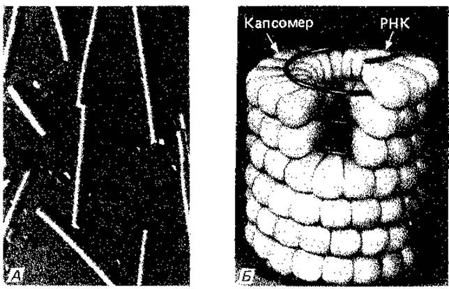
Fig. 4.4. Tobacco mosaic virus. A. Electron micrograph after oblique shadowing with carbon and platinum; 65,000×. (Photo by N. Frank.) B. Model. (Karlson, Kurzes Lehrbuch der Biochemie, Stuttgart, Thieme, 1980.)
Tobacco mosaic virus. This is a classic example of a virus with helical symmetry. It is easily isolated from the expressed juice of infected plants. The particles are rod-shaped, 18 nm in diameter (Fig. 4.4, A). This rod-like nucleocapsid consists of approximately 2,100 capsomeres arranged in a helical pattern to form a hollow cylinder. Each capsomere consists of a single polypeptide chain (comprising 158 Amino Acids with a determined sequence). An RNA chain, also following a helical path, is embedded in the wall of the hollow cylinder between the capsomeres (Fig. 4.4, B).
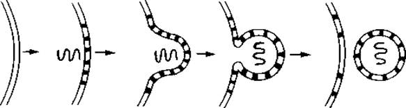
Influenza virus. Influenza virus particles have a diameter of 110 nm (Fig. 4.5, A). Similar to the tobacco mosaic virus, the nucleocapsid has a helical structure, but instead of being rod-shaped, it is tightly coiled multiple times (Fig. 4.5, B). The nucleocapsid is surrounded by an envelope—a fragment of the host Cell membrane from which the virion emerged. The outer surface of the envelope bears spikes that facilitate the adsorption of the virion onto The surface of a new host cell; these spikes contain mucoproteins and the enzyme neuraminidase. This enzyme cleaves one component, N-acetylneuraminic acid, from the mucoproteins of the infected cell, which likely plays a role in thinning the mucus lining the nasopharyngeal epithelial Cells. Viral Replication occurs intracellularly. Virion release resembles a budding process, during which the outer envelope of the viral particle is formed from the host cell membrane, potentially modified by the incorporation of virus-derived Proteins (such as neuraminidase).
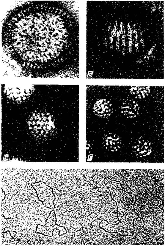
Fig. 4.5. Various animal-pathogenic viruses. A. Influenza A2 virus. B. Nucleocapsid of influenza A2 virus; a contrasting agent, phosphotungstic acid, has penetrated a particle with a ruptured envelope, making the nucleocapsid visible. C. Adenovirus; its particles clearly display an icosahedral shape. D. SV40 virus. This tumor virus is considerably smaller than the adenovirus. It also has an icosahedral shape but appears spherical. E. SV40 viral DNA; its double-stranded circular molecules have a contour length of approximately 1.6 µm (the ends of broken circles are visible on the right and left). A–D: 250,000×, negative staining (phosphotungstic acid); E: 65,000×. (Photos A, B, D, E by N. Frank; C by N. Gelderblom, Max Planck Institute for Virus Research, Tübingen.)
There are many different strains of the influenza virus. The specific tissue targeted by the virus depends on its host cell Specificity and the receptor Properties of the cells. The virus can disrupt cellular METABOLISM or even cause cell death. Additionally, it acts as an antigen, stimulating Antibody production in the host Organism. The viruses responsible for major influenza epidemics differ from one another in virulence and pathogenicity.
Non-enveloped polyhedral viruses. Many viruses that appear spherical actually possess a polyhedral geometry. Most commonly, this is an icosahedron—a polyhedron bounded by 20 equilateral triangles and having 12 vertices (Figs. 4.5, C and 4.6). The capsid of an icosahedral virus consists of two types of capsomeres: pentons, made up of five protein monomers (protomers), located at the vertices; while the remaining surface of the facets and edges is formed by hexons, composed of six protomers. The construction of the capsid from capsomeres follows crystallographic principles; accordingly, the smallest icosahedral capsid must consist of 12 pentons, and the next size up consists of 12 pentons and 20 hexons. Some viruses comprise 252 or even 812 capsomeres.

Fig. 4.6. Potential Structural elements of the capsid in icosahedral viruses. Two types of capsomeres are illustrated: hexagonal (hexon) and pentagonal (penton). The former consists of six protomers, and the latter of five, each comprising one or two polypeptide chains. On the right is an icosahedron composed of 12 pentons and 30 hexons, i.e., 42 capsomeres.
Numerous viruses are built on the icosahedral principle, including poliovirus, foot-and-mouth disease virus, Adenoviruses (Fig. 4.5, C), and the oncogenic SV40 virus (Fig. 4.5, D).
The fact that the viral capsid is composed of A large number of identical subunits becomes clear when we consider that the nucleic acid mass in many viruses is very small. The DNA or RNA chains are so short that the information they contain is only sufficient to code for a few polypeptide chains, most of which perform enzymatic Functions during viral reproduction within the host cell. Building the capsid from numerous identical subunits ensures maximum efficiency with a minimum amount of genetic material.
Enveloped polyhedral viruses. An icosahedron surrounded by an envelope is the characteristic Morphology of the causative agents of chickenpox, shingles, and herpes simplex.
The icosahedral capsid of the herpes virus consists of 162 capsomeres. The outer envelope undoubtedly originates from the inner nuclear membrane of the host cell. Herpes viruses replicate within the Cell Nucleus; the capsids of new viral particles acquire an envelope from the nuclear membrane, "bud off" from The Nucleus, and are transported outward via The Endoplasmic reticulum system.
Chickenpox is a relatively mild childhood disease. The virus infects the Upper Respiratory Tract, spreads via the bloodstream throughout the body, and ultimately settles in the Skin, where it causes vesicle formation. Shingles (herpes zoster) occurs in partially immune individuals As a result of the reactivation of the varicella-zoster virus. Thus, both conditions are caused by the exact same virus.
Poxviruses. Poxviruses are the largest of the animal-pathogenic viruses. Their particle structure differs markedly from the four types described above. They contain DNA, protein, and several Lipids, which is why they are sometimes referred to as complex virions (Table 4.1). Particles of smallpox (variola) and vaccinia (cowpox) viruses appear as rounded blocks. They consist of an internal body containing double-stranded DNA, a double protein-containing layer, elliptical protein bodies, and an outer membrane, all tightly bound by surrounding threads. These viral particles are highly resistant to desiccation and are consequently extremely infectious. Smallpox naturally infects only humans and monkeys. The cowpox virus can also infect cattle, rabbits, and sheep. Both viruses share common Antigens. Therefore, people are prophylactically vaccinated with the cowpox virus, which is harvested from cattle and causes very mild symptoms in humans. Such active vaccination induces The production of Antibodies that also confer Immunity against smallpox.
Last update: 13/08/2026
Editorial and Educational Adaptation: This material has been compiled based on the primary/original source text. The project team performed an editorial review, corrected technical inaccuracies, structured sections, and adapted the content for an educational format.
What was processed:
- elimination of formatting defects (OCR errors, structural breaks, corrupted characters);
- editorial organization of content;
- standardization of terminology in accordance with academic sources;
- verification of factual statements against the original source text.
All mentions of the author, publication year, and origin of the primary text have been preserved in accordance with the source.