The Evolution of Viruses - Zhdanov, V. M. 1990
Special Section
Influenza Viruses
The example of Influenza clearly illustrates the previously mentioned concept that the evolution of RNA structures occurs a million times faster than that of DNA structures. Indeed, over a span of 10 years, influenza Viruses undergo an evolutionary journey equal to—if not exceeding—that traversed by primates over 10 million years, which culminated in The Emergence of humans. To properly contextualize these evolutionary rates, however, it must be noted that over the course of 10 million years, primates went through approximately 500,000 generations, whereas influenza viruses went through about 5,000 generations in 10 years; consequently, their actual evolutionary rate is only 10-fold higher. From the vast body of literature on orthomyxoviruses, we will briefly review only the core data necessary for understanding the pathways of influenza virus evolution [Kilbourne E., 1978; Burnet F., 1979].
Influenza viruses (Orthomyxoviridae) form two genera: Influenzavirus (influenza viruses A and B) and the genus of influenza C virus. Orthomyxoviruses isolated from Arthropods that remain unclassified are also known. Virions are spherical (though native preparations often show filamentous or irregular forms) with a diameter of 80–120 nm. The nucleocapsid is a tightly wound helix with a diameter of 7 nm and a length of 50–130 nm across its 8 fragments. It is surrounded by an inner membrane (matrix), forming the core of the virion. The outer lipid envelope contains trimers of hemagglutinin and tetramers of neuraminidase, which are surface Glycoproteins. Hemagglutinin molecules are 14 nm long and 4 nm in diameter. Neuraminidase molecules feature a HEAD measuring 4×8.5 nm and a stalk measuring 10×4 nm.
The Genome of influenza viruses consists of 8 segments of single-stranded negative-sense RNA with the following sizes: PB1 — 2341 NUCLEOTIDES, PB2 — 2341, PA — 2233, HA — 1778, NP — 1565, NA — 1413, M — 1027, and NS — 890 nucleotides. The number of nucleotides and their molecular mass vary among different viral strains and differ significantly between type A and B viruses. Influenza C viruses possess 7 RNA segments and lack the neuraminidase Gene. The designated Proteins have the following molecular masses: 96,000 (PB1), 87,000 (PB2), 85,000 (PA), 50,000–60,000 (NP), 48,000–63,000 (MA); hemagglutinin subunits (with the additional carbohydrate molecular mass indicated in parentheses) — 36,000 (11,500) and 27,000 (1,300). Two genes encode two proteins each: M1 (27,000) and M2 (15,000), NS1 (25,000) and NS2 (12,000). Naturally, these values vary across different viruses.
Table 8. Genome segments and proteins of influenza A virus
Genome segment number |
Encoded protein |
Total nucleotides / coding |
Molecular mass, X106 |
Note |
|
1 |
PB2 |
2341 |
2277 |
||
2 |
PB1 |
2341 |
2271 |
81–94 |
|
3 |
PA |
2273 |
2148 |
||
4 |
HA |
1742–1772 |
1686–1701 |
75–80 |
HA1 49,000– |
58,000 |
|||||
HA2 25,005– |
|||||
30,000 |
|||||
5 |
NP |
1565 |
1494 |
55–65 |
|
6 |
NA |
1409–1465 |
1359–1407 |
55–70 |
|
7 |
M1 |
1027 |
756 |
21–27 |
|
M2 |
1027 |
291 |
15 |
||
8 |
NS1 |
890 |
690–711 |
25 |
|
NS2 |
830 |
363 |
12 |
||
Thus, 8 RNA fragments encode the synthesis of 10 proteins (Table 8) [Zhilinskaya I. N., 1983; McCauley J., Mahy B., 1983].
All influenza virus genes have been sequenced repeatedly, and three-dimensional structural models have been generated for hemagglutinin and neuraminidase. Data on nucleotide sequences are presented in a series of studies dedicated to the polymerase complex genes [Winter G., Fields S., 1982; Fields S., Winter G., 1983], hemagglutinin [Hauptmann R. et al., 1983], nucleoprotein [Steuler H. et al., 1985], neuraminidase [Fields S. et al., 1981], matrix [Winter G., Fields S., 1980], and non-structural [Lamb R. et al., 1980] proteins.
The three-dimensional Structure OF THE hemagglutinin molecule turned out to be complex (Fig. 24) [Wilson J. et al., 1981; Wiley D. et al., 1981]. As seen in Fig. 24, two antigenic determinants are located on the sides of the receptor pocket: the first is a loop (amino acid residues 140–146), the second is a helix (residues 187–196), the third is situated at the Base of the globular region of the hemagglutinin molecule near the disulfide bond of amino acid residues Cys 52 and Cys 277, and finally, the fourth is formed at the level of the globular structures. Mutations in these sites are responsible for the antigenic drift of hemagglutinin. Glycosylation also occurs at strictly defined locations. Naturally, the antigenic sites of hemagglutinins from different serogroups are arranged differently. Furthermore, the oligopeptide of the small subunit of the hemagglutinin molecule (amino acid residues 1–11 from the N-terminus) exhibits antigenic properties and plays a crucial role in virion penetration into The Cell [Atassi M., Webster R., 1983].
Class="center">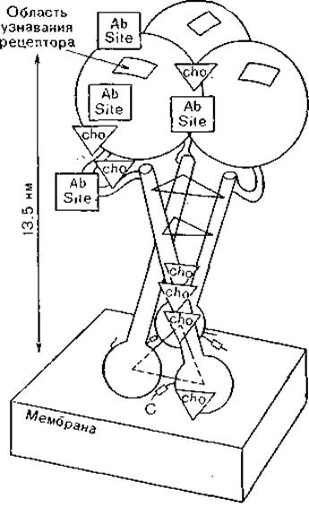
Fig. 24. Structure of the hemagglutinin trimer.
cho — attachment sites for carbohydrate chains, Ab site — antigenic sites. The dashed line indicates the hydrophobic peptide cleaved by bromelain.
Similar studies on neuraminidase [Varghese J. et al., 1983; Colman P. et al., 1983] demonstrated that this glycoprotein is a tetramer featuring a globular head, resembling a four-petaled flower anchored by a thin stalk embedded in The Lipid Bilayer of the outer envelope. The enzymatic active center is located at the center of each petal, with two antigenic sites positioned close to it and two others at some distance. Seven segments interact with Antibodies.
Penetration of virions into susceptible Cells occurs following attachment to receptors. In this process, not only do Different types of orthomyxoviruses (A, B, and C, respectively) possess distinct receptors, but these receptors also differ between Human and Animal influenza A viruses [Rogers G., Paulson J., 1983]. This is due to variations in the composition of sialooligosaccharides and lipid components within cell membranes, as well as the correspondingly different conformation of the "receptor pocket" on the hemagglutinin molecule. Attachment to cell Membrane Receptors occurs via interaction with this "receptor pocket," leading to The formation of an endocytic vacuole, which brings the virions inside the cell. Here, partial uncoating of the virion is completed, and its core is transported toward The Nucleus. A second uncoating stage takes place at the nuclear membrane—specifically, the removal of the M protein—allowing the functionally active nucleocapsid (ribonucleoprotein) to enter the nucleus.
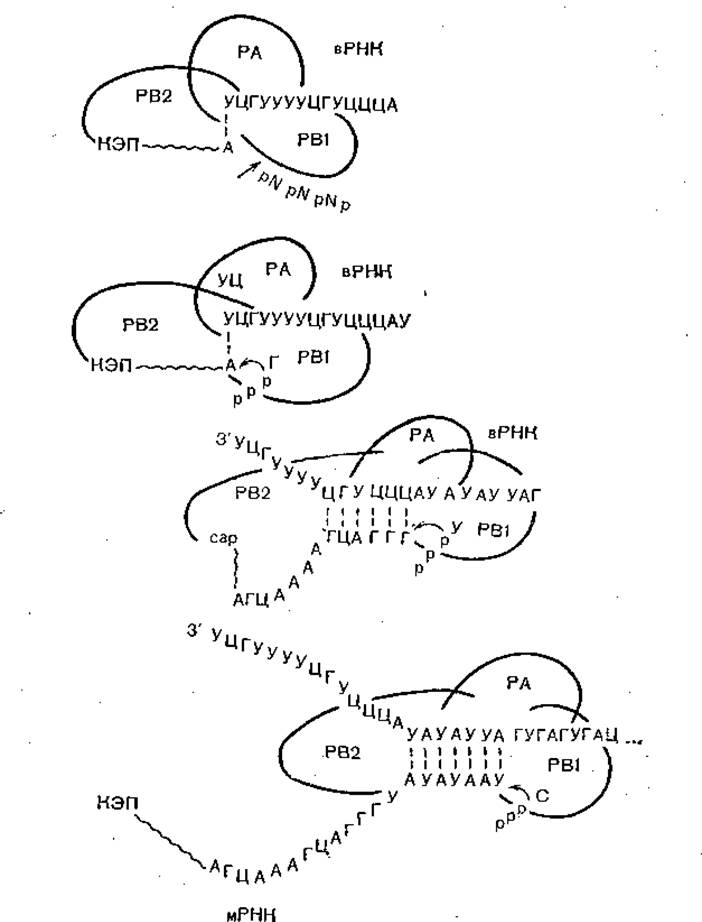
Fig. 25. Model of the function, interaction, and movement of the three P proteins during the Initial Stages of influenza virus mRNA synthesis. The vRNA and mRNA sequences pertain to the NP gene. The upper portion illustrates the binding of PB2 to the capped cellular RNA and Cleavage at a purine site (usually A), after which PB1 catalyzes the extension of the primer on the vRNA template. All three proteins move together along the template.
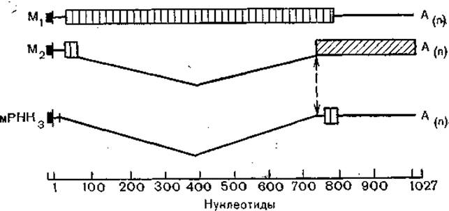
Fig. 26. Structure of the three mRNAs of the type A influenza virus 7th gene. Thin lines denote non-coding regions, and curved lines denote introns. The initial structure is a cellular-derived cap primer.
Gene METABOLISM/31.html">Transcription begins here as well, involving the polymerase complex (PA, PB1–PB2 proteins) alongside the NP protein (Fig. 25). The virus induces the synthesis and Processing of cellular mRNAs, from whose molecules the PB2 protein "snips off" the cap structure along with the adjacent 10–13 nucleotides. These serve as the primer for mRNA synthesis. The Messenger RNA is then transported to the Cytoplasm to direct the Synthesis of the corresponding protein on Ribosomes.
Transcription of the genome fragments proceeds in a unique manner. Unlike Replication, during which a complete complementary strand followed by daughter strands of each fragment is synthesized, transcription yields only the coding portion of the strand. In this process, the Proteins of the polymerase complex and NP migrate into the nucleus [Briedis D. et al., 1981], where progeny ribonucleoproteins are assembled [Davey J. et al., 1985]. As previously mentioned, transcription of segments 7 and 8 (M and NS, respectively) produces two proteins each (Fig. 26). Viral particle assembly takes place at the cellular membranes, into which hemagglutinins and neuraminidase are embedded, and release from the cell occurs via budding, a mechanism typical of enveloped viruses.
To understand THE ORIGIN OF influenza viruses, they should be examined in conjunction with other negative-sense genome viruses: paramyxo-, rhabdo-, bunya-, and Arenaviruses. All of them, with the exception of Rhabdoviruses, infect warm-blooded animals, and many are transmitted by Blood-sucking arthropods. Rhabdoviruses infect both animals and plants. The genomic strategies of these viruses are generally similar. Their genome is non-infectious and requires a polymerase—which must be present within the virions—for its functioning. These properties are common to all 5 groups of viruses under consideration. Ortho- and Paramyxoviruses are morphologically the closest to one another, yet their genome structures differ: linear in paramyxoviruses and segmented in orthomyxoviruses. In this sense, bunya- and arenaviruses are closer to the latter, although their virion architecture differs, as does that of the bullet-shaped rhabdoviruses. Viruses from all 5 groups infect higher animals and higher plants, with insects frequently acting as vectors. Unlike double-stranded RNA viruses, among which transitional forms appear to exist, no such forms are found here. Consequently, it remains unclear whether these viruses shared a common ancestor in the past and subsequently diverged into the 5 groups that survive and thrive today, or if each group emerged de novo independently of the others. The presence of viral polymerases in all considered virus groups seemingly Supports a common origin. This is further supported by the subunit structural similarity of the envelope glycoproteins in ortho- and paramyxoviruses, even though their polymerases are quite distinct. We should also note that viruses in all 5 groups possess outer envelopes, and their ribonucleoproteins are organized according to a helical Symmetry type. The method of nucleic acid Homology analysis is of little utility here, because over millions, tens of millions, or perhaps even hundreds of millions of years of evolution, RNA may have diverged to such an extent that no discernible traces of homology remain for many viruses, even within the same viral group. Therefore, specialized studies are required to resolve the question of a possible common origin for negative-sense genome viruses. Furthermore, it remains entirely unclear which subcellular structures originally gave rise to negative-sense genome viruses.
During replication, which also takes place in the cell nuclei, the entire strand of the RNA segment is transcribed: a plus-strand is synthesized first, followed by the synthesis of minus-strands of progeny RNAs on this template. Nucleocapsid assembly is carried out in the nucleus, while the glycoprotein is transported to The cell membrane post-assembly and becomes embedded within it. The M protein participates in virion formation, and the resulting virions exit the cell via a budding mechanism.
Influenza represents a unique infection with no parallel among other infectious diseases. Each year with the onset of cold weather, morbidity rates begin to rise. The number of causative agents approaches 200. Among them are adeno-, paramyxo-, corona-, rhino-, reo-, and enteroviruses, Mycoplasmas, chlamydiae, streptococci, staphylococci, pneumococci, and others. Against this "Background noise," usually in December—though sometimes earlier (November) or later (January, February)—influenza epidemics break out, affecting 6–8% to 30–35% of an urban population over 3–4 weeks, a country's population over 1.5–2 months, and a hemisphere over 2–3 months. Today, influenza epidemics occur almost annually, rolling like a giant wave from the Northern Hemisphere to the Southern (May–August) and from the Southern to the Northern (November–February). At present, influenza is a truly global infection not only because it is widespread across all nations, but primarily because it is an infection affecting humanity as a whole.
The cause of this peculiar Epidemiology of influenza lies in the unique Variability of the virus's surface Antigens—hemagglutinin and neuraminidase—which are responsible for Immunity against influenza. This variability, in turn, largely depends on the segmented Nature of the influenza virus genome.
Three serological variants of influenza viruses are known: A, B, and C. Of these, only influenza A virus exhibits a pandemic spread. Influenza B virus causes more localized epidemics, while influenza C virus causes sporadic illnesses. All subsequent Discussion will focus on influenza A virus, and the potential origins of influenza B and C viruses will be addressed only briefly. Factual data on influenza can be found in monographs and guidebooks [Zhdanov V. M. et al., 1957; Zhdanov V. M., Gaidamovich S. Ya., 1982].
Until recently, conflicting opinions existed regarding immunity to influenza. On one hand, The high frequency of recurrent influenza illness and the almost annual recurrence of epidemics caused by the same viral type seemed to suggest that acquired immunity is short-lived. On the other hand, the "original antigenic sin" phenomenon in influenza has long been known—namely, the lifelong persistence of an immunological dominance toward the serological variant of the virus encountered during one's first "meeting" in early childhood. As will be shown below, studying this phenomenon has made it possible to determine the Etiology of past influenza epidemics that occurred before the Discovery of the influenza virus in 1933. Definitive Conclusions regarding the durability of acquired immunity were enabled by observations from 1977–1978, when, after a 20-year absence, the influenza A (H1N1) virus "returned." The resulting epidemic wave swept almost exclusively through individuals under the age of 20. Thus, immunity to influenza is quite durable; rather than any "weakness" in immunity, it is the extraordinary, unparalleled variability of the influenza virus that drives recurrent illnesses and epidemics.
These variations are of two types: antigenic shifts, which involve the emergence of a virus with novel hemagglutinin and/or neuraminidase antigens, and antigenic drift, characterized by the gradual Modification of the hemagglutinin and neuraminidase antigenic structures. Antigenic shifts have occurred several times, each time triggering a pandemic spread of influenza. In 1918, the "Spanish" influenza virus (H1N1) emerged, causing the most devastating pandemic in history, which claimed approximately 20 million lives in just 1.5 years. Never before or since has an influenza outbreak been accompanied by such extraordinarily high mortality. In 1957, a novel "Asian" influenza virus (H2N2) arose and began to spread rapidly, causing a pandemic that affected roughly 2 billion people. Prior to its appearance, its predecessor (the H1N1 virus) "vanished," ceasing to circulate among the human population. The new "Hong Kong" virus (H3N2) appeared 11 years later, spawning a pandemic "wave" that struck about 1.5 billion people; simultaneously, its predecessor (the H2N2 virus) ceased circulating. In 1977, as noted earlier, the H1N1 virus returned and disseminated globally. Although the epidemic caused by this virus (like all preceding ones) originated in China, it was designated as USSR/1977 because it was first identified by Soviet scientists when the influenza outbreak was imported into the USSR from China via Japan. Unlike previous shifts, the H3N2 virus did not disappear from human Circulation this time, and both viruses continue to circulate today (as of 1988), annually triggering epidemic "waves."
Upon establishing circulation within the human population, a novel "shifted" virus undergoes gradual alterations in its antigenic structure, a process known as antigenic drift. This drift can be quite pronounced. For instance, the H1N1 virus that emerged in 1918 underwent such substantial drift over 39 years that its original intermediates (H0N1) and final descendants (H1N1)—based on the old nomenclature (Hsw 1N1)—came to be regarded as shift variants. Viruses of the Hong Kong Lineage underwent a roughly equivalent evolution within 11 to 12 years (Table 9), indicating a much stronger selective pressure from herd immunity—which drives the Selection of viral mutants with altered antigenic structures—than existed 50 years prior. Table 9 displays the serum titers neutralizing corresponding viruses isolated between 1968 and 1981. As shown, sera against ancestral viruses fail to neutralize the terminal viruses of the respective lineages, and conversely, sera against terminal viruses do not react with the original strains.
Table 9. Antigenic drift of H3N2 viruses from 1968 to 1981
Virus |
Serum Titer |
||||||
1 |
2 |
3 |
4 |
5 |
6 |
7 |
|
A (Hong Kong) 68 |
1280 |
320 |
160 |
20 |
20 |
0 |
0 |
A (England) 72 |
320 |
1280 |
640 |
80 |
20 |
10 |
0 |
A (Port Chalmers) 75 |
160 |
160 |
2560 |
240 |
160 |
0 |
10 |
A (Victoria) 75 |
80 |
320 |
480 |
2560 |
320 |
40 |
160 |
A (Texas) 77 |
20 |
40 |
80 |
960 |
1280 |
80 |
320 |
A (Hong Kong) 79 |
10 |
10 |
20 |
20 |
480 |
1280 |
640 |
A (India) 81 |
0 |
0 |
0 |
10 |
320 |
320 |
5120 |
Note. Reciprocal titer values are presented.
Sequencing of the M and NS genes from swine influenza virus A (swine) Iowa 15130 and human virus A/PR 8/34, both sharing the antigenic formula H1N1, along with an estimation of the evolutionary rate (nucleotide substitutions), enabled the extrapolation of the time of emergence of their common ancestor to between 1915 and 1920, which aligns with the 1918 pandemic [Nakajima K. et al., 1984].
While the drivers of antigenic drift are more or less understood, the mechanisms of antigenic shifts began to clarify following The Study of numerous animal influenza viruses, all of which belong to influenza A group. These viruses have been isolated from domestic animals (pigs, horses, camels, calves, chickens), numerous species of wild mammals, and especially birds. In recent years, they have also been recovered from open bodies of Water. Historically, animal viruses were designated by specific prefixes—sw (swine), eq (equine), av (avian)—but since all human influenza viruses were ultimately derived from animals as well, the revised nomenclature (specifically for hemagglutinin and neuraminidase) implements a continuous numbering system devoid of References to human or animal origins (Table 10) [World Health Organization, 1980].
It is evident that an intensive exchange of influenza A viruses occurs within the biosphere between animal and human reservoirs. At the same time, serological findings indicate that the cessation of circulation of "disappearing" viruses represents their complete exit from the human population and, quite frequently, their spillover into animal populations. This is precisely what occurred, for instance, with the "Spanish" influenza virus, which persisted among swine (hence its former designation Hsw1N1).
Table 10. Nomenclature of hemagglutinin and neuraminidase subtypes of influenza A viruses
1980 Nomenclature |
1971 Nomenclature |
1980 Nomenclature |
1971 Nomenclature |
|
Hemagglutinins |
Neuraminidases |
|||
H1 |
H0, H1, Hsw1 |
N1 |
N1 |
|
H2 |
H2 |
N2 |
N2 |
|
H3 |
H3, Heq2, Hav7 |
N3 |
Nav2, Nav3 |
|
H4 |
Hav4 |
N4 |
Nav4 |
|
H5 |
Hav5 |
N5 |
Nav5 |
|
H6 |
Hav6 |
N6 |
Nav1 |
|
H7 |
Heq1, Hav1 |
N7 |
Neq1 |
|
H8 |
Hav8 |
N8 |
Neq2 |
|
H9 |
Hav9 |
N9 |
Nav6 |
|
H10 |
Hav2 |
|||
H11 |
Hav3 |
|||
H12 |
||||
H13 |
||||
It has already been noted that influenza viruses possess a segmented genome, which in turn underlies The phenomenon of genetic recombination, or reassortment, upon cellular coinfection by two distinct viruses. For instance, coinfecting cells or chick embryos with two viruses possessing the antigenic formulas H1N1 and H3N2 results in progeny that include not only the parental H1N1 and H3N2 viruses but also their recombinants, H1N2 and H3N1. Recombinants between human and animal influenza viruses are not only readily generated in experimental settings but are also widespread in nature. Thus, following the emergence of the H1N1 virus in 1977, recombinants (reassortants) between it and the H3N2 virus were detected within two years—specifically, viruses containing the HA, NA, M, and NS genes from the H1N1 virus and the P1, P2, P3, and NP genes from the H3N2 virus. In several instances, recombinants with the H3N1 antigenic formula were even isolated. It is also important to highlight that the "evolution" of hemagglutinin from 1950–1957 and 1977–1983 followed entirely different pathways [Raymond F. et al., 1986], despite originating from the exact same gene in both cases.
As previously mentioned, the Hong Kong influenza virus emerged in 1968. Five years prior, viruses containing H3 antigens (designated as Hav7 and Heq2 under the 1971 nomenclature) were isolated from ducks in Ukraine and horses in Miami. Analyses using modern research techniques (gene sequencing, Monoclonal Antibodies, oligopeptide mapping, etc.) revealed striking similarities between H3 and Hav7 antigens (via gene sequencing) and between H3 and Heq2 antigens (using monoclonal antibodies). Given that these viruses were isolated in 1963—5 years prior to the appearance of the Hong Kong influenza virus—it is highly probable that gene recombination events between human and animal influenza viruses serve as the source of novel shift variants. Furthermore, even the USSR/1977 virus, whose antigenic properties closely resemble those of viruses circulating in the 1950s (strain A/Fort Warren/50), proved to be closely related in most genes while its 8th (M) gene was entirely distinct.
The molecular evolution of influenza C viruses between 1947 and 1983 was investigated. The Evolution of the NS and HA genes proved to be distinct and independent, indicating gene reassortment during the epidemic process. Compared to influenza A viruses, influenza C viruses exhibited a higher frequency of nucleotide substitutions in the NS gene [Buonagurio D. et al., 1986]. Comparative Analysis of the NS gene of influenza virus and the VP1 gene of poliovirus demonstrated that the mutation rate of the former was 1.5×10⁻⁵, whereas that of the latter was less than 2.1×10⁻⁶. Thus, the high mutation rate in influenza virus may account for its rapid evolution [Parvin J. et al., 1986].
The Nature of these mutations is distinguished by the fact that dominant strains do not represent milestones along a "main highway," but rather represent variants of a branched diversification pattern in which ancestral strains can be lost, creating the illusion of a linear evolutionary trunk.
The study of human and animal influenza virus genes using oligopeptide and oligonucleotide mapping alongside nucleic acid sequencing has led, firstly, to the Conclusion that these viruses constitute a unified, albeit highly heterogeneous, viral population with a shared gene pool (facilitated by reassortment capabilities); and secondly, to the Construction of a genealogical tree for hemagglutinin genes. These genes form two distinct subgroups. One comprises the H1 and H2 genes alongside 6 other genes from avian influenza isolates; the other comprises the H3 gene alongside 3 other genes likewise derived from avian influenza viruses [Air G., 1981]. Divergence between these two groups likely occurred long ago, yet human influenza virus genes remain closely related to many avian influenza genes (Fig. 27). Figure 28 presents the results of a more detailed analysis of influenza A hemagglutinin variants Webster R. et al., 1982]. This dendrogram demonstrates divergence even within hemagglutinin subtypes, confirming the evolutionary complexity of the "endemic" H3 strain.
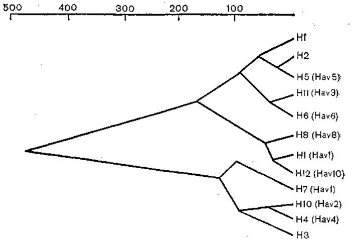
Fig. 27. Dendrogram of evolutionary relationships among type A influenza virus hemagglutinins. The abscissa represents relative values.
Consequently, it can be hypothesized that avian influenza viruses serve as the evolutionary source of human influenza viruses. In birds, influenza manifests as an intestinal infection; the virus replicates within the intestinal mucosa, frequently induces a septic process, and is transmitted via the alimentary route through cloacal contents. The circulation of avian influenza viruses is particularly intensive among migratory colonial species, where mutual exchange of diverse viruses fosters frequent and intensive recombination. Mammals may also become embroiled in this epizootic process. For instance, in 1980 along the Atlantic coast of North America, several hundred seal carcasses were found bearing an avian influenza virus that was typically of low virulence to birds. Could a similar event have occurred in 1918, when an animal-adapted recombinant of low virulence proved catastrophic to humans? And might the renowned English sweating sickness of the 16th century likewise have been an analogue of lethal seal influenza or human Spanish flu? Unfortunately, one can only speculate.
Domestic poultry are frequently drawn into the epizootic cycle of wild bird influenza. Particularly illustrative are studies of Peking ducks exported to Hong Kong from mainland China. Several hundred influenza A strains encompassing nearly all known influenza subtypes, alongside strains with previously undocumented antigenic formulas, were isolated.
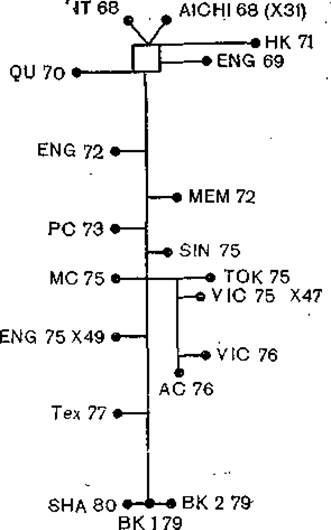
Fig. 28. Relationships among Hong Kong subtype strains based on minimum mutation distances between HA1 coding regions. Mutation counts are expressed relative to the NT68-BK179 distance (73 mutations).
It is reasonable to assume that human influenza began to establish itself as an anthroponosis with the formation of large settlements and The Development of migration patterns. Much like many other anthroponoses, the emergence of human influenza was accompanied by a shift in transmission mechanisms: influenza immediately established itself as an airborne-droplet infection. Its cradle was likely Southeast Asia, given that every recorded pandemic since 1889 originated in China. More than a third of humanity resides within this relatively confined geographic area, migratory routes of colonial waterbirds pass through Southeast Asia where they interact with domestic poultry and mammals, and finally, endemic protein malnutrition heightens human susceptibility to influenza viruses.
Having transitioned into an anthroponotic infection, human type A influenza did not completely sever ties with its natural reservoirs; every major influenza epidemic—particularly in the modern era, with a global human population exceeding 4 billion—is accompanied by a massive spillover of a subset of the viral population into domestic and wild animal populations. Here, the virus is "conserved" (antigenically preserved) due to the absence of herd immunity pressure, which fails to develop within heterogeneous animal biocenoses, while simultaneously undergoing constant recombination. Consequently, gene combinations may arise that enable the reintroduction of the virus back into the human population. This mechanism underlies antigenic shifts and the subsequent pandemics.
Regarding influenza B viruses, molecular-biological analyses indicate roughly 30% homology (Table 11) between its genome and that of influenza A virus [Kemdirimeda A., 1986]. Unlike the latter, influenza B...
Table 11. Homology among various genes of influenza A and B viruses
RNA segment number |
Protein |
Homology, % |
1; 2; 3 |
Polymerase (PB1) |
61 |
4 |
Hemagglutinin |
|
HA1 |
24 |
|
HA2 |
39 |
|
5 |
Nucleoprotein (NP) |
37 |
6 |
Neuraminidase (NA) |
35 |
7 |
Matrix |
|
M1 |
25 |
|
M2 |
14 |
|
8 |
Nonstructural |
|
NS1 |
9.7 |
|
NS2 |
16.2 |
its ability to infect animals decreased, and consequently, the virus lost the capacity for gene reassortment while retaining the potential for antigenic drift. This likely explains its more moderate spread and the absence of pandemics caused by this virus. It is difficult to draw definitive conclusions about the origin of influenza C virus, especially since its Classification within the family Orthomyxoviridae—which includes influenza A and B viruses—remains a subject of debate.
Modern healthcare is equipped with numerous means to combat influenza, including Vaccines, immunoglobulin preparations, chemotherapeutic agents (rimantadine, virazole), interferon and its Inducers, as well as Antibiotics used to treat and prevent bacterial complications. Nevertheless, The Use of all these measures does not provide radical Prevention of influenza, and the infection remains uncontrolled thus far. Two potential pathways can be envisioned for a radical solution to this problem: either the development of a universal anti-influenza vaccine, or the creation of a complex of chemotherapeutic agents that ensure not only effective Treatment but also the elimination of the infectious agent, and consequently, the eradication of the sources of infection. A universal influenza vaccine should contain not only drift variants of viruses that previously circulated among humans (H1N1, H2N2, and H3N2) but also determinants of known animal influenza viruses and potential future antigenic determinants. The scale of the upcoming work is immense and will take more than a decade to accomplish. Such a vaccine must be engineered using Recombinant DNA technology. An equally long path lies ahead before a universal set of chemotherapeutic agents is developed. However, another scenario is possible—an unexpected and unconventional solution to the problem might be found, much like what happened with poliomyelitis in the early 1950s, when the monolayer tissue culture technique was developed, making it possible to cultivate polioviruses and produce inactivated and live vaccines. Until that happens, an enormous amount of work must continue to find solutions through traditional approaches (vaccines, Chemotherapy). Neither administrative impatience nor voluntaristic leaps will help solve this formidable problem. A great deal of labor and patience lies ahead.
In conclusion, a few words regarding orthomyxoviruses isolated from ticks [Lvov D. K., 1982; Clerk J. et al., 1983]. Like influenza viruses, they possess 7–8 RNA segments, and their 3'-end features characteristic sequences UGGUUG UAUUGUUG; the total molecular mass of their genome is comparable to that of influenza viruses. Furthermore, even their proteins share a similar molecular weight. We currently know too little about them to fully understand their origin and relationship with classical orthomyxoviruses. The widespread occurrence of influenza viruses among warm-blooded animals, particularly birds, makes it entirely plausible that ticks are involved in the circulation of these viruses. Thogoto virus, isolated from ticks in Africa, Asia, and Europe, is similar to orthomyxoviruses. Its genome consists of 7 segments of single-stranded RNA. A similar virus has been isolated in Portugal; when administered to mice, it induces pulmonary lesions resembling those caused by influenza virus [Filipe A., 1986].
Last update: 13/08/2026
Editorial and Educational Adaptation: This material has been compiled based on the primary/original source text. The project team performed an editorial review, corrected technical inaccuracies, structured sections, and adapted the content for an educational format.
What was processed:
- elimination of formatting defects (OCR errors, structural breaks, corrupted characters);
- editorial organization of content;
- standardization of terminology in accordance with academic sources;
- verification of factual statements against the original source text.
All mentions of the author, publication year, and origin of the primary text have been preserved in accordance with the source.