Human Anatomy, Part 1 - K. A. Dyubenko, A. K. Kolomiysev, Yu. B. Chaykovsky 2002
General Part
Human Development and Body Structure
Initial Stages of Human Embryogenesis
Embryology (from the Greek embryon meaning embryo and logos meaning word or science) is the branch of science concerned with The Development of the embryo. It investigates the period of individual development that begins with Fertilization and culminates (in mammals and humans) in the birth of a fetus.
Embryology examines the morphological processes involved in the Transformation of a fertilized egg Cell (zygote) into a complex multicellular Organism comprising four basic tissue types, several dozen cell lineages, Organs, Organ Systems, apparatuses, and body parts.
Knowledge of embryology facilitates a deeper understanding of macroscopic anatomy. When aligned logically with embryological data, anatomical facts regarding the macro- and MICROSTRUCTURE OF THE organism confirm that structural relationships in the Postnatal period of development are the direct consequence of morphogenetic processes taking place within the embryo.
Embryology makes it possible to determine the morphological manifestations and successful realization of the genetic program encoded within the zygote, as well as any failures in this process. Such disruptions manifest as Structural and functional anomalies of embryonic development, which are frequently encountered in clinical practice and can only be fully explained through a solid understanding of embryology.
The process of Human embryonic development, much like that of other vertebrates, is divided into a series of stages characterized by specific Qualitative and quantitative features. The principal stages of Embryogenesis include: fertilization, Cleavage, Gastrulation, tissue formation (histogenesis), organ formation (Organogenesis), as well as the development of organ systems and apparatuses (systemogenesis). In some textbooks and manuals, progamete development—The formation of female and Male Germ Cells—is also classified as part of embryogenesis.
This section of the textbook focuses primarily on the Cytology/cytology/16.html">Early stages of human embryonic development, which begin with fertilization and conclude with the Formation of tissue primordia and extraembryonic organs. Histo-, organo-, and systemogenesis will be covered in the context of The Structure of the respective organs and systems.
Fertilization involves the fusion of male and Female Germ Cells (Fig. 1A), resulting in the formation of a single-celled embryo—the zygote (Fig. 1B)—which contains a diploid chromosome set and combines maternal and paternal heredity. Fertilization takes place in the fallopian tubes and typically lasts about one day.
The zygote begins to divide via mitosis. This process of division is known as cleavage, because as the zygote divides repeatedly, the resulting cells become progressively smaller. Consequently, the overall size of the multicellular embryo initially remains approximately the same as that of the zygote.
The cells produced during cleavage are called blastomeres (from the Greek blastos meaning germ or sprout, and meros meaning part). The cleavage period begins with the division of the zygote into two blastomeres and continues until the formation of a fluid-filled sphere (the blastula). In humans, this period commences roughly one day after fertilization and lasts for approximately 4–5 days.
One of the hallmark features of cleavage in mammalian and human zygotes is The Emergence of blastomeres that differ in size and rate of division. Located peripherally are smaller, lighter blastomeres (micromeres) that divide more rapidly and collectively form the trophoblast, or nutritive primordium (from the Greek trophos meaning feeder). Situated in the center of the embryo are larger, dark blastomeres (macromeres) with basophilic Cytoplasm; these divide more slowly and collectively give rise to the embryoblast, or embryonic primordium.
An embryo lacking an internal cavity is referred to as a morula (from the Latin morus meaning mulberry). Although this term is most characteristic of early chordate embryos (such as amphioxus, which genuinely resembles a mulberry during its initial developmental stages), it is also used to denote the early embryos of higher vertebrates that have not yet formed a cavity and appear as a compact cluster of cells (Fig. 1C).
Class="center">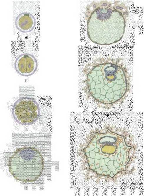
Fig. 1. Schematic representation of the early stages of human embryonic development (after B. M. Patten and B. M. Carlson):
A — fertilized egg cell at the stage of pronuclear approach; B — embryo at the two-blastomere stage; C — embryo at the morula stage; D — blastocyst prior to implantation; E — blastocyst at the stage of implantation (8-day embryo); F — chorion, amniotic, and yolk sacs within a 12-day embryo; G — amniotic and yolk sacs within a 13–14-day embryo
Subsequently, a small fluid-filled cavity appears within the embryo, and it assumes the form of a sphere known as a blastocyst (blastula). The wall of the blastocyst is formed by the trophoblast, while the embryoblast is located on its inner surface (Fig. 1D).
By the middle of the sixth day, the embryo consists of over a hundred cells. During this period, it resides within the Uterus, where implantation takes place (from the Latin implantatio meaning insertion or embedding), which involves the embryo embedding itself into the mucosal lining. The number of blastomeres within the embryo continues to increase during this process.
At the end of the first week of development, implantation (Fig. 1E) prompts the trophoblast to differentiate into a bilayered epithelium, the inner layer of which is called the cytotrophoblast. It is composed of cuboidal cells. Externally lies the plasmodiotrophoblast, also referred to as the syncytiotrophoblast, which is a symplast—a cytoplasmic mass containing numerous nuclei without distinct cell boundaries.
The embedding of the embryo into the uterine mucosa is driven by The activity of the trophoblast, which secretes histolytic Enzymes that cause the local destruction of the inner lining of the uterus—the endometrium. The embryo sinks deep into its tissue, and the endometrium closes over it. Thus, subsequent embryonic development proceeds entirely within the mucous membrane of the uterus.
The trophoblast ensures the Nutrition of the embryo during the early stages of its development, plays an active role in embryonic implantation, and contributes to the formation of a temporary organ—the chorion (Fig. 1E). By interacting with the uterine mucosa, the chorion forms the Placenta, an extraembryonic organ that connects the embryo to the maternal organism. The development, structure, and Functional Significance of the placenta are examined in courses on embryology and Histology.
The cellular elements of the embryoblast give rise to the embryo proper and other extraembryonic organs. A key feature of Human Development is the very early Formation of the extraembryonic mesoderm, which originates from the embryoblast. Cells delaminate from the embryoblast, forming bands that fill the blastocyst cavity and line the inner surface of the trophoblast. The extraembryonic mesoderm rapidly transforms into extraembryonic mesenchyme, resulting in an outer embryonic membrane composed of trophoblastic epithelium and mesenchyme, known as the chorion. As noted above, the chorion forms the embryonic portion of the placenta—the organ responsible for linking the embryo to the maternal organism.
Gastrulation then begins, which is divided into two phases. During the first phase, germ layers are formed, while the second phase establishes the axial complex—defining the general body plan of vertebrates and humans, as well as the embryonic primordium from which various tissues differentiate.
Germ layers are the primary sheets of cells formed during the initial phase of gastrulation. They differ in their spatial arrangement (the outer layer is the ectoderm, the inner is the entoderm/endoderm, and the middle is the mesoderm), cell Size and Structure, and the direction of their future development. The axial complex includes the neural tube, notochord (Spinal Cord precursor), dorsal mesoderm (located on either side of the neural tube and notochord), and the primitive gut. During the second phase of gastrulation, embryonic primordia also form—these are cell complexes derived from the germ layers that give rise to various tissues. Thus, diverse tissues can originate from the very same germ layer.
The morphological manifestation of The first phase of gastrulation, observed in a 7.5-day embryo, is the division of the embryoblast into two layers: an outer layer (epiblast) and an inner layer (hypoblast) (see Fig. 1E).
The epiblast contains the primordia for the ectoderm, embryonic mesoderm, notochord, and embryonic (intestinal) endoderm. The hypoblast serves as the primordium for the extraembryonic or yolk endoderm, representing that portion of the inner germ layer that will later contribute to the formation of a temporary organ—the yolk sac.
Endodermal cells that have separated from the epiblast begin to grow around the cavity formed in the underlying Regions of the extraembryonic mesoderm. Gradually, they take on a cup-like shape whose margins fuse, resulting in the formation of the yolk vesicle. The onset of yolk vesicle formation in this process is analogous to the development of this extraembryonic organ in mammals (see Fig. 1F).
Later, following the onset of implantation, a primary amniotic cavity forms within the epiblast as fluid accumulates and pushes the cells apart. Subsequently, the roof of this cavity (the amniotic vesicle) ruptures and is temporarily isolated from the outside by a patch of cytotrophoblast. Following this, the walls of the amniotic vesicle grow upward and fuse, causing the cavity of the amniotic vesicle to close again, surrounded by epiblast cells. This process is reminiscent of the formation of the so-called amniotic folds in birds and mammals.
From days 9 to 14 of development, the human embryo exhibits the following structure (Fig. 1E): the outer wall, formed by the chorion, consists of extraembryonic mesenchyme and trophoblastic epithelium. As noted previously, the latter includes the cytotrophoblast and syncytiotrophoblast. Within the thickened wall of the embryo, which faces deep into the uterine wall, the extraembryonic mesenchyme contains two communicating vesicles—the amniotic and yolk vesicles. The region of the amniotic vesicle adjacent to the yolk vesicle forms a thickening known as the embryonic disc, from which the body of the embryo develops. A strand of cells, the amniotic stalk, extends from the mesenchyme enclosing the two vesicles toward the chorion; its site of origin corresponds to the caudal end of the embryonic body.
Due to the processes described above, the embryo is provided with three extraembryonic organs—the chorion, which participates in placenta formation, the amnion, and the yolk sac (Fig. 2). The greater part of the amniotic vesicle forms an extraembryonic organ known as the amnion, one of the Functions of which is The production of Amniotic Fluid—an artificial aqueous environment for the developing embryo. The hypoblast gives rise to the yolk vesicle (sac), which in humans and most mammals contains no yolk, yet plays a vital role as the primary hematopoietic organ and participates in the development of primordial germ cells.
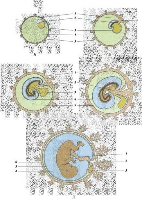
Fig. 2. Changes in the relationships and fetal membranes during early Selection/3.html">Stages of development (after B. M. Patten):
A and B: 1 — amniotic cavity; 2 - embryonic body; 3 - yolk sac; 4 - extraembryonic coelom; 5 - trophoderm; 6 - allantoic stalk; C and D: 1 - chorion; 2 - allantois; 3 - yolk sac; 4 - amnion; 5 - extraembryonic coelom; E: 1 - umbilical cord; 2 - umbilical vessels; 3 - yolk sac; 4 - amnion; 5 - extraembryonic coelom; 6 - chorion
Thus, during human embryonic development, the appearance of A number of extraembryonic organs (chorion, amnion, and yolk sac) precedes the formation of the embryonic body itself, which demonstrates the primary establishment of conditions necessary for the embryo's development.
On the 15th day of intrauterine development, the second phase of gastrulation begins, occurring in the same manner in both birds and placental mammals. The transition from the first phase of gastrulation to the second occurs gradually, combining the Features of the first (formation of germ layers) and the second (formation of the axial organ complex). The external sign of the second phase of gastrulation is the appearance at the caudal end of the embryonic disc of an elongated cell strip known as the primitive streak. At its anterior end, a small cellular elevation forms—the primitive or Hensen's node.
At the apex of the primitive node, a tiny depression arises—the primitive pit. In the central part of the primitive streak, a primitive groove appears as a continuation of the primitive pit. A portion of the outer germ layer material plunges inward through the primitive pit and becomes incorporated into the anterior region of the endoderm, forming the prechordal plate, which gives rise to the epithelial lining of the anterior digestive tract and the first two pairs of somites (segmented regions of the dorsal mesoderm).
The orientation of the primitive streak determines the positioning of the axial organs and, consequently, the general body plan of vertebrates. The diagram illustrating The Cell movements that lead to the formation of the primitive streak and its elongation is shown in Fig. 3A, while the primitive streak itself is depicted in Fig. 3B.
A portion of the epiblast cells migrates through the primitive streak and, embedding themselves in the underlying part of the hypoblast, form the primordium of the intestinal endoderm, which subsequently participates in gut development. The second part of the hypoblast, as mentioned above, goes on to form the yolk endoderm—the inner layer of the yolk sac.
As a result of cell migration from the epiblast through the primitive streak, the embryonic mesoderm also arises, expanding laterally beneath the epiblast. A diagram showing the migration of embryonic mesoderm cells in a cross-section of the embryo is presented in Fig. 3C.
The Water/144.html">Origin of the notochord is closely linked to the formation of the embryonic mesoderm. Its primordium forms as the chordal (or HEAD) process—a cord of cells derived from the embryonic mesoderm that grows forward from the primitive (Hensen's) node between the epiblast and hypoblast. The notochord determines the Location OF THE future Vertebral Column, as the vertebrae form around it, and significantly influences the development of The Nervous System. In higher mammals, remnants of the notochord persist postnatally as The Nucleus pulposus within the intervertebral discs.
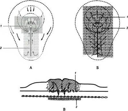
Fig. 3. Diagram of epiblast cell migration and formation of the primitive streak (A), mesoderm, and endoderm (B, C) (after A. G. Knorr):
A: 1 - Hensen's node; 2 - primitive streak; B: 1 - chordal process; 2 - Hensen's node; C: 1 - mesenchymal cells; 2 - intestinal endoderm
Following the formation of the notochord, the upper layer of Cells of the embryonic disc transforms into the ectoderm. Under the Influence of the notochord or chordamesoderm on the overlying ectoderm, the latter thickens and transforms into the neural plate, then into an elongated neural groove, and subsequently into the neural tube, which gives rise to both the central and peripheral nervous systems.
At the end of the second to the beginning of the third week of embryonic development, the fourth extraembryonic organ—the allantois—begins to form. Initially, it appears as a blind-ending outgrowth of the yolk sac that grows into the amniotic stalk. Later (after the formation of the gut), the allantois originates from the anterior wall of the hindgut. Following the formation of the neural tube, the mesodermal layers lying lateral to it and the notochord undergo segmentation. The segmented portions of the dorsal mesoderm are called somites. The lateral PARTS OF THE mesoderm, which later come to lie in the ventral part of the embryo and are therefore termed ventral mesoderm, split into two layers: an outer layer contacting the ectoderm (parietal layer) and an inner layer associated with the endoderm (visceral layer). The somites are connected to the ventral mesoderm by thin cellular strands called intermediate mesoderm. Subsequently, the somites divide into three parts: dorsolateral (dermotome), medioventral (sclerotome), and intermediate (myotome). The appearance of somites marks the beginning of the somite period of embryonic development. On the 21st day, the embryo has 2–3 pairs of somites; on the 23rd day, 10 pairs; on the 25th day, 14 pairs; on the 27th day, 25 pairs; and by the end of the 5th week, 43–44 pairs.
The spaces between the germ layers and main organs are filled with embryonic Connective Tissue—mesenchyme, which consists of spindle-shaped or stellate cells that contact one another via cellular processes and are embedded in an amorphous Extracellular matrix. Mesenchyme is formed mainly from the mesoderm, although other germ layers, including the ectoderm, also contribute to its development (M. F. Kashchenko). The main derivatives of the germ layers and mesenchyme are presented in Table 1. (More detailed information can be found in histology textbooks.)
Table 1. Main derivatives of the germ layers and mesenchyme
|
Germ layer |
Embryonic primordium |
Tissue derivatives |
|
Ectoderm |
Neuroectoderm |
Nervous Tissue of the central and peripheral nervous systems and Sensory Organs |
|
Surface ectoderm |
Epithelium of the Skin and its Appendages (hair, sebaceous and Sweat Glands) |
|
|
Prechordal plate |
Epithelium of: - Oral Cavity and Esophagus - respiratory organs |
|
|
Endoderm |
Yolk |
Epithelium of the yolk sac |
|
Intestinal |
Epithelium of: |
|
|
- Stomach and intestines - gastric and intestinal glands - Pancreas - Liver |
||
|
Mesoderm |
Somites |
Dermatome - connective tissue of the skin Sclerotome - cartilaginous and osseous tissue of the Axial Skeleton |
|
Mesenchyme |
Nephrotome |
Epithelium of: - Kidneys - urinary tracts - reproductive tracts |
|
Splanchnotome |
Epithelium of serous membranes (mesothelium) All types of connective tissue Non-Striated muscle tissue of mesenchymal origin |
When the visceral layer of the ventral mesoderm comes into contact with the intestinal endoderm, the body of the embryo begins to separate from the extraembryonic organs via body folds. This process starts with the ventral bending of the embryo's margins, resulting in a dorsal convexity. Subsequently, the curved margins of the embryo in the head and caudal regions begin to deepen. The expansion of these delimiting folds leads to the formation of the lateral and ventral walls of the embryo, as well as the emergence of the so-called vitelline stalk, which connects it to the yolk sac. As the body folds deepen, the intestinal endoderm and the visceral layer of the ventral mesoderm roll into a tube, leading to the formation of the gut. Meanwhile, just as the latter incorporates the intestinal endoderm, the yolk sac contains the yolk endoderm. The allantois, which previously appeared as an outpocketing of the yolk sac, now originates from the hindgut.
As the amniotic membrane expands, the volume of the amnion increases, approaching and fusing with the chorion. The amniotic stalk thereby shifts from the caudal region to the ventral wall of the embryo and joins with the amnion, forming the umbilical cord. The allantois, which previously penetrated the amniotic stalk, is partially incorporated into the umbilical cord. In the course of these transformations, the yolk sac moves away from the embryo, while its connection to the gut is maintained by the tubular vitellointestinal duct. The latter, along with the allantois, is also located within the umbilical cord. Subsequently, the allantois and the vitellointestinal duct, which form part of the umbilical cord, undergo regression.
Last update: 08/08/2026
Editorial and Educational Adaptation: This material has been compiled based on the primary/original source text. The project team performed an editorial review, corrected technical inaccuracies, structured sections, and adapted the content for an educational format.
What was processed:
- elimination of formatting defects (OCR errors, structural breaks, corrupted characters);
- editorial organization of content;
- standardization of terminology in accordance with academic sources;
- verification of factual statements against the original source text.
All mentions of the author, publication year, and origin of the primary text have been preserved in accordance with the source.