CYTOLOGY, HISTOLOGY, EMBRYOLOGY - 2008
Chapter 4. SPECIAL HISTOLOGY
4.6 Skin and Its Appendages
The Skin (from Lat. cutis) plays a vital role in maintaining the essential Functions of the animal body. It protects the Organism against mechanical, physical, and chemical damage. The epidermis, particularly its cornified layer, acts as a barrier against pathogenic microorganisms. Furthermore, the skin performs excretory and thermoregulatory functions, participates in Water-Salt METABOLISM, stores Blood, water, and other substances, synthesizes vitamin D, and serves as a major sensory receptor field.
The Structure of mammalian skin exhibits species-specific features and specialized formations such as hair, glands, hooves, and claws, whereas in birds, it forms combs, feathers, and other structures.
The skin develops from two embryonic germ layers: the epidermis originates from the embryonic ectoderm, while the dermis (corium) and subcutaneous layer are formed by mesenchyme derived from dermatomes.
STRUCTURE OF THE Skin. The skin consists of three main layers: the epidermis, the dermis, and the subcutaneous adipose tissue.
The epidermis is a Cytology/practical/35.html">Stratified squamous keratinized epithelium. Its thickness and cellular composition vary depending on the animal's species and breed, as well as other factors. In general, the epidermis is thicker in areas with less hair coverage.
The epidermis comprises five layers of epidermal Cells (keratinocytes): the stratum basale, stratum spinosum, stratum granulosum, stratum lucidum, and stratum corneum.
The stratum basale is the deepest layer, resting on the basement membrane, and is referred to as the germinative (cambial) layer. Here, new cells are produced via mitosis and migrate into the overlying stratum spinosum. The basal cells are nearly cylindrical in shape with basophilic Cytoplasm containing Ribosomes, polyribosomes, and other Organelles. The boundary between the basal epidermal cells and the papillary layer of the dermis is irregular, which increases their contact surface area. The epidermal Cells of the basal layer and the overlying stratum spinosum are interconnected by desmosomes and attached to the basement membrane by hemidesmosomes. Interspersed among the basal cells are another type of dendritic cells known as melanocytes. Originating from the neural crest, they produce the pigment melanin.
The stratum spinosum (prickle Cell layer) consists of several rows of polyhedral cells. Numerous spine-like processes of adjacent cells are interconnected by desmosomes, with narrow intercellular clefts separating The Cell boundaries. The cytoplasm of the cells in both the basal and spinosum layers contains numerous tonofibrils that extend into the intercellular spaces. Alongside spinate keratinocytes, Langerhans cells are present, characterized by 2-5 dendritic processes, a lobulated Nucleus, and pale cytoplasm containing rod-shaped structures and granules. Langerhans cells are considered to be epidermal macrophages.
The stratum granulosum is formed by 2-4 tightly packed layers of cells; their cytoplasm contains keratohyalin granules and keratosomes (spherical lamellar granules). Keratosomes contain Lipids and hydrolytic Enzymes. The lipids penetrate the intercellular space, thereby providing a barrier against transepidermal water loss to the body surface.
The stratum lucidum is characterized by a small thickness and a homogeneous appearance. The cytoplasm of cells in this layer contains eleidin, a transformation product of the keratohyalin found in the granular layer. In the stratum lucidum, cell boundaries are indistinct, and nuclei are absent.
The stratum corneum is formed by the layers of underlying cells that have completed The process of keratinization, which is associated with cell death and accompanied by the periodic shedding of this layer. The outermost cells gradually slough off and are replaced by new ones originating from deeper layers. On the epidermal surface, each keratinocyte transforms into a single scale that gradually desquamates. The cytoplasm of these scales is bounded by a cornified envelope and contains keratin fibrils. The detachment of the scales is believed to be influenced by keratosomes—modified Lysosomes of the superficial epidermal cells.
The dermis (corium). It is subdivided into the papillary and reticular layers.
The papillary layer lies directly beneath the epidermis, separated from it by the basement membrane. It is composed of loose Connective Tissue, which forms papillae extending into the epidermal ridges. The papillary layer contains a significant number of cells—predominantly fibroblasts, macrophages, mast cells, and bundles of smooth Muscle cells that are occasionally associated with hair follicles. This layer is rich in a branching microcirculatory blood vessel network, nerve fibers, and nerve endings. The reticular layer is formed by dense irregular connective tissue arranged in bundles that run in various directions, interweaving to form a meshwork. The thickness of these bundles varies depending on the body region, skin thickness, as well as the animal's species, breed, sex, and age. Interspersed between individual Collagen fiber bundles are layers of loose connective tissue containing various cells, Blood Vessels, and nerves. The hair roots, Sebaceous Glands, and Sweat Glands are located within the reticular layer of the skin.
The subcutaneous layer (hypodermis) consists of loose connective tissue. It connects the skin to deeper Tissues and Organs, allows for its mobility relative to underlying body parts, serves as a fat reservoir, and cushions the skin against environmental impacts. Fat accumulation in the subcutaneous layer limits heat loss. In addition to adipocytes, the subcutaneous layer contains fibroblasts. This layer is absent in the eyelids, Lips, nasal plane (speculum), and certain other areas of the body.
The blood supply provides nourishment to the Skin and Its Appendages and participates in thermoregulation. Arterial vessels penetrate the skin from the suprafascial arterial network, which forms a deep arterial plexus at the level of the lower boundary of the reticular layer. A dense arterial network is formed at the level of the secretory units of the sebaceous glands, from which vessels penetrate the papillary layer to establish a microcirculatory bed reaching the epidermis.
Innervation of the skin. As an Organ of Touch, the skin contains a vast network of nerve fibers that converge into a wide-meshed subcutaneous plexus. This plexus gives rise to another nerve network within the thickness of the dermis. The densest nerve plexus is located in the papillary layer, and individual nerve fibers reach as far as the epidermis. Encapsulated corpuscles are also situated in the subcutaneous layer.
By origin, these nerves are spinal and autonomic, and functionally they are classified into receptors (which transmit impulses to the Central Nervous system), secretory nerves (which efferently innervate Skin glands), vasomotor nerves (which supply blood vessels), and motor nerves associated with arrector pili Muscles.
Skin glands. It should be noted that the topography and structure of skin glands vary among different mammals.
Sebaceous glands. Their secretion, sebum, is used to lubricate the skin and hair, rendering them elastic. Sebaceous glands are located in the reticular layer of the skin adjacent to the papillary layer.
Sebaceous glands are invariably situated close to the hair follicles, between their roots and the arrector pili muscle. These are simple alveolar glands; in horses and dogs, they are branched, alveolar-tubular glands. The excretory duct is short, lined with Stratified Epithelium, and opens into the hair canal (infundibulum). The secretory units are formed by a single layer of
squamous epithelium, interspersed with unspecialized, mitotic cells located near the basement membrane. Closer to the center of the secretory unit, larger cells appear, and their cytoplasm contains small lipid droplets. Gradually, the process of lipid synthesis intensifies within them; simultaneously, the cells increase in size, migrate toward the excretory duct, and transform into sebum. The secretion of sebaceous glands occurs via a holocrine mechanism (Fig. 124).
Class="center">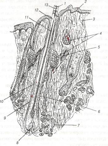
Fig. 124. Structure of horse skin.
1-hair shaft; 2-epidermis; 3-dermis (corium); 4-sebaceous gland; 5-arrector pili muscle; 6-sweat gland duct;
7-secretory portion of the sweat gland; 8-hair papilla; 9-ROOT sheath; 10-connective tissue hair follicle; 11-medulla of the hair; 12-cortex of the hair; 13-cuticle of the hair.
Sweat glands. Structurally, they are simple tubular glands; their secretory portions are located within the reticular layer of the dermis, significantly deeper than the sebaceous glands, and end blindly.
In horses, dogs, pigs, and to a lesser extent in other animal species, these blind tubules coil into glomeruli.
The walls of the sweat glands consist of a single layer of cuboidal epithelium, surrounded externally by myoepithelial cells of ectodermal origin. The secretory portion gradually transitions into an excretory duct, and the wall into a bi-layered epithelium; the excretory duct opens into the hair funnel above the duct of the sebaceous glands. These glands produce secretion via an apocrine mechanism—in the form of cytoplasm from the glandular cells; it is more concentrated and has a specific odor.
In cattle, specialized sweat gland-like glands are found in the skin of the nasolabial plane; in pigs, in the skin of the snout; and in rabbits, in the chin. They produce a serous secretion.
In carnivores, the anal sac contains tubular glands. In horses, simple or branched tubular glands, similar to the glands of carnivore footpads, are located on the frog. Branched sebaceous and tubular glands that produce a yellowish-brown secretion are situated in the skin of the inguinal pouches of sheep, while sebaceous and sweat glands are found in the wall of their interdigital pouch. Branched alveolar cornified glands in goats are located at the Base of the horns.
Last update: 07/08/2026
Editorial and Educational Adaptation: This material has been compiled based on the primary/original source text. The project team performed an editorial review, corrected technical inaccuracies, structured sections, and adapted the content for an educational format.
What was processed:
- elimination of formatting defects (OCR errors, structural breaks, corrupted characters);
- editorial organization of content;
- standardization of terminology in accordance with academic sources;
- verification of factual statements against the original source text.
All mentions of the author, publication year, and origin of the primary text have been preserved in accordance with the source.