Cytology, General Histology and Embryology - V. K. Napkhanyuk 2002
Tissues
Epithelial Tissues
Stratified Epithelium
Stratified epithelia belong to covering epithelia that form the epidermal layer of the Skin, line the Oral Cavity, Esophagus, rectum, and Urinary Organs.
Depending on their morphofunctional characteristics, stratified epithelia are divided into several groups:
— stratified non-keratinized epithelium — squamous, cuboidal, columnar;
— stratified keratinized squamous epithelium;
— transitional epithelium.
Stratified non-keratinized squamous epithelium (epithelium stratificatum squamosum noncornificatum) covers the outer surface of the cornea and lines the Oral Cavity and esophagus.
Stratified non-keratinized squamous epithelium consists of three layers: basal, spinous (intermediate), and squamous (superficial).
The basal layer is formed by columnar epithelial Cells resting on the basement membrane. These cells include stem cells capable of mitotic division.
The spinous layer is formed by irregularly shaped polygonal cells that possess processes extending between the apical PARTS OF THE basal layer cells.
The layer of squamous cells is located superficially and consists of dying cells.
Stratified keratinized squamous epithelium (epithelium stratificatum squamosum cornificatum) covers the skin surface, forming its epidermis, where epithelial cells undergo transformation into cornified scales, a process known as keratinization. Keratinization is accompanied by the synthesis and accumulation of a specific protein, keratin.
The epidermis of the skin on the fingers, palms, and soles consists of 5 layers:
— the basal (germinal, cambial) layer, formed by cylindrical epithelial cells whose Cytoplasm is capable of synthesizing specific Proteins that form tonofilaments. This layer contains stem cells capable of division and, through The process of differentiation, migrating to the upper layers;
— the spinous layer, formed by polygonal cells interconnected by numerous desmosomes. At the sites of desmosomes, small spike-like projections (spines) form on The Cell surface;
— the granular layer, formed by flattened cells whose cytoplasm contains tonofibrils and keratohyalin granules;
— the clear (lucid) layer, formed by flat cells whose cytoplasm contains eleidin (a complex of keratohyalin and tonofibrils). Eleidin strongly refracts light;
— the cornified layer, formed by cornified scales filled with keratin and air bubbles.
Transitional epithelium (epithelium transitionale) lines the Urinary Tract (renal calyces, Ureters, Urinary Bladder), the walls of which stretch significantly when filled with urine. Transitional epithelium consists of several cell layers:
— the basal layer, formed by small, rounded (dark) cells;
— the intermediate layer, formed by polygonal cells;
— the superficial layer, formed by very large cells, often with two or three nuclei, dome-shaped or flattened depending on the state of the wall.
Slides for study
Slide 11. Stratified non-keratinized squamous epithelium of the cornea (Fig. 37).
Class="center">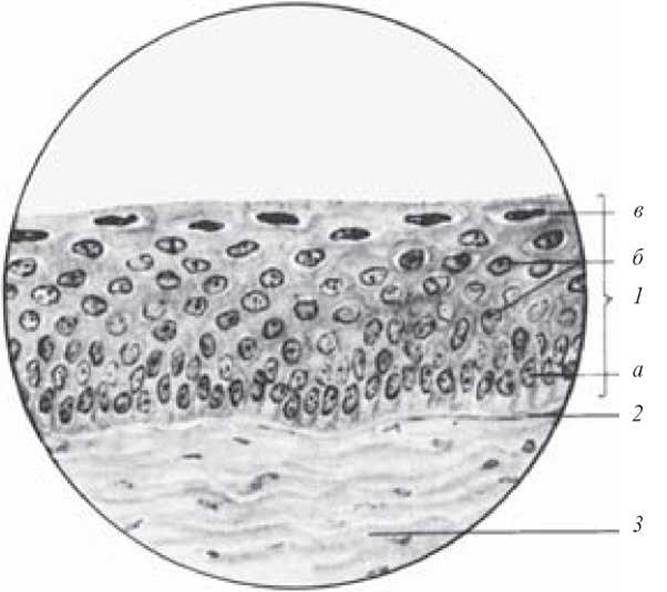
Fig. 37. Stratified non-keratinized squamous epithelium of the cornea. Hematoxylin and eosin staining. x 800:
1 — epithelium (a — basal layer; b — spinous layer; c — squamous layer); 2 — basement membrane; 3 — Connective Tissue
Low magnification. Examine the slide. Under this magnification, locate the stratified epithelium on the outer surface of the cornea.
High magnification. The basement membrane is clearly visible, supporting a single layer of low prismatic cells known as the basal layer. The nuclei of the basal cells are oval and oriented vertically. Above the basal layer lie several tiers of irregularly shaped cells with cytoplasmic projections, forming the spinous layer. The nuclei of these cells are rounded. The outermost region consists of several layers of cells forming the superficial squamous layer. Their nuclei are flattened and aligned parallel to the epithelial surface. Draw the specimen.
Label on the drawing: 1) epithelium: a — basal layer; b — spinous layer; c — squamous layer; 2) basement membrane; 3) connective tissue.
Slide 12. Stratified keratinized squamous epithelium of the finger skin (Fig. 38).
Low magnification. Examine the slide. Under this magnification, locate the epidermis of the finger skin.
High magnification. The basal layer is clearly visible, formed by cells resting on the basement membrane; above it lies the spinous layer, whose cells feature small cytoplasmic projections that interconnect them. The granular layer stands out by its dark staining; its cells are flattened and contain keratohyalin granules in the cytoplasm that stain dark purple. The lucid layer appears pale and homogeneous on the slide. The outermost layer is the cornified (horny) layer, represented by dying cells (cornified scales). Draw the specimen.
Label on the drawing: 1) epithelium: a — basal layer; b — spinous layer; c — granular layer; d — lucid layer; e — cornified layer; 2) connective tissue.
Slide 13. Transitional epithelium of the urinary bladder (Fig. 39).
Low magnification. Examine the slide. Under this magnification, locate the transitional epithelium on the inner surface of the urinary bladder.
High magnification. Small basal cells are clearly visible, some of which lie directly on the basement membrane to form the basal layer, while others are displaced toward the rows above to form the intermediate layer. The superficial layer consists of large, pear-shaped cells with intensely pink cytoplasm. Draw the specimen.
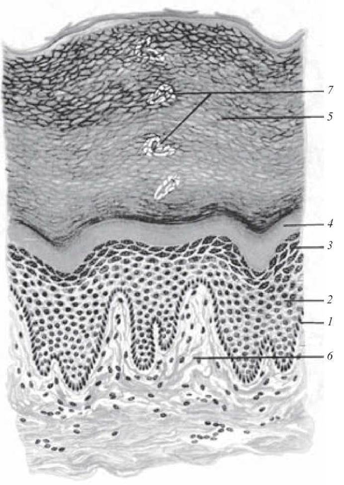
Fig. 38. Stratified keratinized squamous epithelium of the finger skin. Hematoxylin and eosin stain. × 600:
1 — basal layer; 2 — spinous layer; 3 — granular layer; 4 — lucid layer; 5 — cornified layer; 6 — connective tissue; 7 — sweat gland excretory duct
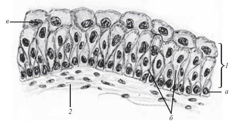
Fig. 39. Transitional epithelium of the urinary bladder. Hematoxylin and eosin stain. × 600:
1 — epithelium (a — Cells of the basal layer; b — cells of the intermediate layer; c — cells of the superficial layer); 2 — connective tissue
Label on the drawing: 1) epithelium: a — cells of the basal layer; b — cells of the intermediate layer; c — cells of the superficial layer; 2) connective tissue; 3) Blood vessel.
Last update: 10/08/2026
Editorial and Educational Adaptation: This material has been compiled based on the primary/original source text. The project team performed an editorial review, corrected technical inaccuracies, structured sections, and adapted the content for an educational format.
What was processed:
- elimination of formatting defects (OCR errors, structural breaks, corrupted characters);
- editorial organization of content;
- standardization of terminology in accordance with academic sources;
- verification of factual statements against the original source text.
All mentions of the author, publication year, and origin of the primary text have been preserved in accordance with the source.