Human Anatomy and Physiology (with Age-Related Features of the Child's Body) - Sapin, M. R., & Sivoglazov, V. I. 2002
The Doctrine of the Viscera (Splanchnology)
Digestive System
Oral Cavity
The oral cavity is divided into two sections: the oral vestibule and the oral cavity proper. The oral vestibule is bounded by the Lips and Cheeks on the outside, and by the Teeth and Gums on the inside. The vestibule opens to the exterior via the oral fissure. The oral fissure is bounded by the lips, which are covered externally by Skin and lined internally by a mucous membrane. Therefore, three parts are distinguished in the lips: the external surface (cutaneous part), the internal surface (mucous part), and the transitional zone, which is covered by a thin layer of keratinized stratified squamous epithelium and lacks mucous glands and Hair.
The oral cavity proper is located internal to the teeth and gums and communicates with the vestibule through the spaces between the dental crowns and the gap between the third molar and the anterior margin of the mandibular ramus. The upper wall of the oral cavity is formed by the hard palate and the soft palate, both covered with a mucous membrane. The soft palate, or palatine velum, adjoins the hard palate posteriorly and terminates in the uvula. Laterally and inferiorly, the palatine velum continues into two pairs of arches (the posterior palatopharyngeal arch and the anterior palatopharyngeal arch), between which the paired palatine tonsil is located. The floor of the oral cavity is formed by the oral Diaphragm, consisting of the paired mylohyoid Muscle, upon which the Tongue rests. Extending onto the Inferior surface of the tongue, the mucous membrane forms its frenulum. On both sides of the frenulum, at the apex of the sublingual caruncles, the ducts of the Salivary Glands open.
Class="center">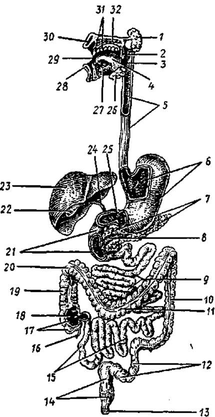
Fig. 39. Diagram of The Structure of the Digestive System:
1 — Cytology/practical/97.html">Parotid salivary gland, 2 — soft palate, 3 — Pharynx, 4 — tongue, 5 — Esophagus, 6 — Stomach, 7 — Pancreas, 8 — pancreatic duct, 9 — jejunum, 10 — descending colon, 11 — transverse colon, 12 — sigmoid colon, 13 — external anal sphincter, 14 — rectum, 15 — ileum, 16 — vermiform Appendix, 17 — cecum, 18 — ileocecal valve, 19 — ascending colon, 20 — right (hepatic) colic flexure, 21 — duodenum, 22 — Gallbladder, 23 — Liver, 24 — common Bile duct, 25 — pyloric sphincter of The Stomach, 26 — submandibular gland, 27 — sublingual gland, 28 — lower lip, 29 — oral cavity, 30 — upper lip, 31 — teeth, 32 — hard palate
The oral cavity communicates with the pharyngeal cavity via the fauces, which are bounded superiorly by the soft palate, laterally by the palatine arches, and inferiorly by the ROOT of the tongue.
In a newborn infant, the oral cavity is small in size, and the vestibule is separated from the oral cavity proper by the gingival margin. The lips are thick, and the transitional zone is narrow. The cheeks are rounded, with a well-developed fat pad. After four years of age, part of this fat pad atrophies, and its posterior portion extends behind the masseter muscle. The hard palate is flattened, and its mucous membrane is sparse in glands. The soft palate is relatively wide and short, lying almost horizontally. However, it does not reach the posterior wall of the pharynx, which ensures unobstructed breathing during suckling.
Tongue
The tongue is formed by Striated Muscle tissue covered with a mucous membrane. It participates in suckling, swallowing, and speech articulation, and also serves as The ORGAN OF TASTE. The Role of the tongue in a newborn and infant during maternal suckling is exceptionally vital. Consequently, the tongue of a newborn and infant is relatively thicker and wider.
The lateral margins of the tongue bound it anteriorly at the apex and posteriorly at the root. The body of the tongue lies between the apex and the root. Its upper surface is referred to as the dorsum of the tongue.
The mucous membrane of the tongue is covered with non-keratinized stratified squamous epithelium. The mucous membrane of the dorsum and margins of the tongue forms numerous papillae: filiform, fungiform, vallate (surrounded by a trench), and foliate papillae. Filiform papillae are the most numerous, giving the dorsum of the tongue a velvety appearance. These papillae are about 0.3 mm long and contain nerve endings that perceive Touch sensations.
Fungiform papillae are fewer in number than filiform ones; their length is 0.7–1.8 mm and their diameter is 0.4–1 mm. Vallate papillae, numbering 7–12 with a diameter of 2–3 mm, are located at the border between the dorsum and the root of the tongue. Each papilla is surrounded by a narrow, deep trench, which in turn is bounded externally by a mucosal fold. Taste buds—groups of specialized receptor taste Cells—are located within the epithelium On the surface of the fungiform and vallate papillae. Taste buds are also present in the foliate papillae situated on the lateral surfaces of the tongue.
There are no papillae on the mucous membrane of the root of the tongue; its surface is uneven due to accumulations of lymphoid tissue in its lamina propria, which form the lingual tonsil.
The Muscles of the tongue are divided into two groups: extrinsic and intrinsic. Extrinsic muscles of the tongue (genioglossus, hyoglossus, and styloglossus) originate on the cranial bones and insert into the tongue. These muscles perform the Movements of the tongue. Intrinsic muscles are not attached to bones and alter the shape of the tongue.
The intrinsic muscles of the tongue consist of bundles of longitudinal, transverse, and vertical fibers that interweave with each other and with the fibers of the extrinsic muscles. All muscles of the tongue are innervated by fibers of the Hypoglossal nerve (XII pair of Cranial Nerves).
Teeth
Humans have two successively replacing sets of teeth: deciduous (temporary) and permanent. The teeth are lodged in dental alveoli.
An adult has 32 permanent teeth, whereas a child has 20 deciduous teeth. Each tooth comprises a crown, a neck, and a root (Fig. 40). The crown projects above the gingiva (gum). The neck lies at the border between the root and the crown, where the mucous membrane of the gums contacts the tooth. The root is located within the alveolus and terminates in an apex, which features a small aperture through which Blood Vessels and nerves enter the tooth. Inside the tooth is a cavity filled with dental pulp, which is rich in vessels and nerves. Each tooth has one (incisors, canines), two, or three roots (molars). The roots of the teeth are firmly fused to The surface of the dental alveoli via the periodontium.
Teeth are constructed primarily of dentin, which is covered by enamel in the region of the crown and by cementum in the region of the root. Enamel consists mainly of inorganic salts (96–97%), among which calcium phosphate and calcium carbonate predominate, along with about 4% calcium fluoride. Dentin contains approximately 28% organic matter (predominantly Collagen) and 72% inorganic matter (calcium phosphate, magnesium, calcium fluoride).
In its composition, cementum approximates bone, containing 29.6% organic matter and 70.4% inorganic matter (primarily calcium phosphate and calcium carbonate).
Based on the shape of the crown, the following tooth types are distinguished: incisors, canines, premolars, and molars. Incisors have a chisel-shaped crown and a single root. Canines feature a crown with two cutting edges and a tubercle on the lingual surface, as well as a single root. Premolars are located posterior to the canines; their crowns have cusps on the masticatory surface, and they have a single root. Molars possess a cuboidal crown with several cusps on the masticatory surface, and two or three roots. The occlusion of the teeth is termed the bite (occlusion), during which the upper and lower teeth firmly contact each other, with the upper incisors typically overlapping the lower incisors.
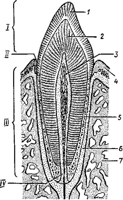
Fig. 40. Diagram of tooth structure:
1 — enamel, 2 — dentin, 3 — dental pulp, 4 — gingiva, 5 — cementum, 6 — periodontium, 7 — bone. I — clinical crown of the tooth, II — neck of the tooth, III — root of the tooth, IV — root canal
The number of teeth is conventionally designated by a dental formula, which is expressed as a fraction. In the numerator, the first digit indicates the number of incisors, the second represents canines, the third denotes premolars, and the fourth stands for molars on one side of the upper jaw, while the denominator indicates the corresponding teeth on the lower jaw. An adult has 32 teeth, and the dental formula is represented as follows:

Eruption of the deciduous teeth begins at 6—7 months of age. The medial lower incisors erupt first. The eruption of deciduous teeth is completed by the beginning of the child's 3rd year of life. There are 20 deciduous teeth, and their dental formula is as follows:

These numbers also indicate the number of deciduous teeth in each half of the jaw: two incisors, one canine, and two molars. Among the permanent teeth, the lower teeth erupt earliest—the first molar and the medial incisor. The timing of eruption for deciduous and permanent teeth is presented in Table 7.
Table 7 Average timing of tooth eruption
|
Tooth name |
Deciduous teeth, months |
Permanent teeth, years |
|
Medial incisor |
6—8 |
6-8 |
|
Lateral incisor |
7-9 |
7-9 |
|
Canine |
16-20 |
9-12 |
|
First premolar |
10—12 |
|
|
Second premolar |
— |
10-12 |
|
First molar |
12-15 |
6-7 |
|
Second molar |
20-24 |
11-13 |
|
Third molar |
12-26 |
Salivary glands
Minor glands (labial, buccal, lingual, and palatine) are located in the mucosa, submucosa, and within the buccinator muscle. The ducts of three pairs of major salivary glands—the parotid, submandibular, and sublingual glands—also open into the oral cavity. The parotid glands, lingual glands, and the Glands of the vallate papillae secrete a protein (serous) fluid. The palatine and posterior lingual glands secrete mucus. The submandibular, sublingual, labial, buccal, and anterior lingual glands produce a mixed secretion (both serous and mucous).
The parotid gland weighs 20—30 g and is covered by a well-defined Connective Tissue capsule. It is located on the lateral surface of the face, anterior and inferior to the auricle; posteriorly, it extends into the retromandibular fossa, while anteriorly it partially overlaps the masseter muscle. The excretory duct of the gland pierces the buccinator muscle and opens onto the lateral wall of the oral vestibule at the level of the second upper molar.
The submandibular gland, weighing 13—16 g, is located quite superficially within the submandibular triangle. The gland is enclosed in a Dense connective tissue capsule, and its excretory duct opens on a papilla beside the lingual frenulum.
The sublingual gland, weighing about 5 g, is narrow, elongated, and situated on the upper surface of the oral diaphragm; its capsule is poorly developed. The gland has a main duct (the major sublingual duct) that opens via a common orifice with the duct of the submandibular gland, as well as several minor ducts that open along the sublingual fold.
Last update: 10/08/2026
Editorial and Educational Adaptation: This material has been compiled based on the primary/original source text. The project team performed an editorial review, corrected technical inaccuracies, structured sections, and adapted the content for an educational format.
What was processed:
- elimination of formatting defects (OCR errors, structural breaks, corrupted characters);
- editorial organization of content;
- standardization of terminology in accordance with academic sources;
- verification of factual statements against the original source text.
All mentions of the author, publication year, and origin of the primary text have been preserved in accordance with the source.