Anatomy of the Vessels and Nerves of the Head and Neck (Angioneurology) - Chornokulskyi S.T. 2009
Nerves of the Head and Neck
Hypoglossal nerve
[XII] (n.hypoglossus) (Fig. 47)
It has 1 Nucleus — the hypoglossal nucleus (nuc. n. hypoglossi). The axons of this nucleus emerge On the surface of the Medulla Oblongata between the olive and the pyramid as 10–15 rootlets. Merging into a single trunk, the nerve leaves the cranial cavity through the hypoglossal canal and, arching downwards, courses towards the submandibular triangle. Here, it forms the upper boundary of Pirogov's triangle and penetrates the ROOT of the Tongue, giving off lingual branches (rr. linguales) that innervate all the Muscles of the tongue. The hypoglossal nerve has communicating branches with the V, VII, and X Cranial Nerves and innervates the orbicularis oris Muscle (via anastomosis with the VII cranial nerve).
*The issue has not yet been fully investigated.
Class="center">
Fig. 46. Accessory nerve (schematic):
A — nuclei and rootlets of the XI cranial nerve:
1 - ambiguous nucleus (nuc. ambiguus);
2 - portion of the ambiguous nucleus belonging to n. accessorius;
3 - Nucleus of the accessory nerve (nuc. n. accessorii);
4 - spinal root (radix spinalis);
5 - cranial root (radix cranialis);
6 - Vagus nerve (n. vagus);
B — trunk and external branch of the XI cranial nerve:
1 - intracranial part of the XI cranial nerve;
2 - external branch of the accessory nerve (r. externus n. accessorii)
Fibers from the anterior rami of the first two cervical Spinal Nerves (C1 and C2) join the hypoglossal nerve and branch off within its arch as the superior root (radix superior).
Fibers from the anterior rami of the second and third cervical spinal nerves form the inferior root (radix inferior), which joins the superior root to form the cervical loop (ansa cervicalis). The cervical loop innervates all Infrahyoid muscles and the geniohyoid muscle (m. geniohyoideus) (see Fig. 47).
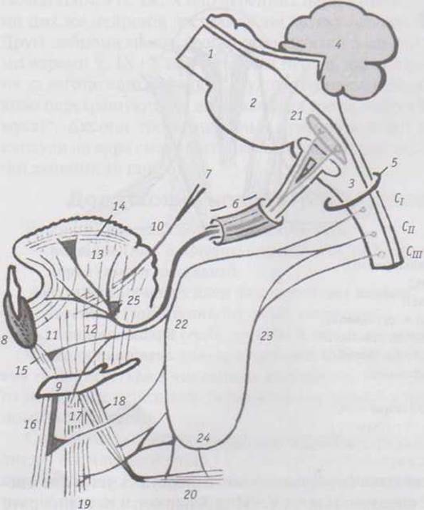
Fig. 47. Hypoglossal nerve (schematic):
1 - Midbrain (mesencephalon);
2 - Pons;
3 - medulla oblongata;
4 - Cerebellum;
5 - foramen magnum;
6 - hypoglossal canal (canalis n. hypoglossi);
7 - styloid process (processus styloideus);
8 - section of the Mandible;
9 - Hyoid bone (os hyoideum);
10 - styloglossus muscle (m. styloglossus);
11 - genioglossus muscle (m. genioglossus);
12 - hyoglossus muscle (m. hyoglossus);
13 - vertical muscle of tongue (m. verticalis linguae);
14 - superior longitudinal muscle (m. longitudinalis superior);
15 - geniohyoid muscle (m. geniohyoideus);
16 - sternohyoid muscle (m. sternohyoideus);
17 - thyrohyoid muscle (m. thyreohyoideus);
18 - superior belly of omohyoid muscle (venter superior m. omohyoidei);
19 - sternothyroid muscle (m. sternothyreoideus);
20 - inferior belly of omohyoid muscle (venter inferior m. omohyoidei);
21 - nucleus of hypoglossal nerve (nuc. n. hypoglossi);
22 - superior root of cervical loop (radix superior ansae cervicalis);
23 - inferior root of cervical loop (radix inferior ansae cervicalis);
24 - cervical loop (ansa cervicalis);
25 - lingual branches of hypoglossal nerve (rr. linguales n. hypoglossi)
CI, CII, CIII — upper cervical segments of the Spinal Cord
Last update: 11/08/2026
Editorial and Educational Adaptation: This material has been compiled based on the primary/original source text. The project team performed an editorial review, corrected technical inaccuracies, structured sections, and adapted the content for an educational format.
What was processed:
- elimination of formatting defects (OCR errors, structural breaks, corrupted characters);
- editorial organization of content;
- standardization of terminology in accordance with academic sources;
- verification of factual statements against the original source text.
All mentions of the author, publication year, and origin of the primary text have been preserved in accordance with the source.