MODERN BOTANY - P. RAVEN - 1990
SECTION V. STRUCTURE AND DEVELOPMENT OF THE ANGIOSPERM BODY
CHAPTER 21. THE ROOT: PRIMARY STRUCTURE AND DEVELOPMENT
Class="center">Fig. 21-1. Portion of a dicotyledonous root, showing the spatial relationships among the root cap, the root Hair zone, and the emergence sites of lateral roots originating deep within the parent root. New root hairs form immediately behind the elongation zone at approximately the same rate at which older ones die off.

Root Systems
The first root of a plant, which originates in the embryo, is generally called the primary root. In gymnosperms and dicots, it develops into the taproot, growing straight down and producing lateral roots. The oldest of these lateral roots are located near the root crown (the junction between root and stem), whereas the youngest are closest to the root tip. Such a root system, comprising a taproot and lateral roots, is known as a taproot system (Fig. 21-2, A).
In monocots, the primary root is typically short-lived, and The Root System develops instead from adventitious roots arising from the stem. Together with their associated lateral roots, these form a fibrous root system, in which all elements are more or less equally developed (Fig. 21-2, B). A taproot system generally penetrates deeper into the soil than a fibrous system; however, the shallow spread of a fibrous system and the tenacity with which it binds surrounding soil particles make plants with such roots particularly valuable for forming turf and preventing soil erosion.
Fig. 21-2. Two Types of Root systems: A — taproot system of dandelion (Taraxacum officinale); B — fibrous system of a grass.

The depth and lateral spread of root systems depend on several factors, including soil moisture, Temperature, and composition. The bulk of the so-called "feeder roots," which actively participate in the uptake of water and mineral salts, is located within the top 1 meter of soil—specifically, in the upper 15 cm for most trees, which is typically the horizon richest in organic matter. Certain species, such as spruces, beeches, and aspens, rarely form deep taproots, whereas others, notably oaks and many pines, typically have well-developed taproots, making these species sensitive to transplanting. The record depth for root penetration is held by the desert mesquite (Prosopis juliflora), whose roots were encountered at a depth of 53.3 m during excavation work 30 km southeast of Tucson, Arizona, in 1960. Roots of tamarisk and acacia were discovered in Egypt at a depth of 30 m during the construction of the Suez Canal. Tree roots generally extend laterally beyond the radius of the tree crown. The root system of corn often reaches a depth of about 1.5 m and spreads roughly 1 m in all directions from the plant. Alfalfa (Medicago sativa) roots can penetrate to depths of 6 m or more.
A living plant must maintain a balance between the total surface area of its photosynthetic organs and that of its water- and mineral-absorbing surfaces. In a young, newly established plant, the latter typically far exceeds the former, though this ratio tends to reverse with age—a factor that gardeners must take into account. Even with the utmost care during transplanting, the balance between shoot and root is inevitably disrupted, as most fine feeder roots are broken when the plant is lifted from the soil. Pruning the shoots helps to restore this equilibrium.
One of the most detailed studies of shoot and root surface areas was conducted on a four-month-old rye plant (Secale cereale). The total surface area of its root system, including root hairs, reached 639 m2—approximately 130 times greater than that of the shoot. Most remarkably, these roots were distributed within a soil volume of only about 6 liters.
Origin and Growth of Primary Tissues
The growth of many roots appears to be a continuous process, halting only under unfavorable conditions such as drought or low temperatures. In the soil, roots advance along the path of least resistance, frequently occupying the spaces left by previously decayed and dead predecessors.
The root tip is covered by a thimble-like root cap (Figs. 21-1, 21-3, and 21-4)—a mass of Cells that protects the apical meristem and facilitates the root's passage through the soil. As the root grows and pushes the root cap forward, cells at the periphery of the cap slough off, releasing a mucilage that coats the root and eases its sliding motion between soil particles. Concurrently, the apical meristem produces new root cap cells. Their lifespan (from formation to sloughing) ranges from four to nine days, depending on the length of the cap and the plant species.
This mucilaginous substance is a highly hydrated polysaccharide, likely of pectic nature, secreted by the outer Cells of the root cap. It accumulates in dictyosome vesicles, which fuse with Cell/30.html">The Plasma Membrane and release their contents into the space between the Plasmalemma and The Cell wall. Ultimately, the mucilage reaches The surface of The Cell wall, where it forms small droplets.
Fig. 21-3. Longitudinal sections of an onion root tip (Allium cepa). A. Primary Meristems can be distinguished immediately behind the apical meristem. B. Fragment of the apical meristem. Compare its Structure with that observed in a corn root (Fig. 21-4).
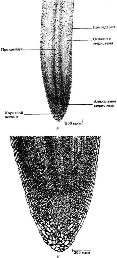
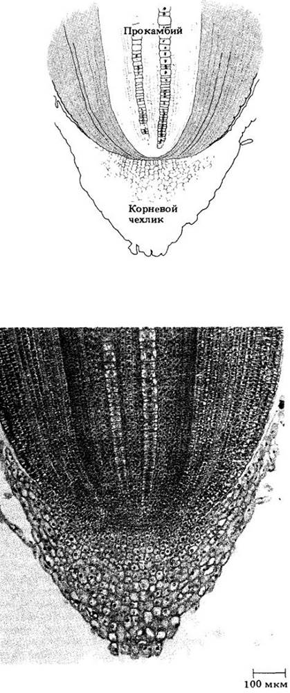
Fig. 21-4. Apical meristem of a corn root tip (Zea mays). Note the three distinct layers of initials. The lower layer gives rise to the root cap, the middle layer to the protoderm and ground meristem (cortex), and the upper layer to the procambium, i.e., the vascular cylinder.
The root cap also performs other vital functions, notably regulating the root's response to gravity (geotropism or gravitropism; see Chapter 25).
Root Growth Zones
Alongside the root cap, the most characteristic structural feature of the root tip is the arrangement of longitudinal cell rows originating in the apical meristem. This meristem consists of relatively small (10 to 20 µm in diameter) polyhedral cells—the initials and their immediate derivatives (p. 16)—characterized by dense Cytoplasm and prominent nuclei (Figs. 21-3 and 21-4). The arrangement and number of initials vary noticeably.
Two MAIN TYPES OF apical Organization have been described in the roots of seed plants. In one type, the root cap, vascular cylinder (xylem and phloem), and cortex originate from a common group of cells in the apical meristem (Fig. 21-3); in the other, each of these regions can be traced back to an independent cell layer (Fig. 21-4), with the epidermis sharing a common origin either with the root cap or with the cortex.
Although the initial zone of the root apical meristem was once considered a site of active Cell Division, it has now been shown that this is not the case; most divisions occur at some distance from the initials, forming a relatively inactive region known as the quiescent center (Fig. 21-5).
Fig. 21-5. Apical meristem of a corn root tip showing the quiescent center (outlined by the dashed line). To obtain this radioautograph, the root was fed tritium-labeled thymine (3H) for 24 hours. In the actively dividing cells surrounding the quiescent center, this base was rapidly incorporated into nuclear DNA, as evidenced by the dark grain clusters on the radioautograph

The word "relatively" indicates that divisions do still occur within this center under normal conditions. Furthermore, this center is capable of regenerating the peripheral Zones of the meristem if they are damaged. Recently, for example, it has been demonstrated that isolated quiescent centers of corn grown in sterile culture can develop into whole roots, bypassing the callus or wound tissue stage. Another study on the same species revealed a clear correlation between the size of the quiescent center and the structural complexity of the primary Vascular System of the root. These and other investigations highlight the essential role of the quiescent center in root structure formation and development.
The distance from the apical meristem at which most cell divisions take place varies among different species and, within a species, among roots of different ages. The apical meristem together with this adjacent portion of the root is referred to as the zone of cell division (Fig. 21-6).
Fig. 21-6. Diagram of the Cytology/cytology/16.html">Early stages of primary growth in a root tip (cf. Fig. 21-1)

This zone is followed, without a sharp boundary, by the zone of elongation, which is typically only a few millimeters long (Fig. 21-6). Cell expansion within this zone drives the primary elongation of the organ. Further away from the apex, this growth ceases. Because longitudinal growth occurs exclusively near the root tip, only a very limited portion of the root continually pushes its way through the soil.
Beyond the zone of elongation lies the zone of maturation (differentiation) for most primary tissues (Fig. 21-6). Root hairs also develop here, which is why this region of the root is sometimes called the root hair zone (see Fig. 21-1).
It is important to note that the transition from one zone to the next is gradual, without abrupt boundaries. Some cells begin to elongate and differentiate while still in the zone of cell division, whereas others reach maturity within the zone of elongation—for instance, the earliest phloem and xylem elements, which are often disrupted during root elongation due to ongoing stretching.
Near the apical meristem, the protoderm, procambium, and ground meristem can already be distinguished (see Figs. 21-3 and 22-6); these are the primary meristems that differentiate into the epidermis, primary Vascular Tissues, and cortex, respectively (Chap. 19).
Compared to the Internal Structure of the stem, the root structure is relatively simple. This is primarily due to the absence of leaves and, consequently, nodes and internodes, resulting in very little variation in tissue arrangement across different levels.
In the primary growth stage of the root, transverse (Figs. 21-7 and 21-8) and longitudinal (see Fig. 21-3) sections easily reveal three tissue systems: the epidermis (dermal tissue system), the cortex (ground tissue system), and the vascular tissue system. In most roots, the vascular tissue forms a solid cylinder (Fig. 21-7), whereas in some, it forms a hollow cylinder surrounding a pith (Fig. 21-8).
Fig. 21-7. Transverse sections of a buttercup (Ranunculus) root. A. General view of a mature root. B. Detail of an immature vascular cylinder. Note the intercellular spaces in the primary cortex. C. Detail of a mature vascular cylinder. Numerous starch grains are visible in the cortical cells

Fig. 21-8. Transverse sections of a corn root. A. General view of a mature root. Part of a lateral root is visible at the bottom right. The vascular cylinder with its pith stands out clearly. B. Detail of an immature vascular cylinder. C. Detail of a mature vascular cylinder

Epidermis
The epidermis of young roots absorbs water and mineral nutrients; this function is facilitated by root hairs, which are tubular outgrowths of epidermal cells that significantly increase the absorbing surface area of the organ (Fig. 21-9; see also the section on "Mycorrhizae" in Chap. 13). In the previously mentioned study on four-month-old rye plants, it was calculated that a single plant possesses approximately 14 billion root hairs, providing an absorptive surface area of 401 m2 and a total length exceeding 10,000 km.
Fig. 21-9. Root of a colonial bentgrass (Agrostis tenuis) seedling with root hairs. These can reach their final length of 1.3 cm within a few hours. Each individual hair has a relatively short lifespan, but the continuous formation of new hairs and the death of old ones persist as long as the root continues to grow

These hairs are relatively short-lived, are localized primarily in the zone of maturation, and form immediately behind the zone of elongation (see Figs. 21-1 and 21-6) at roughly the same rate as older hairs die off at the upper end of the hair-covered zone. As the root tip pushes deeper into the soil, new root hairs develop directly behind it, providing a surface capable of absorbing newly encountered reserves of water and mineral ions (see Chap. 27). Evidently, it is the young and growing roots that function as feeder roots—that is, they are primarily responsible for the uptake of water and inorganic substances. For this reason, when transplanting, gardeners must pay special attention to moving the root system with as much adhering soil as possible. If a plant is simply pulled out of the ground, the bulk of the feeder roots will be left behind, and such a "transplant" is unlikely to survive.
The epidermal cells of the root, including those bearing root hairs, are parenchymatous and densely packed. The young epidermis is largely covered by a thin cuticle, so the cell wall offers some resistance to the passage of water and minerals. In addition, the surface of many roots is covered by a mucilaginous sheath that promotes closer contact with soil particles. THE ORIGIN OF this sheath is not yet fully understood, but it is believed to be produced, at least in part, by the root cap. Mucilage has been shown to create favorable conditions for the colonization of beneficial Bacteria. It may also affect the availability of soil ions and provide temporary protection for the root against desiccation.
Primary Cortex
As shown in a transverse section (see Fig. 21-7, A), the primary cortex accounts for the bulk of the primary root tissues. Its cells store Starch and other substances but typically do not contain METABOLISM/14.html">Chloroplasts. Roots with intensive Secondary Growth, such as those in gymnosperms and most dicotyledons, shed their primary cortex early. In these plants, the cortical cells remain parenchymatous. In contrast, the primary cortex of monocotyledons persists throughout the life of the root, and many of its cells become lignified, developing secondary walls.
Regardless of the degree of differentiation, this tissue contains numerous intercellular spaces—air cavities that play a vital role in the aeration of root cells (see Fig. 21-7 and 21-8). The cells form numerous contacts with one another, and their protoplasts are interconnected by plasmodesmata. Consequently, substances can move through the cortex from Cell to Cell via protoplasts and plasmodesmata, or through the cell walls.
Unlike the rest of the primary cortex, the cells in its innermost layer are densely packed without any intercellular spaces. This region, the endodermis (see Fig. 21-7 and 21-8), is characterized by the presence of Casparian strips on the anticlinal (i.e., perpendicular to the root surface) cell walls. These are the equatorial Regions of the primary wall impregnated with the lipid-like substance suberin and sometimes also lignified. The protoplast of the endodermal cells fits tightly against the strip (Fig. 21-10 and 21-11). Because endodermal cells are closely packed and the Casparian strips are impermeable to water, all substances entering or leaving the central vascular cylinder must pass through the protoplasts of the endodermal cells—either crossing their plasma membrane or traveling through the numerous plasmodesmata that connect with the protoplasts of adjacent primary cortex and central cylinder cells.
Fig. 21-10. High-power magnification of a section of an immature buttercup (Ranunculus) root, showing Casparian strips in the endodermal cells. Note that the plasmolyzed protoplasts of these cells adhere closely to the strips.
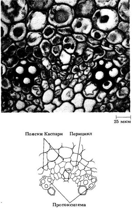
Fig. 21-11. Transverse section of a radial wall between two endodermal cells. The Casparian strip region is stained more intensely than the rest of the wall. The plasma membrane of each plasmolyzed cell can be seen fitting tightly against the wall in the zone of the Casparian strip.

The effectiveness of the Casparian strip as a barrier to the movement of substances through the cell walls of the endodermis has been demonstrated in corn roots absorbing positively charged lanthanum ions, which cannot penetrate cell membranes. Under an Electron microscope, this element is found exclusively in the cortical cell walls, meaning its transport deeper into the root is abruptly and completely halted by the Casparian strips (see Chapter 27 for a more detailed Discussion of The Role of the endodermis in the Movement of water and dissolved substances through the root).
As mentioned above, in roots undergoing secondary growth, the primary cortex and endodermis are shed early. When they persist, a suberin lamella—consisting of alternating layers of suberin and wax—is eventually deposited along the entire inner surface of the endodermal cell walls, followed by Cellulose, which may become lignified (Fig. 21-12). Such modifications in the endodermis begin opposite the phloem strands and spread toward the protoxylem (see Fig. 21-7, A). Opposite the protoxylem, however, some endodermal cells may remain thin-walled and retain their Casparian strips for a long time. These are called passage cells; in most species, they eventually become suberized. In conifers, the modification of cell walls in the endodermis concludes with the deposition of the suberin lamella (Fig. 21-13).
Fig. 21-12. Three-dimensional diagram illustrating three stages of endodermal cell development in the primary root body. A. Initially, the endodermal cell is characterized by the presence of a Casparian strip on its anticlinal walls. B. Next, a suberin lamella is deposited on the inner surface of the entire wall. C. Finally, the suberin lamella is supplemented by a thick, often lignified cellulose layer.

Fig. 21-13. Transmission electron micrograph of suberin lamellae in the walls of two adjacent endodermal cells of a red pine (Pinus resinosa) root. Alternating light and dark layers are visible, believed to be formed by wax and suberin, respectively.

Central Cylinder (Stele)
The central cylinder of the root consists of vascular tissue surrounded by one or more layers of cells, the pericycle (see Fig. 21-7 and 21-8). In a young root, the pericycle is composed of parenchymatous cells with primary walls, which may be supplemented by secondary walls as the plant ages (see Fig. 21-8).
The pericycle performs several important functions. In most seed plants, lateral roots originate within it. In species with secondary growth, it participates in The formation of the cambium and typically gives rise to the first layer of the phellogen. New cells are frequently formed within the pericycle and subsequently become part of it.
In most roots, the inner portion of the central cylinder is occupied by a solid strand of primary xylem that extends outward toward the pericycle in ridges (see Fig. 21-7). Strands of primary phloem are located between these ridges. Thus, the central cylinder of the root is a protostele.
The number of primary xylem ridges varies among species and sometimes even along the axis of a single root. If There are two ridges, the root is termed diarch; if three, triarch; if four, tetrarch (see Fig. 21-7, A, B); and if many, polyarch (see Fig. 21-8). Maturation of the (proto)xylem elements begins beneath the pericycle; the tips of the ridges are typically called protoxylem poles (see Fig. 21-7 and 21-8). Metaxylem (meta- derived from Greek meaning "after"), occupying the inner parts of the ridges and the center of the central cylinder, matures after the protoxylem. The roots of certain monocotyledons (such as corn) possess a pith (see Fig. 21-8), which is regarded by some botanists as potential vascular tissue.
Formation of Lateral Roots
In most seed plants, lateral roots originate in the pericycle. Because they are initiated deep within the parent root, they are termed endogenous, which translates from Greek as "originating from within" (Fig. 21-1 and 21-14).
Fig. 21-14. Three stages of lateral root development in willow (Salix). A. One primordium has already formed (bottom); two others are being initiated in the pericycle (arrows). The central cylinder is still very young. B. Two root primordia push through the cortex. C. One lateral root has emerged at the surface, and another is approaching it.

Divisions in the pericycle leading to the formation of lateral roots occur at some distance from the elongation zone, within partially or fully differentiated root tissues. In angiosperms, cells of both the pericycle and the endodermis participate in The Emergence of the root primordium here, although in many cases the derivatives of the latter are short-lived. As the young lateral root grows, it forces its way through the primary cortex (Fig. 21-14, B), possibly secreting Enzymes that break down the cortical cells in its path. Early in its development, the primordium forms a root cap, an apical meristem, and primary meristems. Initially, the central cylinders of the lateral and parent roots are not connected; they become joined later through the Differentiation of the intervening parenchymatous cells into xylem and phloem elements.
Aerial Roots
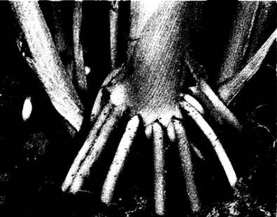
Aerial roots are adventitious roots that develop on above-ground plant organs. In some plants, they provide structural support and are referred to variously as pillar roots, stilt roots, or prop roots (Fig. 21-15). Upon contacting the soil, these roots branch and begin absorbing water and minerals as well. Such roots develop on the trunks and branches of many tropical trees, notably mangrove species (Rhizophora mangle), the banyan (Ficus benghalensis), and certain palms. Aerial roots of another type, such as those of English ivy (Hedera helix), penetrate the surfaces of walls and other objects, anchoring the climbing stem to them.
Fig. 21-15. Prop roots of maize — one of the types of adventitious roots.
Roots require oxygen for Respiration, which is why most plants cannot survive in poorly drained soils lacking air spaces. Some wetland trees develop roots that emerge from the water, serving not only to anchor the plant in the substrate but also to supply oxygen. For example, the root System of the black mangrove (Avicennia germinalis) produces negatively geotropic outgrowths called pneumatophores (respiratory roots) that protrude upward from the mud to ensure necessary aeration (Fig. 21-16). A similar function was once attributed to the "knees" of the bald cypress (Taxodium distichum; see Fig. 18-27), but this view is now disputed.
Fig. 21-16. Pneumatophores (respiratory roots) of the white mangrove (Laguncularia racemosa) emerging from the mud at the Base of the plant.

Specialized Adaptations
Many specialized root adaptations are found in epiphytes—plants that grow on other plants without being parasitic on them. For instance, the multi-layered root epidermis of certain orchids (Fig. 21-17) sometimes serves as the plant's sole photosynthetic organ. This tissue, known as velamen, also provides mechanical protection for the cortex, reduces water loss, and may participate in water absorption.
Fig. 21-17. A. Aerial roots of the orchid Oncidium sphacelatum. B. Cross section of an orchid root showing the multi-layered epidermis (velamen).

The epiphyte Dischidia rafflesiana, sometimes called the "flower pot plant," exhibits a very unusual structure. Some of its leaves are flattened and succulent, whereas others form hollow vessels ("flower pots") that collect debris and rainwater (Fig. 21-18). Ant colonies often inhabit these structures, thereby enhancing the plant's nitrogen Nutrition. Roots originating at the node just above such a modified leaf grow into the "pot," from which they absorb water and mineral nutrients.
Fig. 21-18. The epiphyte Dischidia rafflesiana ("flower pot plant"). A. A leaf modified into a "pot" that accumulates solid particles and rainwater. B. The same leaf opened to reveal the roots growing into it.

Nutrient Storage Adaptations
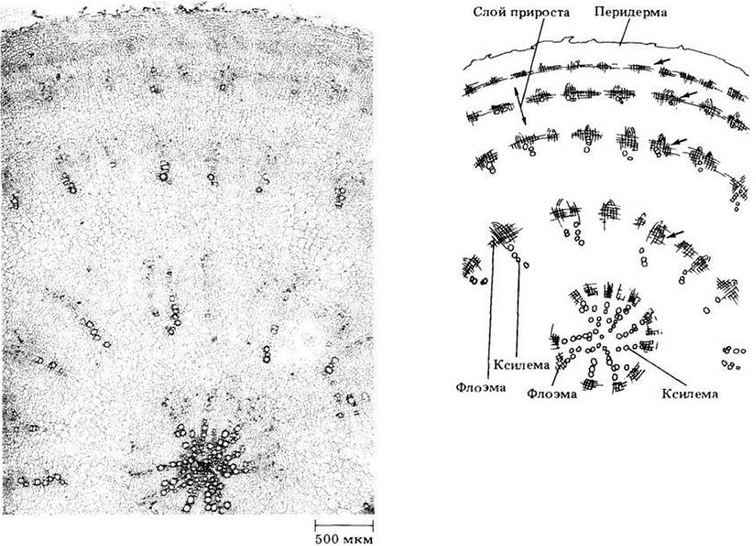
Many roots function as storage organs, and in some plants, they are specifically adapted for this role, becoming fleshy due to the extensive proliferation of parenchyma permeated by vascular tissue. The development of such roots (e.g., in the carrot, Daucus carota) proceeds essentially like that of non-fleshy roots, but with a predominance of parenchymatous cells in the secondary xylem and phloem. The root of the sweet potato (Ipomoea batatas) is structurally similar to that of the carrot, but additional cambial cells differentiate around individual vessels or groups of vessels in the secondary xylem (Fig. 21-19). After producing a few tracheal elements toward the vessels and sieve tubes in the opposite direction, they give rise primarily to storage parenchyma cells on both sides. In the sugar beet (Beta vulgaris), root thickening occurs mainly through The activity of accessory cambia that form in concentric layers around the primary cambium (Fig. 21-20). These layers, which outwardly resemble the growth rings of woody roots and stems, produce xylem inward and phloem outward, with parenchyma predominating in both. The upper portion of most fleshy roots actually develops from the hypocotyl.
Fig. 21-19. Cross sections of a sweet potato root (Ipomoea batatas). A. General view. B. Detail of the xylem with cambium surrounding the vessels.

Fig. 21-20. Cross section of a sugar beet root (Beta vulgaris) with accessory cambial layers (indicated by arrows). The original cambium (in the center of the root) produces relatively little xylem and phloem.
Roots are organs specialized for anchoring the plant in the substrate, as well as for absorption, storage, and conduction of substances. Gymnosperms and dicots typically develop taproot systems, whereas monocots form fibrous root systems. Their extent depends on various factors, but the bulk of absorbing roots is located within the top meter of soil.
The apical meristems of most roots contain a quiescent center; maximum meristematic activity, i.e., the most intensive cell division, is observed at some distance from the apical initials. During primary growth, the apical meristem gives rise to three primary meristems—protoderm, ground meristem, and procambium—which differentiate into the epidermis, cortex, and stele (vascular cylinder), respectively. In addition, the apical meristem produces the root cap, which protects it and facilitates root penetration through the soil.
Many epidermal root cells form root hairs, which significantly increase the root's absorptive surface area. Except for the endodermis, intercellular air spaces are abundant throughout the cortex. The tightly packed endodermal cells feature Casparian strips on their anticlinal walls. Consequently, all substances transported between the cortex and the stele must pass through the protoplasts of the endodermal cells.
The stele consists of primary vascular tissues completely surrounded by the pericycle. The central region of the cylinder is typically occupied by primary xylem with lobes extending toward the periphery and alternating with strands of primary phloem. Lateral roots originate in the pericycle and push outward through the cortex and epidermis.
Last update: 07/08/2026
Editorial and Educational Adaptation: This material has been compiled based on the primary/original source text. The project team performed an editorial review, corrected technical inaccuracies, structured sections, and adapted the content for an educational format.
What was processed:
- elimination of formatting defects (OCR errors, structural breaks, corrupted characters);
- editorial organization of content;
- standardization of terminology in accordance with academic sources;
- verification of factual statements against the original source text.
All mentions of the author, publication year, and origin of the primary text have been preserved in accordance with the source.