MORPHOLOGICAL AND TINKTORIAL PROPERTIES OF BACTERIA - E. L. Zaitseva - 2015
CHAPTER I. MAIN TYPES OF MICROSCOPES
In microbiology, a microscope is used to study both live and dead microorganism Cells in stained and unstained states.
Light Microscope (MBI - 1, 2, 3, 6, 11). All objects are examined in transmitted light using dry and immersion objectives. Resolution is 0.4-0.2 µm. The magnification at a given tube length is equal to the product of the objective and eyepiece magnifications. The minimum is 630 (for an immersion objective) and the maximum is 1350. It is used to study the Morphology, Structure, motility, and tinctorial properties of microorganisms.
Class="center">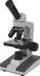
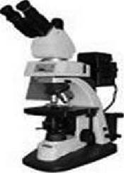
Fluorescent microscope. Uses ultraviolet rays and fluorophores (Dyes capable of emitting light/fluorescing under UV light). It allows microorganisms to be observed in the light and color they emit. Resolution is 0.1 µm. Its enhancement is associated with The Use of short-wave ultraviolet rays. Maximum magnification is up to 3,000x. Advantages include a color image, high contrast, and The ability to examine live specimens. It is used not only to study morphology and tinctorial properties, but also to investigate the life processes of microbial cells.
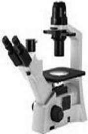
Inverted microscopes (dark-field, phase-contrast). Studies are conducted in transmitted light in a bright or dark field using the phase-contrast method. MBI - 12, 13 are equipped with dedicated stage thermostats and movie cameras. The eyepiece and objective lenses produce an inverted magnified image. This allows for a wide range of microscopic studies: visual observation, photography, use of bright and dark fields in direct and reflected light, direct and oblique illumination, polarized light Cell/15.html">Microscopy, phase-contrast Methods, and luminescence microscopy. Dark-field and phase-contrast observation methods are used to study living microorganism cells.
Electron microscope. Its Operating Principle and design are similar to those of a standard light microscope. The differences are that instead of a light source, it uses an electron wave source (a tungsten wire heated by an electric current), and instead of optical lenses, electromagnetic lenses. Resolution is 0.001 µm. The first intermediate magnification is 130x, from the projection lens 20-200x, overall 2,500-25,000x, and up to 100,000x maximum. It is widely used to study Viruses and the smallest microorganisms. In bacteriology, it is used to study fine structural details.

Stereomicroscope. Provides illumination in direct and oblique transmitted light. Most suitable for large objects (Fungi). Used for studying colonies and mycological cultures.

1. Additional microscope accessories.
AU-12 binocular attachment. Brings microscopy closer to natural Vision conditions. Increases the productivity of the microscopist. Resolution is slightly increased. Compatible with any microscope.
Micrometer: stage and ocular. The stage micrometer and ocular micrometer make it possible to determine the dimensions of Bacteria using calibration grids. Resolution is slightly increased, enabling The Study of microorganisms in their Native State for size determination.
Dark-field condenser for a light microscope. By darkening the central part of the condenser and, consequently, providing side illumination of the object, microorganisms can be seen against a dark Background in the light reflected from them. Resolution is increased. Same as in dark-field microscopy.
Phase-contrast device (objective). In conventional transmitted-light microscopy, it increases contrast and reflection by converting phase shifts of the light wave into amplitude shifts detectable by the human eye. Unlike a dark-field device, it allows the observation of contrasting live, unstained specimens against a bright background. Used for observing living biological objects.
Stage incubator (thermostat stage). Preserves the motility and viability of the specimen. Used for studying Protozoa.
2. Microscopy of a specimen using an immersion objective.
When using immersion objectives, a liquid must be present between the front lens of the objective and the object of study.
The immersion objective provides 90x magnification, provided there is no scattering of the light beam due to medium inhomogeneity during passage. Therefore, to avoid this defect, immersion cedar oil or its substitute, liquid paraffin (mineral oil), is used. Microscopy with the 90x immersion objective is performed with 7x and 10x eyepieces, most frequently with the former.
The Location for the microscope should be chosen away from direct sunlight. Working on a dark-surfaced desk helps reduce eye strain. It is recommended to look into the eyepiece with the left eye without closing the right one. It is more convenient to work with a binocular microscope.
The microscope must be carried with both hands: hold the arm with one hand and the base with the other. The microscope should be protected from shocks and contact with aggressive substances such as acids and alkalis.
The Microscopy Technique consists of several stages:
1. A drop of immersion oil is applied to the prepared and stained smear.
2. While observing from the side, carefully lower the microscope tube to immerse the objective lens into the oil drop. Careless execution can crush either the objective lens or the microscope slide.
3. Once the objective lens touches the oil, further lowering of the tube is carried out using the microscope's fine-adjustment knob until the microobjects come into view in the eyepiece.
4. Examination of the specimen is conducted exclusively by manipulating the fine-adjustment knob and moving the slide.
5. Upon completion of microscopy, the tube is raised, withdrawing the objective lens from the oil drop.
6. Oil is removed from the objective using clean gauze, followed by wiping with a fat solvent—either xylene, alcohol, or chloroform. Leaving oil on the objective lens is strictly prohibited.
7. The revolving nosepiece on the tube is set to the x8 objective, the condenser and tube are lowered into a non-working position, and the microscope is covered with a protective cap.
Principle of the method. Certain biological objects, when illuminated by short-wave rays (blue, violet, ultraviolet), are capable of absorbing them and emitting rays of a longer wavelength (glowing with a yellow-green or orange light). This phenomenon is known as intrinsic or primary fluorescence.
Non-fluorescing objects can be treated with specific fluorochromes to also exhibit fluorescence; however, this is referred to as induced or secondary fluorescence. It is more frequently used in microscopy.
Fluorochromes include acridine yellow, acridine orange, auramine, primuline, thioflavin, congo red, tetracycline, quinine, and other substances (Table 1).
Table 1. Fluorochromes for fluorescence microscopy used in microbiological research
Name of dye |
Characteristics |
Application in bacteriology |
Acridine yellow |
3,6-diamino-2,7-dimethylacridine dihydrochloride |
Detection of gonococci and Mycobacterium tuberculosis |
Acridine orange |
3,6-bis(dimethylamino)acridine hydrochloride |
Universal fluorochrome for counterstaining nuclei and Nucleic Acids |
Auramine |
Tetramethyldiaminobenzophenoniline hydrochloride |
For the detection of acid-fast bacteria (tuberculosis, leprosy), rickettsiae, and certain viruses |
Congo red |
Disodium salt of biphenyl-4,4-bis(azo-2)-aminonaphthalene-4-sulfonic acid |
Additional contrast dye in combination with acridine orange and others |
Neutral red |
2-methyl-3-amino-6-dimethylaminophenazine hydrochloride |
Fluorochrome for the detection of fats and lipoids |
Primuline |
С2Н14N3О3S3Na |
For detecting elementary viral bodies and differentiating living from dead cells |
Thioflavin |
||
Trypaflavin |
Mixture of Hydrochloric acid salts of 3,6-diamino-10-methylacridinium chloride and 3,6-diaminoacridine |
General-purpose fluorochrome |
Fluorescein |
Resorcinphthalein |
Dye for fluorescent analysis; used as a fluorescent protein label |
Fluorescein disodium salt (uranine) |
Dye for fluorescent analysis and microscopy (especially for vital fluorescence microscopy) |
|
Basic fuchsin |
Additional fluorochrome for counterstaining with acridine orange and others |
|
Eosin Y |
Mixture of tetra-dibromofluorescein disodium salts |
For diffuse additional fluorochroming of microscopic preparations |
Advantages of fluorescence microscopy over conventional light microscopy include the following:
1) a color image and high contrast of the studied objects;
2) the ability to study the morphology of living and fixed microbial cells within nutrient media as well as animal and plant Tissues;
3) the feasibility of investigating cellular microstructures that selectively absorb various fluorochromes acting as specific cytochemical indicators;
4) the capability to examine both transparent and opaque living specimens;
5) the study of functional and morphological cellular changes;
6) the detection and localization of individual microbes and viruses;
7) the use of fluorochromes in immunological reactions and for enumerating bacteria in samples with low microbial content.
Staining of preparations for fluorescence microscopy. Specimens for fluorescence microscopy are prepared and fixed in the same manner as for conventional microscopy. They are stained with special dyes—fluorochromes—applied as weakly concentrated solutions (1:500–1:1,000,000). Solutions are prepared using distilled Water, isotonic sodium chloride solution, or buffer mixtures. The prepared dyes should be stored in dark Glass bottles, protected from light exposure.
The electron microscope is used to examine objects and structures that lie beyond the resolution Limits of the optical light microscope. The resolving power of modern electron microscopes is 3–40 Å, with an average working magnification of 100,000x. Structurally, the electron microscope is analogous to the light microscope. However, illumination of the specimen is provided by a beam of electrons generated by a tungsten filament heated by an electric current.
The specimen under study is placed between electromagnetic coils that function as projection and objective lenses. An electromagnetic coil acting as a condenser lens focuses the electron beam onto the specimen, after which the electrons pass into the "objective lens," which forms the primary image of the object. This image can be viewed on an intermediate screen. A portion of the electrons, passing through the aperture of the "objective lens," strikes the "projection lens." The magnified image produced by this lens is projected onto a fluorescent screen—a metal plate coated with a thin layer of zinc sulfide or zinc sulfide with cadmium selenide. Upon impact by the electron beams, the zinc sulfide particles begin to glow. The finer their dispersion, the sharper the structural details of the object. By replacing the screen with a photographic plate, the image of the object can be captured permanently.
Preparations for electron microscopy are placed on special grids coated with an extremely thin film (substrate). The total thickness of the preparation and the substrate must not exceed 2500 A.
The contrast of the object is achieved either by shadowing it with heavy metals (chromium, gold, palladium) or by Treatment with various contrasting agents such as phosphotungstic acid and uranyl acetate.
When investigating the Morphological Characteristics of microbial cells under an electron microscope, whole cells are examined. When studying cell ultrastructure, thin sections are prepared. The thickness of the sections must not exceed 800-900 A.
Last update: 13/08/2026
Editorial and Educational Adaptation: This material has been compiled based on the primary/original source text. The project team performed an editorial review, corrected technical inaccuracies, structured sections, and adapted the content for an educational format.
What was processed:
- elimination of formatting defects (OCR errors, structural breaks, corrupted characters);
- editorial organization of content;
- standardization of terminology in accordance with academic sources;
- verification of factual statements against the original source text.
All mentions of the author, publication year, and origin of the primary text have been preserved in accordance with the source.