Practical Course in Zoology: Study Guide - T. A. Dauda 2014
Protozoa
Sarcomastigophora
Class Flagellata
The Class Mastigophora is characterized by the presence of flagella, which serve as Organelles of locomotion. In a few species, temporary or permanent pseudopodia may also be present alongside flagella.
Representatives of certain flagellate groups possess chromatophores containing chlorophyll. These forms, much like true green plants, are capable of Photosynthesis in the light, meaning they exhibit an autotrophic type of Nutrition. Other flagellates, like all animals, are heterotrophs. Finally, there are species that combine both Types of nutrition, thus being mixotrophic. Depending on the type of nutrition, the class Mastigophora is divided into 2 subclasses.
1. Phytomastigina (capable of photosynthesis).
2. Zoomastigina (animal flagellates with a heterotrophic type of METABOLISM).
The size and shape of flagellates vary considerably: oval, cylindrical, spherical, bottle-shaped, etc. Flagella (1, 2, 4, 8, or more—up to several hundred) originate from the anterior pole of the body. In some zoomastigotes, a flagellum may run along the body and fuse with its surface, forming an undulating membrane (most commonly seen in parasitic species).
Some species from the subclass Phytomastigina form colonies, which are the result of incomplete Cell Division. These colonies vary both in shape (dendritic, spherical, etc.) and in their mode of development.
Order Euglenoidea
Green euglena (Euglena viridis).
Materials and equipment. Numerous species of the genus Euglena are found in freshwater bodies rich in organic matter. Some euglena species can be cultured in artificial media, such as Knop's medium (distilled H2O — 1000 ml, MgSO4 — 0.25 g, Ca(NO3)2 — 1.0 g, KH2PO4 — 0.25 g, KCl — 0.12 g, FeCl3 — traces). Euglenas develop best when organic substances are added to mineral media. Specifically, they thrive on meat broth. Test tubes or small flasks can be used as culture vessels for euglenas.
As a representative of euglenoids for study, one can use either a live culture of Euglena viridis or slides with fixed euglenas.
Required items: Microscope; Glass slides and coverslips; pipettes; strips of filter paper; Water from a stagnant pond (green in color) or aquarium; test tubes or beakers containing a live euglena culture; slides with fixed euglenas.
Task. Examine a drop of water containing euglenas and observe the locomotion of this protozoan. Observe the stigma (photoreceptor eyespot), chromatophores, paramylon grains, and the contractile vacuole reservoir. Make a drawing of the green euglena. Pay attention to other flagellates found in the same drop.
Preparation and examination of the slide. Place a small drop of water with euglenas onto a glass slide using a pipette and cover it with a coverslip.
Examine the drop of water with euglenas under low magnification of the microscope, noting The Nature of their movement—forward progression and rotation. Locate a sluggish specimen of green euglena that differs in size from the others, and examine it under high magnification using a 15x eyepiece. To observe the Structure more clearly, you can slow down the movement of the euglenas by drawing off the water with strips of filter paper. To do this, place the strips on the slide along the sides of the coverslip. Remove the strips once you notice the euglenas' movement slowing down.
Note that the euglena (see Fig. 9) has a spindle-shaped body; it is pointed at the posterior end and rounded at the anterior, while the outer layer of Cytoplasm forms a delicate pellicle. The elasticity of the pellicle allows the euglena to temporarily change its body shape. Under the microscope, it is clearly visible that as it shortens, it becomes thicker; when elongated, it becomes thinner and more slender.
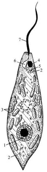
Fig. 9 Euglena (Euglena viridis):
1 — nucleus; 2 — paramylon grains; 3 — chromatophores; 4 — eyespot; 5 — contractile vacuole; 6 — contractile vacuole reservoir; 7 — flagellum.
The green color is due to the presence of oval or rounded chromatophores in its protoplasm containing the green pigment, chlorophyll. Locate and draw the chromatophores. Thanks to chlorophyll, the euglena is capable of photosynthesis. The products of photosynthesis are stored in the cytoplasm as colorless grains of the carbohydrate paramylon—a reserve nutrient and structural material similar in chemical composition to starch. The Nucleus, located closer to the posterior end of the body, is usually not visible as it is masked by chromatophores and paramylon grains, but it shows through them as a light patch. Reduce the illumination by slightly turning the microscope mirror or lowering the substage condenser, and locate the Hair-like cytoplasmic outgrowth at the anterior end of the euglena—the flagellum, by means of which it moves.
Observe the water currents caused by the beating of the flagellum. Examine the bright red spot at the Base of the flagellum—the stigma (eyespot), which Functions as a light-sensitive organelle. Locate the light area adjacent to the stigma, which is the contractile vacuole reservoir; it communicates with the external environment via a narrow canal opening to the outside at the anterior end of the euglena's body.
Make a drawing of the green euglena and label its structural details.
The nutrition of euglena is based on its ability to synthesize CARBOHYDRATES (paramylon) from water and carbon dioxide in the light. Essential mineral salts are absorbed osmotically from the surrounding environment through the pellicle. In the dark, euglenas feed on preformed Organic compounds, just like all animals. This type of nutrition is called mixed, or mixotrophic. Under unfavorable conditions (a drop in water Temperature, drying up of the water body, etc.), the euglena encysts: it sheds its flagellum, rounds up, and secretes a thick protective wall around itself against adverse conditions, in other words, forming a cyst.
Volvox (Volvox sp.). Volvox is frequently found in ponds and lakes during the summer months. The most widespread species is Volvox aureus, while V. globator is somewhat less common.
Materials and equipment. For study, it is best to use living material. If unavailable, formalin-fixed material may be used.
Required items: microscope; glass slides and coverslips; pipettes; dissecting needles; test tubes or beakers containing a live volvox culture, or slides with fixed volvox specimens.
Task. Examine a drop of water containing volvox (if live volvox is unavailable, use fixed material). Observe and sketch 1–2 colonies. Pay attention to the shape and STRUCTURE OF THE colony. Examine the somatic and generative Cells, as well as the daughter spheres inside the volvox.
Preparation and Study of the slide. Transfer a drop of water with volvox from a test tube onto a glass slide using a pipette. Examine the colonies—consisting of A large number of individuals (500–20,000)—under low magnification without placing a coverslip over the drop. Note that volvox forms a hollow sphere 0.5–2.0 mm in diameter. The wall of the sphere consists of a gelatinous matrix in which individual individuals of the colony are embedded. Smaller spheres—daughter colonies—are sometimes visible within the cavity of the parent sphere (Fig. 10).

Fig. 10 Volvox:
A — Volvox globator, section of the body wall with Gametes; B — V. aureus, general view (six daughter spheres inside the parent sphere); 1 — macrogametes; 2 — microgametes; 3 — vegetative individual.
The cells forming the colony are not all identical. To examine their structure, cover the preparation with a coverslip and gently press it with a dissecting needle (crushing the volvox). Observe it under high magnification while adjusting the fine adjustment screw. The vast majority of the colony members are small vegetative, or somatic, cells that ensure the movement, nutrition, and growth of the volvox (Fig. 11).
Examine them: they have a pear-shaped form; each possesses a chromatophore, nucleus, stigma, contractile vacuoles, and two flagella. The coordinated action of the flagella of all somatic cells drives the rotational and forward movement of the colony, which slowly "rolls" through the water. The cells are interconnected by cytoplasmic bridges. Locate a small number of generative cells in the colony, which perform a reproductive function. In some volvox specimens, daughter spheres are visible within the cavity of the parent sphere; these develop from generative cells through multiple divisions and are released into the cavity of the parent volvox, after which the parent volvox eventually dies. From a small portion of the generative cells, Germ Cells arise: some develop into macrogametes (egg cells), while others undergo multiple divisions to form microgametes. Upon the fusion of the gametes, a zygote (fertilized egg) is formed, which gives rise to a new colony.

Fig. 11 Volvox wall under high magnification:
1 — vegetative individual; 2 — generative individual; 3 — chromatophores;
4 — nucleus; 5 — cytoplasmic bridge connecting individual volvox cells; 6 — flagella.
Sketch the parent volvox colony with daughter spheres and a section of the volvox body wall containing reproductive cells.
Trypanosome (Trypanosoma sp.). Trypanosomes are parasitic protozoa found in the Blood of many vertebrates and humans. For instance, Trypanosoma lewisi is frequently found in the blood of the brown rat (Norway rat). Another species, T. equiperdum, is the CAUSATIVE AGENT OF dourine in horses, donkeys, and mules.
Materials and equipment. Among the numerous species of trypanosomes parasitizing the blood of vertebrates, Trypanosoma lewisi is the most accessible for study, as it is very commonly found in the blood of live rats. To study live trypanosomes, take a drop of blood from a live rat, cover it with a coverslip, and examine it. Under high magnification, the movement of trypanosomes is clearly visible in the Blood Plasma. Fixed and stained blood smear preparations from infected animals are more commonly used in laboratory practicals.
Required equipment: a microscope with an immersion objective; cedar oil; slide preparations of blood smears from an infected horse or brown rat.
Task. Examine and sketch a stained blood smear containing trypanosomes—the causative agents of dourine in horses, donkeys, and mules.
Study of the slide. Take the prepared slide containing trypanosomes. Under high magnification (40x objective, 15x eyepiece), examine the blood smear to observe numerous pale pink, anucleate red Blood Cells (erythrocytes), as well as a smaller number of larger white blood cells (leukocytes and lymphocytes) with dark blue nuclei. Pay attention to the slender, spindle-curved trypanosomes with red-stained flagella; the trypanosomes are located in the plasma spaces between the white and red blood cells. Sketch the blood smear with trypanosomes.
Examine one of the trypanosomes in the blood smear using an immersion objective to study its structure. To do this, place a drop of cedar oil directly onto the slide and lower the immersion objective into it. Note the body shape of the trypanosome: it is tapered at both ends, ribbon-like, and adapted for movement through the plasma between blood cells. Their ecological niche has profoundly shaped the Morphology of these parasites (Fig. 12). The nucleus is oval, stains dark blue in the preparation, and is located closer to the anterior end of the body. Locate the pinkish-red kinetoplast situated near the posterior end of the trypanosome, from which the flagellum originates. In the preparation, the flagellum stains dark red and runs along the edge of a thin, transparent fold of the membrane (the undulating membrane), which connects it to the body of the trypanosome. At the anterior end of the body, the flagellum hangs free. Trypanosomes move by means of flagellar beats and undulations of the undulating membrane.
Sketch the general appearance and structural details of the trypanosome. Trypanosomes are characterized by the absence of digestive organelles, as they inhabit the host's blood plasma and absorb pre-formed nutrients. Their nutrition occurs via osmosis.

Fig. 12 Trypanosome (Trypanosoma sp.):
1 — kinetoplast; 2 — nucleus; 3 — undulating membrane; 4 — flagellum.
Last update: 13/08/2026
Editorial and Educational Adaptation: This material has been compiled based on the primary/original source text. The project team performed an editorial review, corrected technical inaccuracies, structured sections, and adapted the content for an educational format.
What was processed:
- elimination of formatting defects (OCR errors, structural breaks, corrupted characters);
- editorial organization of content;
- standardization of terminology in accordance with academic sources;
- verification of factual statements against the original source text.
All mentions of the author, publication year, and origin of the primary text have been preserved in accordance with the source.