Biochemistry - Chemical Reactions in Living Cells, Vol. 1 - D. Metzler 1980
The molecules we are made of
Nucleic acids
Double helices
One of the most remarkable discoveries in biology this century was made in 1953, when James Watson and Francis Crick established that DNA is a double helix composed of two antiparallel polynucleotide chains1. The most crucial feature of the proposed Structure was the pairing of bases from opposite strands via hydrogen bonding. Hydrogen Bonds (indicated by dashed arrows in Fig. 2-21) can form only when adenine pairs exclusively with thymine (two hydrogen bonds) and cytosine with guanine (three bonds) throughout the Introduction/20.html">DNA Structure. Consequently, The nucleotide sequence of one strand is complementary to, yet distinct from, The sequence of the other. It became almost immediately apparent that The base sequence within a DNA strand encodes Genetic information. The complementarity of the two strands provides a remarkably straightforward mechanism for Gene Replication across successive Cell divisions. According to this mechanism, the two DNA strands separate, and a new complementary strand is synthesized along each template, yielding two DNA molecules—one for each daughter cell. The fundamental validity of this model is now well established.
Class="center">
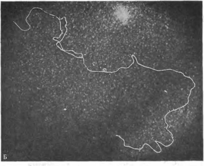
FIG. 2-23. A. DNA double helix; B-form. (Arnott S., Hukins D. W. L., JMB, 81, 93—105, 1975.) B. Electron micrograph of a bacterial virus DNA molecule (bacteriophage T7) caught in the act of replication. The viral DNA appears as a long (~14 µm) duplex rod containing approximately 40,000 Base Pairs. A small replication "eye"—the region where DNA duplication is underway—is clearly visible. DNA Synthesis initiates at a specific site located 17% of the molecular length from one end of the duplex. Uranyl acetate staining; negative contrast. (Courtesy of T. Wolfson and D. Dressler.)
The geometry of The Double Helix is illustrated in Fig. 2-23, A. In fibers, DNA molecules can adopt two Conformations: A and B. The A-form1 is characterized by base-pair planes tilted by approximately 20° relative to the normal to the helix axis, whereas in the B-form, which occurs at high humidity, the planes are nearly perpendicular to the axis (backbone torsion angles are ω = 155°, ξ = 36°, θ = —146°, φ = —96°, and ψ = 46°) [65]. Intracellular DNA is generally believed to exist predominantly in the B-form.
1 This structure was derived through molecular modeling combined with X-Ray Diffraction data obtained by Wilkins and Franklin from oriented DNA fibers. For this discovery, Watson, Crick, and Wilkins were awarded the Nobel Prize in 1962.
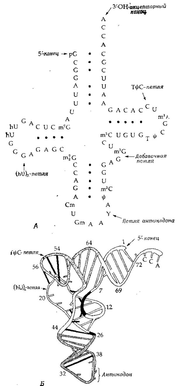
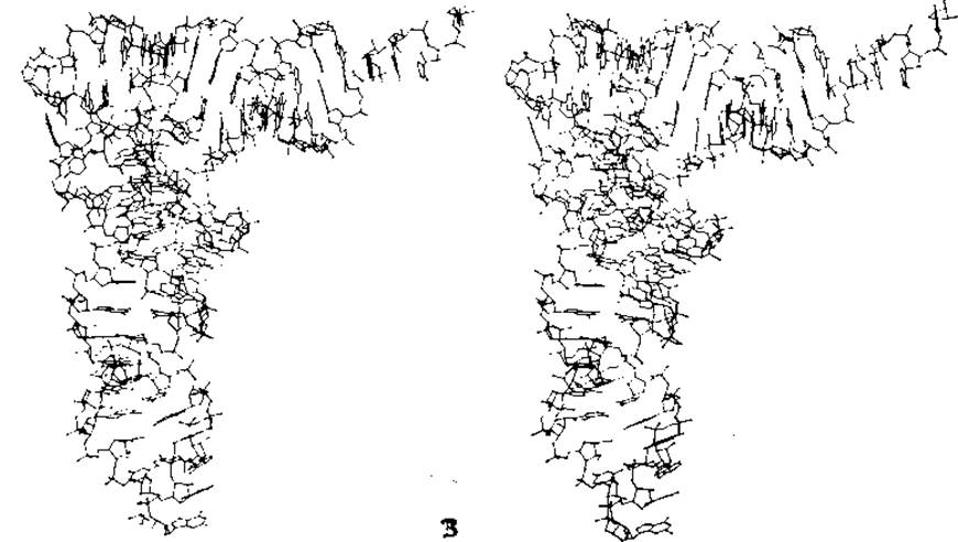
FIG. 2-24. Structure of Yeast phenylalanine Transfer RNA. A. Nucleotide sequence forming the traditional "cloverleaf" structure. B. Schematic diagram of the polynucleotide chain folding. The ribose-phosphate backbone is depicted as a continuous ribbon; straight bars connecting different segments represent hydrogen-bonded base pairs, while unpaired bases are shown as short bars. The TΨC loop is highlighted with dots, and the anticodon loop with vertical hatching. Tertiary-structure interactions are indicated by solid black bars. (Quigley G. J., Rich A., Science, 194, 796—806, 1976.) C. Stereoview of yeast phenylalanine tRNA based on X-ray Diffraction Analysis. The acceptor stem with its protruding 3'-terminal ACCA sequence is on the right, and the GAA anticodon is in the lower right. The guanine ring at the very bottom of the figure is particularly prominent. The plane of the middle adenine of the anticodon is oriented strictly perpendicular to the page, as is that of the "hypermodified" Y base (see Fig. 15-10), which lies directly above the anticodon with its side chain pointing away from the viewer. Directly preceding the anticodon in the 5'-half of the molecule are two unpaired bases (C and U). The 2'-hydroxyl groups of cytidine and the anticodon guanosine are methylated. Moving upward along the anticodon loop, we encounter two base triplets that follow both Watson-Crick and Hoogsteen pairing schemes. Guidelines for viewing such stereopigures are provided in the caption to Fig. 2-6. (Courtesy of A. Rich.)
Neglecting the slight Asymmetry of base pairs, one can observe that in the double helix, the two nucleotide chains are related by a 2-fold axis of Symmetry. This symmetry element, arising from the antiparallel arrangement of the strands, makes the DNA molecule appear identical from both ends—whether viewed by a human inspecting the model or by an enzyme interacting with the molecule. In reality, the two strands are not identical, and genetic information is read from specific regions exposed On the surface within the major groove (Fig. 2-23, A).
1 Strictly speaking, we are dealing here with a family of closely related conformations.
Some key dimensions of the double helix are as follows (Fig. 2-23): its diameter, defined by the distance between phosphorus atoms, is exactly 2.0 nm. The pitch of the helix is 3.4 nm, with ten base pairs per turn. Thus, the distance between adjacent base-pair planes is 0.34 nm, which roughly corresponds to the sum of the Van der Waals radii of the aromatic rings (Table 2-1). Consequently, the bases are stacked centrally within the helix. A typical gene of 1,000 base pairs spans a DNA segment approximately 340 nm in length (Fig. 2-23, B).
As befits a master template designed for accurate copying, the DNA double helix is exceptionally stable. Despite its great length, it is rarely cleaved under physiological conditions. This structural stability is maintained by several factors: (1) hydrogen bonding between bases; (2) van der Waals attraction between the parallel-stacked planar bases; (3) numerous oxygen atoms on the molecular surface—both charged and neutral—capable of forming hydrogen bonds with Water or surrounding specific Proteins; and (4) the capacity to form various superhelical structures (see below).
As noted above, DNA can transition into the paracrystalline A-form (characterized by tilted base orientations and 11 base pairs per turn). This suggests that this conformation may be just as biologically significant as the B-form. Although RNA molecules are typically single-stranded, they frequently form hairpin loops—double-stranded segments adopting the A-form [71]. The B-conformation is precluded in RNA by the presence of 2'-hydroxyl groups on the ribose sugar. It is also believed that Cells form transient DNA-RNA hybrid double helices, which likewise appear to be restricted to the A-form. Notably, the A-form differs from the B-form in possessing a relatively wide axial cavity (~0.8 nm in diameter) and a deeper major groove [71a]. Unlike the structure shown in Fig. 2-23, the base-pair planes in the A-form do not intersect the helical axis.
The structure of low-molecular-weight tRNA has been studied in the greatest detail. All these molecules contain double-stranded regions stabilized by hydrogen bonds to form three hairpins, occasionally supplemented by a fourth (the "cloverleaf"). The three-dimensional structure of one such tRNA was solved by X-ray crystallography [72—74] (Fig. 2-24). The irregularity and complexity of its folding place it on par with Globular proteins. Of particular note is the anticodon (base triplet) located at the bottom of the figure, whose structure ensures precise pairing with the three bases of the codon that specifies a particular amino acid—in this case, phenylalanine.
Last update: 06/08/2026
Editorial and Educational Adaptation: This material has been compiled based on the primary/original source text. The project team performed an editorial review, corrected technical inaccuracies, structured sections, and adapted the content for an educational format.
What was processed:
- elimination of formatting defects (OCR errors, structural breaks, corrupted characters);
- editorial organization of content;
- standardization of terminology in accordance with academic sources;
- verification of factual statements against the original source text.
All mentions of the author, publication year, and origin of the primary text have been preserved in accordance with the source.