Genetics - A. V. Sivolob 2008
The Nature of Genetic Material
DNA Structure
The Double Helix
Two polynucleotide chains (primarily DNA, but in certain cases also RNA or hybrid RNA-DNA) can combine into a single double-stranded Structure (duplex), the scheme of which is presented in Fig. 1.4. Such an association occurs under a strict condition: specific nitrogenous bases must face each other—A opposite T (or U), G opposite C. This principle of complementarity, formulated by Watson and Crick (James D. Watson, Francis H. C. Crick), is driven by The formation of specific Hydrogen Bonds between the exocyclic groups of these bases: two bonds in the A-T pair, and three in the G-C pair (Fig. 1.5). THE PRINCIPLE OF complementarity is key to understanding the functioning of Nucleic Acids.
Class="center">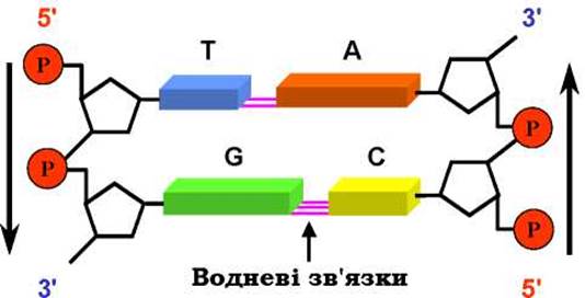
Fig. 1.4. Scheme of the combination of two dinucleotides into a double-stranded structure
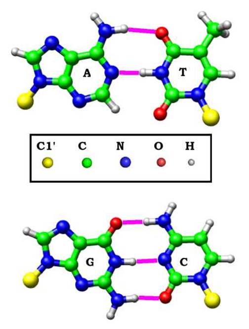
Fig. 1.5. Complementary Base Pairs within DNA with hydrogen bonds between the bases
The two chains within the duplex run in opposite directions (are antiparallel), with the sugar-phosphate backbones (which interact well with Water) located on the outside, and the base pairs on the inside of this structure. As a result of interactions between the planes of neighboring base pairs (base-stacking interactions), the polynucleotide chains twist around each other into a double helix (Fig. 1.6). Two grooves of different sizes—a major and a minor groove—are formed between the backbones On the surface of the helix, into which specific exocyclic groups of nitrogenous bases, not involved in forming complementary hydrogen bonds, are "exposed".
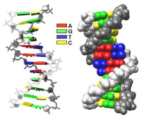
Fig. 1.6. Two Variants of the DNA double helix representation.
The structure of a DNA dodecamer in crystals is shown (Protein Data Bank structure code 355D). The image was generated using UCSF Chimera software (http://www.cgl.ucsf.edu/chimera)
Under physiological conditions, the double helix is a fairly stable structure, and it is in this form that DNA exists in living systems. Various RNA molecules are generally single-stranded, but individual self-complementary regions of RNA often also form double-Helical structures within a single molecule.
Nucleic acid Double helices exhibit considerable structural polymorphism, which depends on the base pair sequence, the type of pentose sugar, and external conditions. The structure shown in Fig. 1.6 is the so-called B-form of DNA: a right-handed helix with ~10.5 base pairs per turn. It is in this form that DNA exists under physiological conditions in vivo.
Another form—the A-form (also a right-handed helix, ~11 base pairs per turn, with a significant tilt of the base pair planes relative to the helical axis)—is adopted by DNA only in vitro under certain conditions far removed from physiological ones. However, it is precisely in the A-form that RNA double helices exist under physiological conditions (due to the substitution of deoxyribose with ribose). Furthermore, DNA can transition into the A-form, or a conformation close to it, when complexed with Proteins.
Yet another form of the double helix—the Z-form—is a left-handed helix and is formed exclusively by alternating poly(GC) sequences (where G and C alternate within the chain). Such sequences are present in natural DNAs, but the transition to the Z-form occurs in vitro under conditions very far from physiological. The Biological Significance of the Z-form remains somewhat unclear, although proteins with a high affinity specifically for this form have been discovered, meaning they can induce the B→Z transition in vivo.
The STRUCTURE OF THE main physiological B-form of DNA is not completely regular. The conformational Features of the double helix (the degree of twist, local bends, groove dimensions, etc.) significantly depend on the base pair sequence—one might say that the sequence carries information about the Structural Features of DNA (much like an Amino Acid Sequence determines the three-dimensional structure of a protein). In addition, the base pair sequence determines variations in double helix stability (dictating how easily the polynucleotide chains can be separated) and conformational flexibility (the ability of the helix to undergo deformations—bending, changes in helical periodicity, etc.). These sequence-dependent features of double helix structure and its potential for conformational changes form The basis of the mechanism by which proteins specifically recognize DNA sequences.
Last update: 11/08/2026
Editorial and Educational Adaptation: This material has been compiled based on the primary/original source text. The project team performed an editorial review, corrected technical inaccuracies, structured sections, and adapted the content for an educational format.
What was processed:
- elimination of formatting defects (OCR errors, structural breaks, corrupted characters);
- editorial organization of content;
- standardization of terminology in accordance with academic sources;
- verification of factual statements against the original source text.
All mentions of the author, publication year, and origin of the primary text have been preserved in accordance with the source.