Human Anatomy, Part 1 - K. A. Dyubenko, A. K. Kolomiyytsev, Yu. B. Chaykovsky 2002
Special Part
Splanchnology, splanchnologia [the study of viscera] — Major Digestive Glands
Variants of the relative positioning of the cystic duct and common hepatic duct
The following anatomical variations in the relative positions of the cystic and common hepatic ducts are distinguished (Fig. 259):
- typical arrangement;
- short cystic duct;
- short common hepatic duct;
- cystic duct crossing anterior to the common hepatic duct;
- cystic duct crossing posterior to the common hepatic duct;
- cystic and common hepatic ducts located side by side at a certain distance;
- separate entry of the common hepatic and cystic ducts into the duodenum (Bile reaches the Gallbladder via Luschka's ducts).
Class="center">
Fig. 259. Variations in the relative positions of the cystic and common hepatic ducts (diagram) (after S. S. Mykhailov):
A - typical arrangement; B - short cystic duct; C - short common hepatic duct; D - cystic duct crossing anterior to the common hepatic duct; E - cystic duct crossing posterior to the common hepatic duct; F - cystic and common hepatic ducts located side by side; G and H - cystic and common hepatic ducts emptying separately into the duodenum
Occasionally, all three ducts empty separately into the duodenum. Cases are also observed where the bile duct communicates with an accessory pancreatic duct. These anatomical variations are of great significance when analyzing the causes of BILE AND PANCREATIC juice flow into the duodenum and during surgical interventions on the biliary ducts.
Blood supply. The gallbladder receives its blood supply from the cystic artery, a. cystica (a branch of a. hepatica propria). This artery is of vital surgical importance during cholecystectomy (holecystectomia). Calot's triangle (tr. Calot) serves as a landmark for locating and ligating this vessel (Fig. 260). Its boundaries are: to the right — the cystic duct, ductus cysticus; to the left — the common hepatic duct, ductus hepatis communis; superiorly — the Base of the Liver. It contains the proper hepatic artery, a. hepatica propria, the cystic artery, a. cystica, and the cystic duct Lymph node. Venous drainage from the gallbladder is carried out by 3 to 4 Veins located along its surfaces, which drain into the intrahepatic Branches of the portal vein.
Lymphatic vessels. The wall of the gallbladder (mucous and serous membranes) contains networks of lymphatic vessels (Fig. 261). There is also a plexus of lymphatic capillaries in the submucosa. These capillaries anastomose with the superficial Vessels of the liver. Lymph drainage is directed into the hepatic Lymph Nodes, nodi lymphatici hepatici, located near the neck of the gallbladder in the porta hepatis and along the common bile duct, as well as into the lymphatic bed of the liver.
Innervation. The gallbladder is innervated by the hepatic nerve plexus, pl. hepaticus, formed by branches of the celiac plexus, anterior Vagus nerve, phrenic nerves, and branches of the gastric nerve plexus.
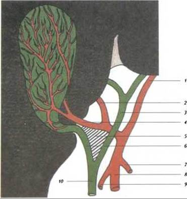
Fig. 260. Blood supply to the gallbladder (diagram) (after S. S. Mykhailov):
1 - left branch of the proper hepatic artery; 2 - right branch of the proper hepatic artery; 3 - common hepatic duct; 4 - cystic artery; 5 - Calot's triangle (tr. Calot); 6 - cystic duct; 7 - right gastric artery; 8 - common hepatic artery; 9 - gastroduodenal artery; 10 - common bile duct

Fig. 261. Networks of lymph capillaries and postcapillaries of the serous membrane of a newborn's gallbladder. (Prepared by O. A. Sushko)
Last update: 08/08/2026
Editorial and Educational Adaptation: This material has been compiled based on the primary/original source text. The project team performed an editorial review, corrected technical inaccuracies, structured sections, and adapted the content for an educational format.
What was processed:
- elimination of formatting defects (OCR errors, structural breaks, corrupted characters);
- editorial organization of content;
- standardization of terminology in accordance with academic sources;
- verification of factual statements against the original source text.
All mentions of the author, publication year, and origin of the primary text have been preserved in accordance with the source.