Human Anatomy - Kotsan I. Y. 2009
Vessels of the Lesser (Pulmonary) Circulation
Veins of the Systemic Circulation
System of the Inferior Vena Cava
The INFERIOR VENA CAVA (v. cava inferior) is the largest vein in The Human Body. It lies retroperitoneally alongside the aorta, to the right of it (Fig. 216). The inferior vena cava is formed at the level of the IV lumbar vertebra by the merger of the right and left common iliac Veins, slightly inferior to the bifurcation of the aorta into the corresponding iliac Arteries. From its origin, it ascends to the right of the Abdominal Aorta, passes through the caval opening in the Diaphragm into the thoracic cavity, and empties into the right atrium.
The tributaries of the inferior vena cava are divided into parietal (parietal) and visceral.
Parietal tributaries collect Blood from the upper and posterior walls of the Abdominal cavity. These include the following veins.
1. Lumbar veins (vv. lumbales), three or four on each side, lie on the posterior abdominal wall and collect blood from body regions corresponding to the branching of the lumbar arteries. Frequently, the two upper lumbar veins drain into the azygos vein rather than the inferior vena cava. The lumbar veins on each side anastomose via the ascending lumbar vein (v. lumbalis ascendens). Blood drains into the lumbar veins from the vertebral venous plexuses via spinal branches.
2. Inferior phrenic veins (vv. phrenicae inferiores), in pairs, accompany the corresponding arteries and empty into the inferior vena cava immediately below the diaphragm.
Visceral tributaries of the inferior vena cava collect blood from the paired abdominal Organs and the Liver. These include the following veins.
1. Testicular (ovarian) vein (v. testicularis (ovarica)) is paired and originates from the posterior border of the Testis (or the hilum of the Ovary) as multiple veins that entwine the corresponding artery, forming the pampiniform plexus (plexus pampiniformis), which in males is a component of the Spermatic Cord. Merging together, these small veins form a single venous trunk on each side. The right testicular vein empties directly into the inferior vena cava at an acute angle, while the left empties into the left renal vein at a right angle. This anatomical feature impedes blood outflow and accounts for the higher incidence of left-sided Varicocele compared to the right.
2. Renal vein (v. renalis) is paired, runs horizontally from the renal hilum, and empties into the inferior vena cava at the level of the intervertebral disc between the First and Second lumbar vertebrae. The left renal vein is longer than the right and crosses anterior to the abdominal aorta along its course.
3. Suprarenal (adrenal) vein (v. suprarenalis) is paired. It is a short, valveless vessel emerging from the hilum of the Adrenal gland. The left suprarenal vein (v. suprarenalis sinistra) empties into the left renal vein, whereas the right suprarenal vein (v. suprarenalis dextra) drains into the inferior vena cava.
4. Hepatic veins (vv. hepaticae), numbering 3 to 4, are located within the liver parenchyma and empty into the inferior vena cava where it courses along the posterior surface of the liver (at the level of the groove for the vena cava). The hepatic veins carry blood away from the liver, where it arrives via the portal vein and hepatic artery.
Class="center">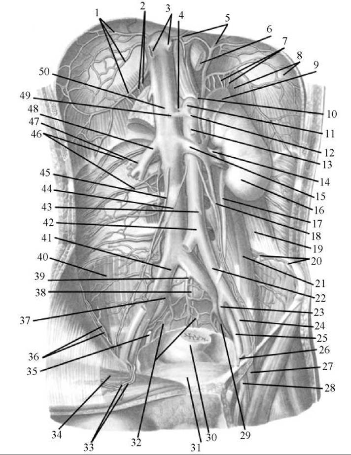
Fig. 216. Inferior vena cava and abdominal aorta (anterior view)
1 — right inferior phrenic veins; 2 — right suprarenal veins; 3 — hepatic veins; 4 — left gastric artery; 5 — left inferior phrenic veins; 6 — Esophagus; 7 — left suprarenal veins; 8 — diaphragm; 9 — left suprarenal gland; 10 — left inferior phrenic artery; 11 — celiac trunk; 12 — splenic artery; 13 — superior mesenteric artery; 14 — left renal artery; 15 — left renal vein; 16 — left Kidney; 17 — Ureter; 18 — left testicular vein; 19 — quadratus lumborum Muscle; 20 — IV lumbar artery and veins; 21 — psoas major muscle; 22 — left common iliac artery; 23 — left internal iliac artery; 24 — left external iliac artery; 25 — left external iliac vein; 26 — inferior epigastric artery; 27 — femoral artery; 28 — femoral vein; 29 — left internal iliac vein; 30 — rectum; 31 — Urinary Bladder; 32 — sacral venous plexus; 33 — inferior epigastric artery and vein; 34 — rectus abdominis muscle; 35 — ureter; 36 — deep circumflex iliac artery and vein; 37 — right internal iliac vein; 38 — median sacral artery; 39 — median sacral vein; 40 — iliacus muscle; 41 — right common iliac vein; 42 — abdominal aorta; 43 — inferior mesenteric artery; 44 — right testicular artery; 45 — right testicular vein; 46 — lumbar artery and vein; 47 — right renal artery; 48 — right renal vein; 49 — common hepatic artery; 50 — inferior vena cava
Last update: 08/08/2026
Editorial and Educational Adaptation: This material has been compiled based on the primary/original source text. The project team performed an editorial review, corrected technical inaccuracies, structured sections, and adapted the content for an educational format.
What was processed:
- elimination of formatting defects (OCR errors, structural breaks, corrupted characters);
- editorial organization of content;
- standardization of terminology in accordance with academic sources;
- verification of factual statements against the original source text.
All mentions of the author, publication year, and origin of the primary text have been preserved in accordance with the source.