Human Anatomy - Kotsan I. Ya. 2009
Vessels of the Lesser (Pulmonary) Circulation
Veins of the Systemic Circulation
Veins of the Upper Extremity
The Veins OF THE upper limb are divided into superficial and deep ones. They communicate with each other through A large number of anastomoses and contain numerous Valves.
The superficial (subcutaneous) veins of the upper limb (vv. superficiales membri superioris) are more developed than the deep ones. They lie within the subcutaneous adipose tissue of the upper limb and collect Blood from the Skin, subcutaneous tissue, and the venous plexuses located within them. The largest superficial venous trunks, which include the cephalic and basilic veins, originate from the dorsal venous network of the hand (rete venosum dorsale manus) (Fig. 215).
The cephalic vein (v. cephalica) appears as a continuation of the first dorsal metacarpal vein, originating from the radial part of the dorsal venous network of the hand. It ascends initially along the radial border of the forearm, then shifts to its anterior surface at the junction of the lower and middle thirds, and reaches the cubital fossa. Here, via the median cubital vein (v. mediana cubiti), it anastomoses with the basilic vein and continues into the arm. In the arm, the cephalic vein runs in the lateral bicipital sulcus, and more superiorly, in the groove between the deltoid and pectoralis major Muscles. Upon reaching the clavicle, it pierces the clavipectoral fascia and empties into the axillary vein. For a long time, it was believed that phlebotomy of this vein drained blood from the HEAD (relieving headaches during bloodletting). Hence its name — the "cephalic" (main) vein.
The basilic vein (v. basilica) is a continuation of the fourth dorsal metacarpal vein, originating from the ulnar side of the dorsal venous network of the hand. From there, it passes to the ulnar side of the anterior surface of the forearm and runs toward the cubital fossa, where it anastomoses with the cephalic vein via the median cubital vein. Afterwards, the basilic vein ascends along the medial bicipital sulcus into the arm and, at approximately the middle of the arm, empties into one of the brachial veins. It was named the "basilic" vein because it was historically frequently incised to treat Liver diseases, which were considered of primary importance.
The median cubital vein (v. mediana cubiti) is an obliquely positioned anastomosis connecting the cephalic and basilic veins in the elbow region. It originates from the cephalic vein in the upper third of the forearm, runs inferiorly to superiorly and medially, crosses the cubital fossa obliquely, and empties into the basilic vein. The median cubital vein is of great practical significance as it serves as a site for intravenous drug administration, blood transfusions, and blood sampling for Laboratory tests.
The median cubital vein does not always exist as a single trunk. Frequently, In addition to the cephalic and basilic veins, the median antebrachial vein (v. mediana antebrachii) is located in the forearm. In the anterior cubital region, it empties into the median cubital vein or divides into two branches that independently flow into the cephalic and basilic veins.
The deep veins of the upper limb (vv. profundae membri superioris) accompany the Arteries and share the same names. Each artery is accompanied by two veins. The only exceptions are the veins of the digits and the axillary vein. All deep veins of the upper limb have numerous tributaries in the form of small veins that collect blood from the bones, muscles, joints, ligaments, and other deep structures of the upper limb. The deep veins feature numerous anastomoses among themselves and with the superficial veins.
In the region of the palmar surface of the hand, the deep veins form two venous arches: superficial and deep.
The superficial palmar venous arch (arcus venosus palmaris superficialis) is poorly developed, lies near the superficial arterial arch, and receives the palmar digital veins (vv. digitales palmares).
The deep palmar venous arch (arcus venosus palmaris profundus) consists of two anastomosing veins that receive the paired palmar metacarpal veins (vv. metacarpales palmares). The deep and superficial palmar venous arches continue into the deep veins of the forearm — the paired ulnar and radial veins (vv. ulnares et vv. radiales), which accompany the corresponding arteries. In the region of the cubital fossa, the ulnar and radial veins join to form two brachial veins (vv. brachiales) that accompany the brachial artery. The union of the two brachial veins forms the axillary vein (v. axillaris), which lies medially and anteriorly to the corresponding artery in the axilla, partially covering it. Passing beneath the clavicle, it continues further as the subclavian vein (v. subclavia). Apart from the aforementioned cephalic vein, The most significant tributaries of the axillary vein are the lateral thoracic vein (v. thoracica lateralis), which receives the thoracoepigastric veins (thoracoepigastricae) and delicate veins branching off from the I-VII posterior intercostal veins.
Class="center">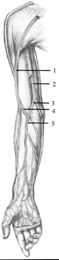
Fig. 215. Superficial (subcutaneous) veins of the right upper limb
1 — cephalic vein; 2 — basilic vein; 3 — median basilic vein; 4 — median cephalic vein; 5 — median cubital vein
Last update: 08/08/2026
Editorial and Educational Adaptation: This material has been compiled based on the primary/original source text. The project team performed an editorial review, corrected technical inaccuracies, structured sections, and adapted the content for an educational format.
What was processed:
- elimination of formatting defects (OCR errors, structural breaks, corrupted characters);
- editorial organization of content;
- standardization of terminology in accordance with academic sources;
- verification of factual statements against the original source text.
All mentions of the author, publication year, and origin of the primary text have been preserved in accordance with the source.