Human Anatomy - Kotsan I. Ya. 2009
Vessels of the Lesser (Pulmonary) Circulation
Veins of the Greater Circulation
Portal Vein System
The portal vein collects Blood from all unpaired abdominal Organs except the Liver: from the gastrointestinal tract, where the absorption of nutrients takes place, which are then transported via the portal vein to the liver for purification and Glycogen storage; from the Pancreas, which supplies Insulin to regulate glucose METABOLISM; and from the Spleen, which delivers blood Cell breakdown products used by the liver to produce Bile. The structural connection of the portal vein with the gastrointestinal tract and its major glands (The Liver and pancreas) is determined not only by their functional relationship, but also by their common developmental origin (genetic relationship).
Class="center">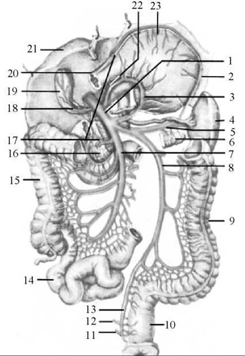
Fig. 217. The portal vein and its tributaries
1 — portal vein; 2 — left gastro-omental (gastroepiploic) vein; 3 — left gastric vein; 4 — spleen; 5 — splenic vein; 6 — tail of the pancreas; 7 — superior mesenteric vein; 8 — inferior mesenteric vein; 9 — descending colon; 10 — rectum; 11 — inferior rectal vein; 12 — middle rectal vein; 13 — superior rectal vein; 14 — ileum; 15 — ascending colon; 16 — HEAD of the pancreas; 17 — right gastro-omental (gastroepiploic) vein; 18 — cystic vein; 19 — Gallbladder; 20 — duodenum (cut); 21 — liver; 22 — prepyloric vein; 23 — Stomach
The portal vein (v. portae) is a thick venous trunk (5–6 cm long, 11–18 mm in diameter) (Fig. 217). It is located within the thickness of the hepatoduodenal ligament, posterior to the hepatic artery and common bile duct, alongside nerves, Lymph Nodes, and vessels. The portal vein is formed behind the pancreatic head by the confluence of two Veins: the superior mesenteric vein and the splenic vein (which receives the inferior mesenteric vein). As it ascends toward the porta hepatis, it receives the right and left gastric veins (v. gastrica dextra et v. gastrica sinistra) and the prepyloric vein (v. prepylorica), which drain blood from the respective PARTS OF THE stomach. Upon entering the porta hepatis, the portal vein divides into right and left branches (r. dexter et r. sinister). Each of these branches subsequently subdivides into segmental branches, and then into progressively smaller branches that transition into interlobular veins. Within the lobules, they break up into wide capillaries, known as sinusoids, which empty into the central vein. The central veins anastomose to form sublobular (collecting) veins, which ultimately converge to form 3–4 hepatic veins that empty into the INFERIOR VENA CAVA. Thus, the blood flowing into the inferior vena cava via the hepatic veins passes through two capillary beds along its pathway: one located in the wall of the digestive tract, where the tributaries of the portal vein originate, and the second in the hepatic parenchyma, represented by the capillaries of its lobules.
Tributaries of the portal vein:
1. The superior mesenteric vein (v. mesenterica superior) is located in the ROOT of the mesentery of the Small Intestine. Its tributaries include: jejunal and ileal veins (vv. jejunales et ileales); pancreatic veins (vv. pancreaticae); pancreaticoduodenal veins (vv. pancreaticoduodenales); ileocolic vein (v. ileocolica); right gastro-omental vein (v. gastroomentalis dextra); right colic vein (v. colica dextra); middle colic vein (v. colica media); and appendicular vein (v. appendicularis). Through these tributaries, the superior mesenteric vein collects blood from the walls of the jejunum, ileum, and their mesentery, the cecum and Appendix, the ascending and transverse colons, and partially from The Stomach, duodenum, and pancreas.
2. The splenic vein (v. splenica) collects blood from the spleen, the gastric fundus, the pancreas, and the greater omentum. It runs along the superior border of the pancreas, inferior to the splenic artery. It is formed in the region of the splenic hilum by numerous splenic veins emerging from the spleen substance. Passing from left to right posterior to the pancreatic head, it unites with the superior mesenteric vein. Its tributaries are pancreatic veins (vv. pancreaticae), short gastric veins (vv. gastricae breves), and the left gastro-omental vein (v. gastroomentalis sinistra).
3. The inferior mesenteric vein (v. mesenterica inferior) is the continuation of the superior rectal vein (v. rectalis superior), which originates from the rectal venous plexus. Ascending from its origin, it receives the sigmoid veins (vv. sigmoideae), which drain the sigmoid colon, and the left colic vein (v. colica sinistra), which collects blood from the descending colon. Posterior to the pancreatic head, the inferior mesenteric vein empties into the superior mesenteric vein, either directly after joining the splenic vein or independently.
Last update: 08/08/2026
Editorial and Educational Adaptation: This material has been compiled based on the primary/original source text. The project team performed an editorial review, corrected technical inaccuracies, structured sections, and adapted the content for an educational format.
What was processed:
- elimination of formatting defects (OCR errors, structural breaks, corrupted characters);
- editorial organization of content;
- standardization of terminology in accordance with academic sources;
- verification of factual statements against the original source text.
All mentions of the author, publication year, and origin of the primary text have been preserved in accordance with the source.