Human Anatomy - Kotsan I. Y. 2009
Urogenital system
Urinary organs
Kidney
The Urinary Organs (organa urinaria) remove unnecessary Metabolic waste products from the body. While some of these substances are eliminated through the Skin, respiratory tract, and digestive canal, the Urinary system serves as the primary excretory pathway. Depending on their specific Functions, urinary organs are divided into two groups: urine-producing organs (the Kidneys) and urine-excreting organs (the Ureters, Urinary Bladder, and Urethra).
The kidney (ren) is a paired organ responsible for Urine Formation and Excretion. It features a bean-like shape, a dark red color, and a firm consistency. In an adult, the kidney measures 10–12 cm in length, 5–6 cm in width, and 4 cm in thickness, with a mass of 150–200 g. The left kidney is slightly longer than the right and occasionally heavier. The kidneys are situated in the lumbar region on the posterior abdominal wall's internal surface, occupying the retroperitoneal space on either side of THE Vertebral Column, roughly spanning from the 11th thoracic vertebra to the 3rd lumbar vertebra. They lie obliquely, with their upper poles positioned closer to the median line than the lower poles. The longitudinal axes of the right and left kidneys intersect to form an inferiorly open angle. The right kidney lies 2–3 cm lower than the left, an Asymmetry caused by the Liver's position on the right side. The upper pole of the left kidney reaches the middle of the 11th thoracic vertebra, whereas the upper pole of the right kidney reaches the lower margin of this vertebra. The lower pole of the left kidney aligns with the upper margin of the 3rd lumbar vertebra, while the lower pole of the right kidney corresponds to its midpoint. Renal positioning also varies individually, categorized as high or low placement. In women, the lower poles of both kidneys frequently Touch the iliac crests.
The kidneys maintain complex anatomical relationships with adjacent organs. The posterior surface of the kidneys, along with their capsules, abuts the Diaphragm, quadratus lumborum, transversus abdominis, and psoas major Muscles, which together form a recess known as the renal bed. The superior pole of each kidney contacts the suprarenal (adrenal) gland. The majority of the anterior renal surface is covered by a layer of parietal Peritoneum and comes into contact with internal viscera. The upper two-thirds of the anterior surface of the right kidney lie against the liver, while the lower third touches the right colic (hepatic) flexure. The descending part of the duodenum adjoins the medial margin of the right kidney. On the left side, the upper third of the anterior surface contacts The Stomach, the middle third touches the Pancreas, and the lower third meets loops of the jejunum. The lateral margin of the left kidney neighbors the Spleen and the left colic (splenic) flexure. Normal topographic placement of the kidneys is maintained by a fixation apparatus comprising the renal bed, renal pedicle, and renal capsules (particularly the adipose capsule and renal fascia). Intra-abdominal pressure, sustained by the contraction of the abdominal wall musculature, also plays a crucial role in anchoring the kidneys.
The kidney presents two surfaces: a convex anterior surface (facies anterior) and a flatter posterior surface (facies posterior); two poles (extremities): a superior pole and an inferior pole (extremitas superior et extremitas inferior); and two margins: a medial margin and a lateral margin (margo medialis et margo lateralis). The lateral margin is convex, whereas the medial margin is concave. Near the center of the medial margin lies a depression known as the renal hilum (hilum renale), through which the renal artery and nerves enter, and the Ureter, renal vein, and Lymphatic vessels exit. These structures collectively form the renal pedicle. The renal hilum leads into a large cavity called the renal sinus (sinus renalis).
Externally, the kidney is enveloped in three layers. Closely investing the kidney is the fibrous capsule (capsula fibrosa), which can be easily stripped away. External to the fibrous capsule lies a layer of adipose tissue known as the adipose capsule (capsula adiposa), which extends through the renal hilum into the renal sinus. The adipose capsule is thickest on the posterior surface of the kidney, forming a distinct fat pad called the pararenal fat body (corpus adiposum pararenale). During rapid weight loss,
when the thickness of the adipose capsule decreases sharply, the kidney may become excessively mobile (floating kidney), a condition that can require surgical fixation (nephropexy) to the diaphragm. Externally, the kidney and its fibrous and adipose capsules are enclosed by the renal fascia (fascia renalis), which consists of two layers: an anterior (prerenal) layer and a posterior (retrorenal) layer. Along the lateral margins of the kidneys, these two layers fuse and transition into the subperitoneal Connective Tissue from which they originated. At the medial margin, the anterior and posterior layers of the renal fascia do not unite but continue separately toward the median line: the anterior layer passes anterior to the renal vessels, aorta, and INFERIOR VENA CAVA to merge with the corresponding layer of the opposite side, whereas the posterior layer passes anterior to the vertebral bodies, anchoring to the lateral aspects of the vertebral column on both the right and left. Near the upper poles of the kidneys, the two fascial layers fuse, restricting superior mobility; near the lower poles, they do not connect and instead blend into the subperitoneal adipose tissue of the iliac fossa. Through Fibrous connective tissue strands, the renal fascia is intimately connected to both the adipose and fibrous capsules of the kidney and transitions directly into the subperitoneal Fascia of the Abdominal cavity.
Structure OF THE kidney. The kidney consists of two main parts: the renal sinus (the renal cavity) and the renal substance (renal parenchyma) (Fig. 170).
Class="center">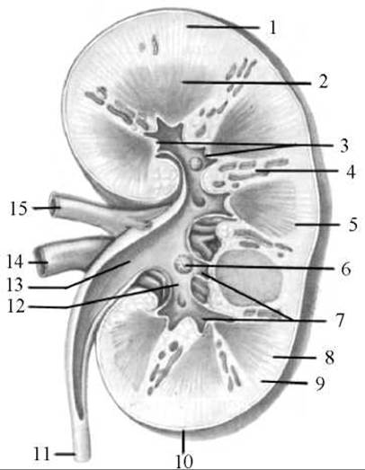
Fig. 170. Frontal section of the kidney
1 — renal cortex (cortex); 2 — renal medulla; 3 — renal papillae; 4 — renal column; 5 — Base of the pyramid; 6 — cribriform area; 7 — minor renal calyces; 8 — pars radiata (cortical labyrinth); 9 — pars convoluta; 10 — fibrous capsule; 11 — ureter; 12 — major renal calyx; 13 — renal pelvis; 14 — renal vein; 15 — renal artery
The renal sinus (sinus renalis) is a continuation of the renal hilum. Its component structures include the minor and major renal calyces and the renal pelvis.
The renal substance (substantia renalis) appears inhomogeneous on cross-section. It comprises two layers that differ in density and coloration: an outer layer—the renal cortex or cortical substance (cortex renalis s. substantia corticalis)—and an inner layer—the renal medulla or medullary substance (medulla renalis s. substantia medullaris). The renal cortex has a yellowish-red color and is formed primarily by renal corpuscles, where urine is produced. The renal medulla is denser than the cortex, exhibits a bluish-red hue, and consists mainly of the tubular systems through which urine flows away.
The renal cortex, which is approximately 5 mm thick, not only forms the surface layer of the kidney but also extends inward between the medullary structures, forming the renal columns (columnae renales). The cortex is heterogeneous, consisting of alternating light and dark regions. The light areas are cone-shaped and appear as rays extending from the medulla into the cortex; these form the radiate part (pars radiata) of the cortex, which contains the straight renal tubules (continuing into the medulla) and the initial segments of the collecting ducts. The dark areas of the cortical substance are known as the convoluted part (pars convoluta). These regions house the renal corpuscles along with the proximal and distal convoluted tubules.
Unlike the cortex, the renal medulla does not form a continuous layer; on cross-section, it appears as discrete triangular zones separated by the renal columns. These zones are called renal pyramids (pyramides renales), numbering between 10 and 15 per kidney. Each renal pyramid features a base (basis pyramidis) directed toward the cortex and an apex (apex pyramidis) pointing toward the renal hilum. The apices of two to three pyramids fuse to form a renal papilla (papilla renalis), which projects into the renal sinus. The surface of the papilla is designated as the cribriform area (area cribrosa) because it is perforated by 15 to 25 papillary foramina (foramina papillaria) through which urine is discharged into the initial segments of the Urinary Tract. At the apex of each pyramid, the renal papilla is embraced by a funnel-shaped cavity called a minor renal calyx (calix renalis minor). Occasionally, a single minor calyx encompasses two or even three renal papillae. Two to three minor calyces unite to form a major renal calyx (calix renalis major). A kidney typically contains 2 to 3 major calyces, which subsequently merge to form the renal pelvis (pelvis renalis), which narrows as it transitions into the ureter. The shape of the renal pelvis may be ampullary, dendritic (branching), or mixed.
The walls of the renal pelvis, major calyces, and minor calyces share an identical structure consisting of three layers: an outer adventitial tunic, a middle muscular tunic, and an inner mucous membrane (mucosa). Smooth Muscle Cells within the wall of the minor calyx at its fornix (the initial region) form a slightly thickened circular layer known as the sphincter of the fornix. This circular muscle layer, together with adjacent nerve fibers, Blood Vessels, and lymphatic vessels, constitutes the fornical apparatus of the kidney. Its physiological role is to regulate the rate at which urine is discharged from the renal tubules into the minor calyces, prevent the backflow of urine, and regulate intrapelvic pressure and the body's Water balance.
Based on its structural and vascular features, the renal substance can be divided into five segments: the superior segment (segmentum superius), which corresponds to the upper pole of the kidney; the anterior superior segment (segmentum anterius superius), located anterior to the renal pelvis; the anterior inferior segment (segmentum anterius inferius), also situated anterior to the renal pelvis; the inferior segment (segmentum inferius), corresponding to the lower pole; and the posterior segment (segmentum posterius), which occupies the middle two-quarters of the organ's posterior half between the superior and inferior segments.
These segments are composed of renal lobes (lobi renales). Each renal lobe comprises a single renal pyramid with its associated overlying cortical tissue, bounded by interlobular (interlobar) Arteries and Veins running within the renal columns. A single renal lobe contains approximately 600 cortical lobules (lobuli corticales). A cortical lobule consists of one central convoluted part flanked by two radiate structures and bounded by adjacent interlobular vessels.
The Structural and functional unit of the kidney is the nephron (nephron). Each kidney contains approximately 1 million nephrons, which are functionally linked with the vascular network. The nephron consists of a renal corpuscle and a renal tubule (tubulus renalis) (Fig. 171).
The renal corpuscle is formed by a vascular capillary glomerulus (glomerulus) enclosed within a deep, cup-shaped glomerular capsule (capsula glomerularis), from which the renal tubule originates. A slit-like capsular space separates the two layers of the glomerular capsule and continues directly into the proximal part of the nephron tubule (pars proximalis tubuli nephroni). This is followed by the nephron loop (ansa nephroni) (Henle's loop), which comprises a descending limb and an ascending limb (pars descendens ansae et pars ascendens ansae). The ascending limb transitions into the distal part of the nephron tubule (pars distalis tubuli nephroni), which empties into a collecting duct (tubulus renalis colligens). Collecting ducts gather urine into papillary ducts (ductus papillarii), which open via papillary foramina onto the renal papilla. The proximal and distal segments of the renal tubule consist of convoluted tubules, whereas its loop consists of straight tubules. The tubular length of a single nephron ranges from 20 to 50 mm, and the combined length of all tubules across both kidneys is approximately 100 km.
Although the glomeruli of all nephrons reside in the renal cortex, they are topographically distributed into two populations: cortical nephrons (nephronum corticale), located predominantly in the outer cortical zone, and juxtamedullary nephrons (nephronum juxtamedullare), situated near the cortico-medullary junction. In juxtamedullary nephrons, the entire tubular system dips deeply into the medulla, whereas in cortical nephrons, only their loops extend into the medullary tissue.
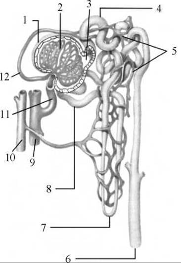
Fig. 171. Diagram of the structure and blood supply of the nephron
1 — glomerular capsule (Bowman's capsule); 2 — glomerulus of the renal corpuscle; 3 — capsular space; 4 — proximal tubule of the nephron; 5 — blood capillaries; 6 — collecting duct; 7 — nephron loop; 8 — distal tubule of the nephron; 9 — artery; 10 — vein; 11 — afferent glomerular arteriole; 12 — efferent glomerular arteriole
Vessels of the kidney. Understanding the Structure and function of the kidney is impossible without a thorough knowledge of its vascular supply.
The renal vascular bed comprises arterial and venous vessels as well as capillaries through which 1,500 to 1,800 liters of blood pass daily. Blood enters the kidney via the renal artery (arteria renalis), which divides at the renal hilum into anterior and posterior branches (ramus anterior et ramus posterior). Within the renal sinus, these branches pass anterior and posterior to the renal pelvis, respectively, and branch further into segmental arteries. The anterior branch gives rise to four segmental arteries—the superior segmental artery (a. segmenti superioris), anterior superior segmental artery (a. segmenti anterioris superioris), anterior inferior segmental artery (a. segmenti anterioris inferioris), and inferior segmental artery (a. segmenti inferioris)—which supply their respective renal segments. The posterior branch continues into the posterior segment of the organ as the posterior segmental artery (a. segmenti posterior). Segmental arteries further divide into interlobar arteries (arteriae interlobares), which course through the renal columns. At the cortico-medullary junction, interlobar arteries give off arcuate arteries (arteriae arcuatae), which arch over the bases of the renal pyramids. In turn, the arcuate arteries give rise to interlobular arteries (arteriae interlobulares), which ascend into the renal cortex, and straight arterioles (arteriae rectae), which descend into the medulla. The straight arterioles branch into capillary networks within the medulla that surround the renal tubules and supply the renal pyramids. Interlobular arteries give off afferent glomerular arterioles (arteriolae glomerulares afferentes), which branch within the renal corpuscles to form the capillary glomeruli of the nephrons. The primary capillaries of the glomerulus reunite to form the efferent glomerular arteriole (arteriola glomerularis efferens), which has a diameter roughly half that of the afferent arteriole. Upon exiting the glomerulus, the efferent arteriole—which still carries oxygenated arterial blood—subdivides once more into a secondary capillary network that entwine the renal tubules, thereby nourishing the renal parenchyma. This specialized branching of an afferent vessel into glomerular capillaries followed by their reunion into an efferent vessel is known as a rete mirabile (wonderful net).
From the secondary capillary network, the blood (now venous) flows into venules, which merge to form interlobular veins (venae interlobulares). From the interlobular veins, blood drains into arcuate veins (venae arcuatae), and then into interlobar veins (venae interlobares). The latter unite to form larger vessels, ultimately creating the renal vein (vena renalis). The renal vein emerges from the renal hilum and empties into the inferior vena cava.
The lymphatic vessels of the kidney accompany the blood vessels, exit the kidney together with them through the hilum, and drain into the lumbar Lymph Nodes.
The kidney is innervated by fibers of the renal plexus, which is formed by Branches of the celiac plexus (sympathetic fibers from the Sympathetic trunk ganglia, and parasympathetic fibers from the Vagus nerve).
Urine formation. Due to the significant blood pressure within the glomerular capillaries of the renal corpuscle (approximately 70–90 mm Hg), all components of Blood Plasma except Proteins and formed elements are filtered from the blood flowing through the capillaries into the capsular space. This filtrate is called primary urine. About 150–170 liters of it are produced per day. Such a large volume of primary urine is generated because approximately 1,700 liters of blood pass through the kidneys daily. The primary urine enters the renal tubules. Within the renal tubules, a process of reabsorption takes place, whereby water and essential substances are reabsorbed from the primary urine back into the blood capillaries surrounding the tubules. As a result, out of 150–170 liters of primary urine, about 1.5 liters of secondary urine are produced over the course of a day. It lacks substances needed by the body. Secondary urine contains protein breakdown products such as urea, uric acid, ammonia, and several others, as well as organic acids and organic salts.
Secondary urine flows through the collecting ducts and papillary ducts into the minor calyces, and then into the major calyces, the renal pelvis, and the ureter.
Last update: 08/08/2026
Editorial and Educational Adaptation: This material has been compiled based on the primary/original source text. The project team performed an editorial review, corrected technical inaccuracies, structured sections, and adapted the content for an educational format.
What was processed:
- elimination of formatting defects (OCR errors, structural breaks, corrupted characters);
- editorial organization of content;
- standardization of terminology in accordance with academic sources;
- verification of factual statements against the original source text.
All mentions of the author, publication year, and origin of the primary text have been preserved in accordance with the source.