Human Anatomy and Physiology (with Age-Related Characteristics of the Child's Body) - Sapin M.R., Sivoglazov V.I. 2002
The Study of the Viscera (Splanchnology)
Urinary System
Urinary Organs
The excretion of Metabolic waste products that cannot be utilized by The Human Body is performed by the digestive Organs, Lungs, Skin, and Urinary system. Salts, Bile pigments, Cholesterol, and Water are eliminated from the Digestive System in feces. The lungs excrete carbon dioxide, other gaseous substances, and water. Water (up to 0.6 L per day), carbon dioxide, various salts, and nitrogenous waste products are excreted through the sweat and Sebaceous Glands of the skin.
Up to 75% of metabolic waste products excreted from the body are eliminated through the Kidneys. Water, salts, and protein breakdown products (urea, uric acid, etc.) are excreted in the urine. The kidneys help maintain the body's acid-base balance (pH), a constant, normal volume of water and salts, and stable osmotic pressure. Thus, the kidneys (along with other organs) ensure the constancy of the body's internal environment (Homeostasis).
The Urinary organs include the kidneys, where urine is formed, as well as the Ureters, Urinary Bladder, and Urethra, which serve as pathways for excreting urine from the kidneys (out of the body).
Kidneys
The human Kidney (a paired organ) is bean-shaped, weighing 120—200 g. The kidneys are located on the posterior abdominal wall on either side of THE Vertebral Column, at the level of the XII thoracic to the I—II lumbar vertebrae. The right kidney lies slightly lower than the left. The Adrenal gland is adjacent to the superior pole. Anteriorly, the kidneys are bordered by the Peritoneum and the Internal Organs located at this level (Stomach, duodenum, colic flexures, Liver, Pancreas, etc.).
The kidney has anterior and posterior surfaces and two borders: a convex lateral border and a concave medial border. On the medial border, There is a depression called the renal hilum, which leads into the renal sinus. The renal artery and nerves enter the kidney through the hilum, while the renal vein and Lymphatic vessels exit. The renal sinus contains the Major and minor calyces, the renal pelvis, and adipose tissue.
Externally, the kidney is covered by a dense fibrous capsule, which is surrounded by an adipose capsule.
On a frontal section of the kidney (Fig. 57), an outer, lighter renal cortex and an inner, darker renal medulla can be distinguished. The cortex contains renal corpuscles, as well as the proximal and distal convoluted tubules. The medulla consists of 7—10 renal pyramids. The base of each pyramid is directed toward the cortex, and the narrowing part—the renal papilla—points toward a minor calyx. Extensions of the cortex, known as renal columns, project between the pyramids.
Class="center">
Fig. 57. Right kidney. Frontal (longitudinal) section. Posterior view: 1 — renal capsule, 2 — renal columns, 3 — renal cortex, 4 — renal medulla (pyramids), 5 — minor renal calyces (opened), 6 — major renal calyx, 7 — Ureter, 8 — renal pelvis, 9 — nerve, 10 — renal artery, 11 — renal vein
The nephron is the morphological and functional unit of the kidney. A nephron consists of a renal capsule and a system of renal tubules; the length of a single nephron is 50—55 mm, and the total length of all nephrons in both kidneys is about 100 km. There are more than 1 million nephrons in each kidney. The beginning of each nephron is a double-walled glomerular capsule (Bowman's capsule), which encloses a glomerulus of Blood capillaries. The capsule, together with the glomerulus, forms a renal corpuscle. A nephron is divided into the glomerular capsule, the proximal convoluted tubule, the Loop of Henle (consisting of descending and ascending limbs), and the Distal convoluted tubule (Fig. 58).
The glomeruli of all nephrons are located in the renal cortex, while their loops lie in the medulla. The distal PARTS OF THE nephron tubules open into collecting ducts that begin in the cortex. The collecting ducts then pass through the medullary pyramids and empty into short papillary ducts, which open into the minor renal calyces.
Between the inner and outer walls lies the capsular space, which continues into the nephron tubules. The inner wall of the capsule is tightly fused with the capillaries of the glomerulus. Thus, between the capillary blood and the lumen of the glomerular capsule, There are two fused walls—the Capillary Wall and the capsular wall. Through these walls, fluid—primary urine—is filtered from the blood into the lumen of the glomerular capsule. Over the course of a day, about 180 l of primary urine are filtered into the capsular spaces of both kidneys.
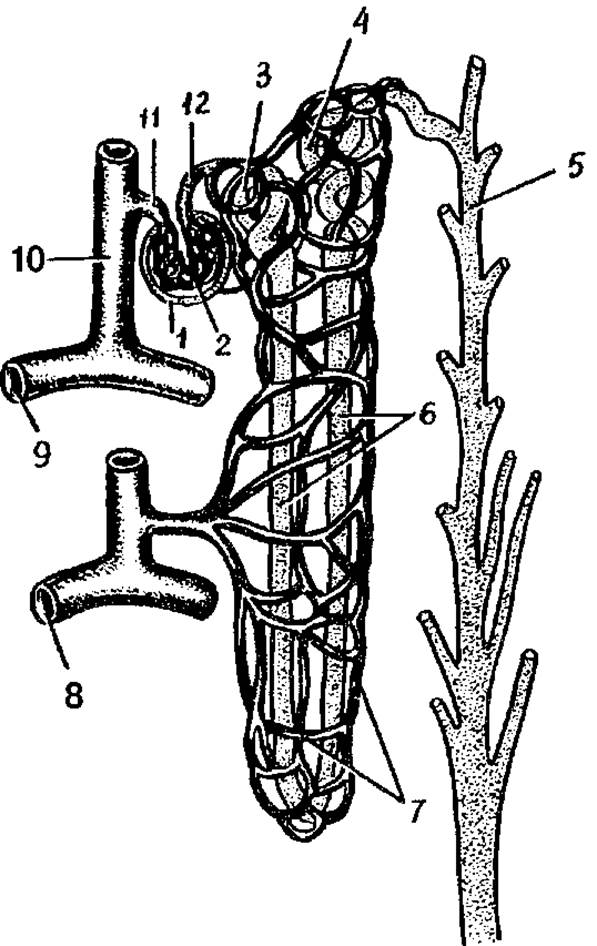
Fig. 58. Structure OF THE nephron and its relationship with Blood Vessels:
1 — glomerular capsule, 2 — glomerulus of the renal corpuscle, 3 — proximal convoluted tubule, 4 — distal convoluted tubule, 5 — collecting duct, 6 — loop of Henle, 7 — peritubular capillary network, 8 — arcuate vein, 9 — arcuate artery, 10 — interlobular artery, 11 — afferent glomerular arteriole, 12 — efferent glomerular arteriole
Afferent arterioles branch off from each interlobular artery, dividing into glomerular capillaries surrounded by the glomerular capsule. From these capillaries arises the efferent glomerular arteriole, which, after leaving the renal corpuscle, branches again into a secondary capillary network surrounding the nephron tubules. From this secondary network, blood drains into venules that continue into interlobular Veins, which then empty into arcuate and subsequently interlobar veins. The latter merge and enlarge to form the renal vein. Thus, there are two capillary systems in the kidneys. One of them—the glomerular capillary bed—is located between two arterioles (forming a so-called "rete mirabile"). The other, typical capillary system lies between the efferent arterioles and venules.
Age-related Features of the kidneys
In newborns and infants, the kidney is rounded. Its surface is lobulated due to its lobular structure, which is associated with the underdevelopment of the renal cortex at this age. The lobular structure of the kidney persists until 2—3 years of age. The length of the kidney in a newborn is 4 cm, and its weight is 12 g. During infancy, the size of the kidney increases approximately 1.5 times, and its weight reaches 37 g. During early childhood, the length of the kidney averages 8 cm, and its weight is 56 g.
In adolescents, the length of the kidney reaches 10 cm, and its weight is 120 g.
Kidney growth occurs mainly During the first year of a child's life. Between 5—9 years and especially 16—19 years, the size of the kidney increases due to The Development of the cortex, which continues until the end of adolescence. The growth of the medulla ceases by age 12. The mass of the renal cortex increases due to the growth in length and width of the convoluted tubules and the ascending limb of the loops of Henle.
The thickness of the renal cortex in an adult increases approximately 4-fold compared to that of a newborn, while the medulla increases only 2-fold.
The fibrous capsule of the kidney becomes clearly visible by 5 years of age, and by 10—14 years, its structure is close to that of an adult. The adipose capsule begins to form only during early childhood, gradually thickening thereafter. By 40—50 years of age, the thickness of the renal adipose capsule reaches its maximum, whereas in old age and senility, it thins out and sometimes disappears.
With age, the Topography of the kidneys changes. In a newborn, the superior pole of the kidney is projected at the level of the upper border of the XII thoracic vertebra, and in infancy (up to 1 year) — already at the level of the middle of the body of the XII thoracic vertebra. The inferior pole of the kidney is at the level of the lower border of the I lumbar vertebra; in a one-year-old child, it is У2 vertebra higher, which is due to the rapid growth of the vertebral column. After 5—7 years, THE POSITION OF the kidneys relative to the spine approaches that of an adult.
In individuals over 50 years of age, especially in elderly and emaciated people, the kidneys may be located lower than in youth. Throughout all periods of human life, the right kidney is located slightly lower than the left.
Renal calyces. Renal pelvis. Ureters
From the nephrons, urine flows through the papillary ducts into the minor renal calyces. The number of minor calyces in a single kidney ranges from 5 to 15. The apices of the renal papillae project into the cavity of the minor calyces; sometimes, the apices of two or three papillae open into a single minor calyx. In this case, the minor calyx surrounds the papilla on all sides, forming the so-called fornix above its apex. The walls of the fornix contain smooth Muscle Cells that form the sphincter of the fornix. The complex of fornical structures, including the sphincter, Connective Tissue, nerves, blood vessels, and lymphatic vessels, is referred to as the fornical apparatus of the kidney. This apparatus plays an important role in The process of urine excretion and prevents its backflow into the renal tubules. Several minor calyces open into a major calyx, of which humans typically have 2—3. The major calyces merge with one another to form a single common cavity—the renal pelvis.
The renal pelvis gradually narrows and transitions into the ureter. The walls of the renal calyces and pelvis consist of a mucosa lined with transitional epithelium, a muscularis, and an adventitia.
The human ureter is a cylindrical tube 6—8 mm in diameter and 25—35 cm in length, located retroperitoneally. The ureter is divided into abdominal and pelvic parts, as well as an intramural part that obliquely penetrates the wall of the urinary bladder.
The mucosa of the ureter is lined with transitional epithelium and is folded, which gives its lumen a star-shaped appearance in cross-section. The muscularis of the ureter consists of three layers: an inner longitudinal, a middle circular, and an outer longitudinal layer. In children, the muscularis is poorly developed. Externally, the ureter is covered by an adventitia.
Urinary bladder
The urinary bladder serves as a reservoir for urine; in adults, it lies in the lesser pelvis behind the Pubic Symphysis. When distended, the bladder projects above the pubis. The capacity of the bladder is up to 500 ml. In men, the rectum, Seminal Vesicles, and Ductus Deferentes lie posterior to the urinary bladder, while in women, the Uterus and Vagina are located there. The posterosuperior surface of the urinary bladder is covered by peritoneum.
The urinary bladder is divided into an apex, body, and fundus. The lower part of the bladder narrows to transition into the urethra. Posterior to the internal urethral orifice lies a triangular area with poorly defined folds—the trigone of the bladder. At the lateral angles of the posterior boundary of the trigone are the ureteric orifices, where the ureters open into the bladder.
The wall of the urinary bladder is composed of a mucosa, submucosa, muscularis, adventitia, and partially serosa (peritoneum). Due to the thick submucosa, the mucosa forms numerous folds that flatten when the bladder fills. The muscularis of the urinary bladder consists of three layers whose bundles interlace: inner and outer longitudinal layers, and a middle circular (transverse) layer. The interweaving of these muscle bundles facilitates uniform contraction of the bladder walls during Micturition, expelling urine into the urethra. In the region of the internal urethral orifice, the circular layer thickens to form the internal urethral sphincter. Fibers of the inner muscular layer of the bladder also surround the ureteric orifices. Contraction of these fibers prevents the backflow of urine from the bladder into the ureters.
The female urethra is a short tube 3—6 cm long, located posterior to the pubic symphysis. The mucosa is folded and lined with pseudostratified epithelium. The myocytes of the wall form two layers: an inner longitudinal layer and a more prominent outer circular layer. The external urethral orifice is located in the Vestibule of the vagina, anterior and superior to the vaginal opening, and is surrounded by the striated external urethral sphincter.
The male urethra will be described below.
Age-related features of the ureters and urinary bladder
In newborns, the ureters have a tortuous course. The length of the ureter reaches 5—7 cm. By the age of 4, its length increases to 15 cm. The muscularis is poorly developed in early childhood.
In newborns, the urinary bladder is spindle-shaped, and in children during the first years of life, it is pear-shaped. During second childhood (8—12 years), the urinary bladder is ovoid, and in adolescents, the capacity of the urinary bladder in newborns is 50—80 ml. By the age of 5, it holds 180 ml of urine, and after 12 years, 250 ml. In newborns, the circular muscle layer in the bladder wall is poorly developed, while the mucosa is well-developed and folded.
...half the distance between the umbilicus and the pubic symphysis; therefore, the urinary bladder in girls at this age does not come into contact with the vagina, and in boys, with the rectum. At the age of 1—3 years, the Cytology/practical/108.html">Fundus of the urinary bladder is located at the level of the upper border of the pubic symphysis. In adolescents and subsequently, the descent of the fundus of the urinary bladder occurs depending on the condition of the Muscles of the urogenital Diaphragm.
Questions for REVIEW AND SELF-control;
1. Describe the parts (regions) into which the kidney is divided.
2. Name the parts of the nephron. In which Regions of the kidney are they located? What is the renal corpuscle?
3. Describe what you know about The structure of the renal calyces, pelvis, and ureter.
4. What is the fornical apparatus of the kidney, and what Functions does it perform?
5. What regions are distinguished in the urinary bladder? What openings does the bladder have, and where are they located?
6. Which organs does the urinary bladder come into contact with in men and women?
7. What volume of urine does the urinary bladder hold in adults and in children of various ages?
Mechanisms of Urine Formation and Excretion
Over a 24-hour period, a person consumes approximately 2.5 L of water, including 1500 ml in liquid form and about 650 ml with solid food. In addition, about 400 ml of metabolic water is produced in the body during the Breakdown of Proteins, fats, and CARBOHYDRATES. Water is excreted from the body mainly through the kidneys (1.5 L per day), as well as through the lungs, skin, and partially with feces.
Urine formation in the kidneys
Urine formation in the nephrons of the kidney occurs in two phases. The first phase is filtration, which involves the Formation of primary urine (glomerular filtrate) in the glomeruli of the nephrons. In the second phase, reabsorption, water and other substances are reabsorbed in the renal tubules, producing concentrated, so-called secondary (final) urine.
In the renal glomeruli, water and dissolved substances are filtered from the renal capillaries into the initial part of the nephrons. Ultrafiltration occurs due to the pressure difference between the glomerular capillaries and the nephron capsule. The blood pressure in the glomerular capillaries is very high, about 60—70 mm Hg (compared to 30 mm Hg in the capillaries of other organs). This high pressure in the glomerular capillaries is maintained by a significant difference in the diameter of the vessels carrying blood to and from the glomeruli. The afferent arterioles of the glomeruli have twice the diameter of the efferent arterioles. Thus, the glomerular capillary network, whose function is to remove substances destined for excretion from the Blood Plasma, is situated between two arterial vessels.
The Blood supply to the kidneys is also remarkable for the volume of blood passing through them. More than 1 L of blood (1.2 L) flows through the kidneys per minute. Over a 24-hour period, up to 1700—1800 L of blood passes through the kidneys. Thus, within 24 hours, the entire blood volume flows through the glomerular capillaries more than 200 times. This blood comes into contact with the inner surface of the capillaries, which have a total surface area of 1.5—2 m2 in the renal glomeruli. Consequently, The amount of primary urine formed reaches 150—180 L per day. This means that for every 10 L of blood flowing through the kidneys, 1 L of primary urine is filtered. Primary urine contains all the components of blood plasma except for high-molecular-weight proteins. It contains Amino Acids, glucose, Vitamins, and salts, as well as metabolic waste products such as urea, uric acid, and other substances.
During the second phase of urine formation—reabsorption—the primary urine, which is similar in composition to blood plasma, flows from the glomerular capsules into the renal tubules. In these tubules, amino acids, glucose, vitamins, and most of the water and salts are reabsorbed from the primary urine back into the blood. Ultimately, up to 1.5 L of secondary (final) urine is produced daily from the 150—180 L of primary urine. The secondary urine travels through the Urinary Tract (renal calyces, renal pelvis, and ureter) to the urinary bladder, from which it is excreted. The tubules reabsorb 99% of the water contained in the primary urine, along with essential solutes. Consequently, secondary urine differs drastically from primary urine. Secondary urine contains no sugar, amino acids, or many of the salts. At the same time, the concentrations of sulfates, phosphates, urea, uric acid, and other substances that are not reabsorbed from the renal tubules back into the blood are sharply increased. For instance, the concentration of urea in secondary urine is 67 times higher than in blood, creatinine is 75 times higher, and sulfates are 90 times higher. The reabsorption of most substances in the renal tubules is an active physiological process that requires energy expended by the epithelial lining and other structures of the tubule walls. It is well known that the kidneys consume a significant portion (over 10%) of the body's total oxygen intake, reflecting the extremely high energy demands of renal function.
When certain substances reach very high concentrations in the blood, a portion of them is not reabsorbed from the primary urine back into the bloodstream. For example, after excessive sugar consumption and the resulting surplus of blood glucose, some glucose remains in the primary urine. Conversely, if there is a deficiency of table salt in the diet, it is not excreted in the urine. In this way, the kidneys regulate the levels of substances in the body, excreting excesses and retaining what is lacking.
In addition to the reabsorption of water and many of its dissolved components, the renal tubules also perform tubular secretion, releasing substances into the urine. These are substances that cannot pass through the "renal filter" on their way from the blood capillaries into the glomerular capsules. They include many medications, particularly certain Antibiotics (such as penicillin), Dyes, and Other Compounds.
Physical and Chemical properties of urine
Urine is a light yellow fluid.
Urine consists of 95% water and 5% solid solutes. These include urea (2%), uric acid (0.05%), creatinine (0.075%), and other substances, including potassium and sodium salts. Over a 24-hour period, 25—30 g of urea and up to 25 g of inorganic substances are excreted from the body in urine. In renal diseases or following brief periods of intense physical exertion, protein may appear in the urine, where it is normally absent. Urinary pH depends on diet. A predominantly meat-based diet results in acidic urine, whereas a vegetable-rich diet leads to alkaline or neutral urine. The presence of blood in the urine (giving it a red or pink color) can result from mucosal damage or hemorrhages within the urinary system. Consuming fresh carrots or beets can also temporarily color the urine pink.
Excretion of urine from the kidneys
Urine formed in the kidneys flows from the renal calyces into the renal pelvis and then into the ureters. Driven by peristaltic contractions of the ureters, urine is carried drop by drop into the urinary bladder, where it accumulates until the bladder is full. During this time, the internal and external urethral sphincters remain contracted, keeping the bladder outlet closed.
Emptying of the urinary bladder occurs reflexively. When urine accumulates in the bladder to a volume of 250—300 mL, it begins to exert noticeable pressure on the bladder walls with a force of about 12—15 cm of water. This pressure triggers the urge to urinate. Nerve impulses generated in the receptors of the bladder walls travel to the micturition center located in the sacral region of the Spinal Cord. From this center, signals are transmitted along parasympathetic pelvic nerve fibers to the bladder walls. These signals cause simultaneous contraction of the bladder wall muscles and relaxation of the urethral sphincters, expelling urine from the bladder. Higher micturition centers are located in the frontal lobes of the cerebral hemispheres, which also regulate THE URINATION PROCESS.
The conditioned reflex that allows for the temporary voluntary suppression of the urge to urinate is developed during childhood training. Newborn infants lack voluntary control over urination. The ability to voluntarily regulate urination begins to develop only toward the end of the first year of life and becomes stable during the second year. The Influence of the Autonomic Nervous system is not limited to urine excretion; nerve impulses can also accelerate or slow down urine formation, as well as increase or decrease the excretion of blood-borne substances in the urine.
Urine formation is also regulated humorally by vasopressin (antidiuretic hormone), which is produced by the neurosecretory Cells of the Hypothalamus and released into the bloodstream via the posterior Pituitary Gland. This hormone enhances the reabsorption of water from the primary urine, thereby increasing the concentration of solutes (salts) in the secondary urine.
In Diseases of the hypothalamus or the posterior pituitary gland, the release of vasopressin into the blood is disrupted, which can increase daily urine output to 20—25 L. A reduction or cessation of urine output can also occur in response to severe pain. Urine formation and excretion are further influenced by fluid intake, consumption of salty foods, and physical labor.
Review and Self-Assessment Questions:
1. Name the phases of urine formation in the kidneys. Describe the characteristics of each phase.
2. List the substances that are filtered from the blood into the urine within the renal corpuscles.
3. What are Primary and secondary urine? What are the differences between them?
4. Describe The Role of the ureters and the urinary bladder in the excretion of urine from the body.
5. Explain the neural and humoral Regulation of urine formation and excretion.
Last update: 10/08/2026
Editorial and Educational Adaptation: This material has been compiled based on the primary/original source text. The project team performed an editorial review, corrected technical inaccuracies, structured sections, and adapted the content for an educational format.
What was processed:
- elimination of formatting defects (OCR errors, structural breaks, corrupted characters);
- editorial organization of content;
- standardization of terminology in accordance with academic sources;
- verification of factual statements against the original source text.
All mentions of the author, publication year, and origin of the primary text have been preserved in accordance with the source.