Review of Medical Physiology - William F. Ganong 2002
Formation and Excretion of Urine
Renal Function and Micturition
Micturition
Anatomical Overview
The smooth Muscle of the Urinary Bladder, similar to that of the Ureters, is arranged in spiral, longitudinal, and circular bundles. Contraction of this muscle, termed the detrusor, is the primary mechanism responsible for emptying the bladder during micturition. Muscle bundles run along both sides of the Urethra; these fibers are sometimes referred to as the internal urethral sphincter, although they do not actually encircle the urethra. Distal to this lies the sphincter urethrae, a voluntary Skeletal Muscle (the external urethral sphincter). The bladder epithelium consists of a Superficial layer of flat Cells and a Deep Layer of cuboidal cells. The Innervation of the urinary bladder is schematically illustrated in Fig. 38-26.
Class="center">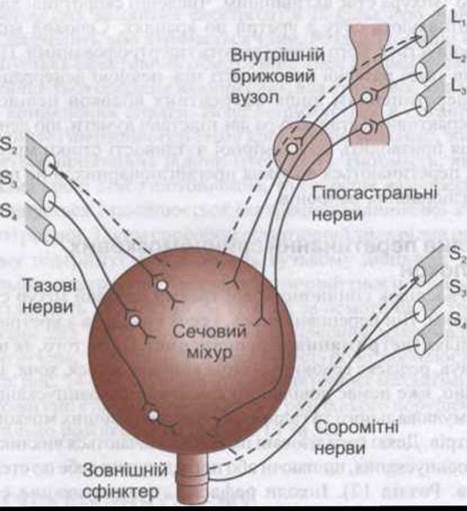
Fig. 38-26. Innervation of the urinary bladder. Sensory nerves are indicated by dashed lines. Parasympathetic innervation is shown on the left, sympathetic innervation in the upper right corner, and somatic innervation in the lower right corner.
Micturition
The physiology of micturition and the pathophysiological mechanisms of its disorders are subjects of ongoing debate. Micturition is a spinal reflex regulated by higher Brain centers and, much like defecation, can be voluntarily inhibited. Urine enters the bladder gradually without causing a sharp increase in intravesical pressure. Like Other types of smooth muscle, the detrusor exhibits pronounced elastic properties: during stretch, tension in its wall does not persist for long. The relationship between intravesical pressure and bladder volume can be investigated by inserting a catheter into the bladder, emptying it, and then recording pressure changes during gradual filling with 50-mL portions of Water or air (cystometry). The plot of intravesical pressure versus fluid volume in the bladder is called a cystometrogram (Fig. 38-27). The curve reflects an initial slight increase in pressure upon the Introduction of the first portions of fluid or air. Subsequent infusions reveal a long segment of the curve that is nearly parallel to the horizontal axis. At a specific volume of infused fluid, a sudden, sharp increase in intravesical pressure occurs, which corresponds to the triggering of the micturition reflex. These three sections of the curve are frequently designated as segments Ia, Ib, and II. The first sensation of bladder fullness occurs when the bladder is filled to approximately 150 mL, and an intense feeling of fullness arises at around 400 mL. The flat segment Ib reflects Laplace's law (see Chapter 30). According to this law, pressure within a spherical container equals wall tension multiplied by two and divided by its radius. As the bladder fills, wall tension increases, but the radius increases proportionally. Consequently, intravesical pressure remains constant during filling.
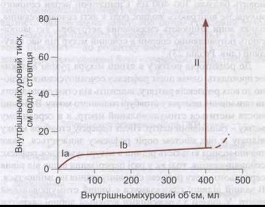
Fig. 38-27. Cystometrogram of a healthy individual. Numbers indicate the three Components of the curve described in the text. The dashed line represents the pressure-volume relationship that would occur in the absence of micturition, corresponding to segment II of the curve (modified and reproduced with permission from Tanagho EA, McAnich JW: Smith's General Urology, 14th ed, McGraw-Hill, 1995).
During micturition, the perineal Muscles and the external urethral sphincter are relaxed, the detrusor contracts, and urine is expelled from the urethra. The bands of smooth muscle on the sides of the urethra play no role in this act; their primary function is to prevent retrograde ejaculation of semen into the urinary bladder during ejaculation.
The Mechanism of voluntary micturition is not yet fully understood. The initial step is the relaxation of the pelvic floor muscles. This alone may provide sufficient stimulation to the detrusor to initiate its contraction. The perineal muscles and the external urethral sphincter can be voluntarily contracted to prevent urine flow through the urethra or to interrupt micturition once it has begun. In adults, The ability to keep the external urethral sphincter contracted until an opportunity to empty the bladder arises is the result of early childhood training. The female urethra is emptied after micturition by the action of gravity. Urine remaining in the male urethra at the end of micturition is expelled by several peristaltic contractions of the bulbospongiosus muscle.
Reflex Regulation
The smooth muscle of the urinary bladder possesses intrinsic contractility. With normal bladder innervation, stretch receptors in the bladder wall trigger a reflex contraction with a lower threshold of excitability than the autonomous contractions inherent to the detrusor. Pelvic nerve fibers constitute the afferent limb of the micturition reflex. Parasympathetic fibers of the bladder form the efferent limb of the reflex and also travel within the pelvic nerves. Integration of the reflex occurs in the sacral segments of the Spinal Cord. In healthy adults, the fluid volume in the bladder that triggers this reflex is approximately 300-400 mL. The sympathetic NERVES OF THE bladder play no role in micturition; however, they initiate detrusor contractions that prevent seminal fluid from entering the bladder during ejaculation (see Chapter 23).
Motor fibers do not project directly to the stretch receptors in the bladder wall; nevertheless, the threshold of the micturition reflex, like that of all stretch Reflexes, depends on excitatory and inhibitory centers in the Brainstem. An excitatory center is located in the pontine region, and an inhibitory center is situated in the Midbrain. Transection of the brainstem just above the Pons lowers the reflex threshold, causing a much smaller bladder volume to trigger micturition, whereas transection above the midbrain level does not alter the reflex. Another excitatory center is located in the posterior Hypothalamus. Individuals with lesions of the superior frontal gyrus exhibit a reduced desire to void and an inability to inhibit micturition once it has started. Animal experiments involving electrical stimulation of various brain regions have demonstrated the presence of other cortical centers that influence micturition. The bladder can be made to contract via voluntary facilitation of the spinal reflex, even when it contains only a few milliliters of urine. Voluntary contraction of the abdominal wall muscles assists in urine expulsion by increasing intra-abdominal pressure, but micturition can also be initiated without straining, even when the bladder is nearly empty.
Disorders of Micturition
There are three MAIN TYPES OF micturition disorders resulting from neural tissue lesions: those due to disruption of afferent nerve pathways from the bladder; those due to disruption of both afferent and efferent pathways; and those due to disruption of excitatory and inhibitory pathways descending from the brain. In each type of voiding dysfunction, the bladder contracts, but these contractions are insufficient to completely empty the bladder, resulting in residual urine retention.
Effects of Deafferentation
When the dorsal roots of the sacral nerves are severed in experimental animals or their conduction is impaired due to various diseases such as tabes dorsalis, all reflex contractions of the bladder cease. The bladder transforms into an overdistended, thin-walled tissue sac largely devoid of muscle tone, although some weak contractions occur due to autonomous Responses of the bladder to stretch.
Effects of Denervation
When both afferent and efferent nerves are destroyed, as in tumors of the cauda equina or filum terminale, the bladder initially becomes flaccid and overdistended. Gradually, however, the muscle of the "decentralized" bladder becomes more active. Numerous contractile waves expel urine from the urethra drop by drop. The urinary bladder shrinks, and its walls become hypertrophied. The reasons for this striking difference between complete denervation and transection of afferent fibers alone remain unknown. The hyperactive state of the bladder suggests that denervation leads to hypersensitivity of the bladder wall, even though preganglionic rather than postganglionic neuronal fibers are transected.
Effects of Spinal Cord Transection
Following spinal cord injury, the urinary bladder becomes flaccid. It overfills, and urine leaks drop by drop from the urethra (overflow incontinence). Once spinal Shock subsides, the micturition reflex returns, although voluntary control of urination and the excitatory and inhibitory influences of higher brain centers are permanently lost. Some paralyzed patients learn to trigger micturition by pinching or stroking their thigh (see Chapter 12). Occasionally, the micturition reflex becomes hyperactive. Bladder capacity decreases, and its wall becomes hypertrophied. This type of bladder is sometimes referred to as a neurogenic bladder. Bladder hyperactivity can be exacerbated by infectious and inflammatory processes in the Urinary Tract.
Last update: 10/08/2026
Editorial and Educational Adaptation: This material has been compiled based on the primary/original source text. The project team performed an editorial review, corrected technical inaccuracies, structured sections, and adapted the content for an educational format.
What was processed:
- elimination of formatting defects (OCR errors, structural breaks, corrupted characters);
- editorial organization of content;
- standardization of terminology in accordance with academic sources;
- verification of factual statements against the original source text.
All mentions of the author, publication year, and origin of the primary text have been preserved in accordance with the source.