Review of Medical Physiology - William F. Ganong 2002
Functions of the Nervous System
Vision
Responses of the Visual Pathways and Cortex
Pathways to the Cerebral Cortex
Ganglion Cell axons provide a detailed spatial PROJECTION OF THE retina onto the lateral geniculate Nucleus (LGN). Each geniculate body has six distinct cell layers (Fig. 8-21). Layers 3 to 6 are composed of small Cells and are therefore termed parvocellular. Layers 1 and 2 contain large cells and are correspondingly called magnocellular. Layers 1, 4, and 6 of each geniculate body receive inputs from the contralateral eye, whereas layers 2, 3, and 5 receive inputs from the ipsilateral eye. Each layer features an exceptionally detailed topographic map of the retina, and all six layers maintain a precise interrelationship, meaning that the receptive fields of cells localized along a perpendicular axis through the layers virtually coincide. Notably, only 10-20% of the impulses originating in the lateral geniculate nucleus actually reach the retina. A significant proportion of additional inputs stems from the visual cortex and other Brain regions. This feedback mechanism involving the visual cortex plays a crucial role in spatial orientation and movement coordination. Two MAIN TYPES OF retinal ganglion cells are distinguished: large ganglion cells (or M cells, from Latin magnus - large), which receive inputs from various cone types and are associated with movement coordination and stereoscopic Vision, and small ganglion cells (or P cells, from Latin parvus - small), which differentiate signals arriving from different cone types and mediate the perception of color, texture, and form. Magnocellular Neurons project to the magnocellular layers of the lateral geniculate nucleus, whereas P cells project to the parvocellular layers (Fig. 8-22).
Class="center">
Fig. 8-20. Response of retinal ganglion cells to light falling on parts of their receptive fields, shown in white. Next to the diagram of each receptive field is the corresponding ganglion cell response diagram, determined by extracellular recording of action potentials. Note that in three out of four cases, the discharge rate increases markedly after the light is turned off (modified from Kandel E, Schwartz JH, Jessell TM [editors]: Principles of Neural Science, 4th ed. McGraw-Hill, 2000).
From the lateral geniculate nucleus, the magnocellular and parvocellular pathways extend to the visual cortex. The magnocellular pathway from layers 1 and 2 (see Fig. 8-21) transmits signals related to motion, depth, and flicker perception. The parvocellular pathway relays signals from layers 3-6 concerning color, form, texture, and fine details of observed objects.
Cells within the interlaminar Regions of the lateral geniculate nucleus also receive inputs from P cells, presumably via dendrites that penetrate the parvocellular layers. They project via a separate component of the parvocellular pathway to the cytochrome oxidase blobs of the visual cortex (see below).
Primary Visual Cortex
Just as ganglion cell axons provide a point-to-point spatial representation of the retina in the lateral geniculate nucleus, the geniculate body similarly projects a point-to-point map onto the primary visual cortex (see Fig. 8-5). A large number of Nerve Cells contact each nerve fiber entering the visual cortex. Like other areas of the neocortex, the visual cortex is composed of six layers (see Fig. 11-2). Axons from lateral geniculate neurons forming the magnocellular pathway terminate in layer 4, specifically in its deepest subdivision, layer 4C. Many axons of the parvocellular pathway also terminate in layer 4C, whereas axons from interlaminar cells terminate in layers 2 and 3.
Layers 2 and 3 of the cerebral cortex contain clusters of cells approximately 0.2 mm in diameter that exhibit a selectively high level of the mitochondrial enzyme cytochrome oxidase. These cell clusters are termed blobs (see Fig. 8-22). They form a distinct mosaic within the visual cortex and are closely linked to color perception. The parvo-
cellular pathway also transmits opponent-color information to the deep portion of layer 4 (see below).
Similar to retinal ganglion cells and lateral geniculate neurons, neurons in layer 4 of the visual cortex respond to The stimulation of their receptive fields with center-surround antagonism—either center-excitation and surround-inhibition, or vice versa. A slit of light covering the center serves as an effective stimulus because it excites the center predominantly while having little effect on the surround. This stimulus lacks any orientation preference and is equally effective regardless of the angle at which it is presented.
The responses of neurons in other layers of the visual cortex differ significantly. So-called simple cells respond to light bars, lines, or edges only when these stimuli possess a specific spatial orientation. If, for instance, a light bar is tilted by as little as 10° relative to its preferred orientation, the response of a simple cell diminishes sharply, and a larger angle of rotation abolishes the response entirely. There are also complex cells, which share with simple cells the property of orientation preference for linear stimuli, yet they are less dependent on the precise retinal Location OF THE stimulus compared to simple cells or layer 4 neurons. Complex cells typically generate a maximal response when a linear stimulus is displaced laterally without altering its spatial orientation. Complex cells presumably receive their inputs from simple cells. If a microelectrode is inserted perpendicularly into the cortical surface and gradually advanced through different layers, the orientation preference of the neurons remains constant. Consequently, the visual cortex, much like the somatosensory cortex (see Chapter 7), is organized into vertical orientation columns. Each Column is roughly 1 mm in diameter. The orientation preferences change systematically from one column to the next. Moving from column to column, the preferred orientation shifts progressively by 5-10° (Fig. 8-23). Therefore, it is likely that the representation of each retinal ganglion cell's receptive field within the visual cortex comprises a set of columns spanning local regions of the visual field that cover the full spectrum of orientation preferences across a complete 360°. Simple and complex cells are referred to as feature detectors because they perceive and analyze specific attributes of a given stimulus. Feature detectors have also been identified in the cortical representations of other sensory modalities.
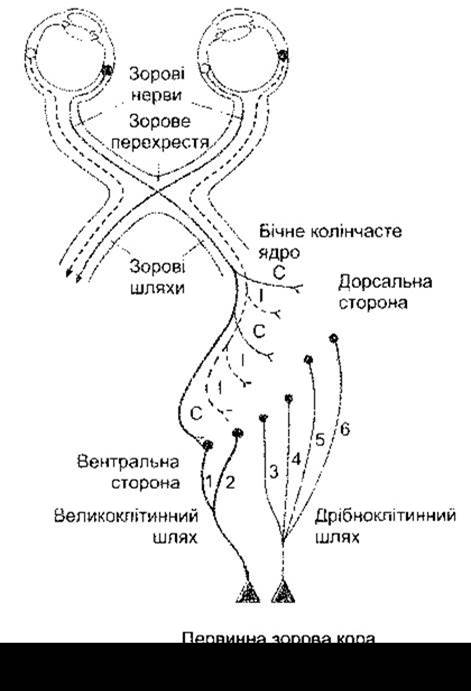
Fig. 8-21. Projections of retinal ganglion cells from the right half of each eye's retina to the right lateral geniculate nucleus and onward to the right primary visual cortex. Note the six layers of the geniculate nucleus. Ganglion P cells project to layers 3-6, whereas ganglion M cells project to layers 1 and 2. Ipsilateral (I) and contralateral (C) eyes project to alternating layers. Interlaminar cells, which project via distinct Components of the parvocellular pathway to the cytochrome oxidase blobs of the visual cortex, are not shown (modified from Kandel E, Schwartz JH, Jessell TM [editors]: Principles of Neural Science, 4th ed. McGraw-Hill, 2000).
Orientation columns can be mapped using radioactive 2-deoxyglucose. The uptake of this glucose derivative is directly proportional to neuronal activity (see Chapter 32). When this labeling technique is applied to animals stimulated with uniformly patterned visual cues, such as vertical lines, the brain reveals a large number of intricately arranged, evenly distributed orientation columns spanning a broad area of the visual cortex.

Fig. 8-22. Organization of visual pathways. Ganglion M cells project to the magnocellular laminae of the lateral geniculate nucleus, while ganglion P cells project to the parvocellular laminae; P cells also establish contact with interlaminar cells, presumably via dendritic extensions.
Another hallmark of the visual cortex is the presence of ocular dominance columns. Individual geniculate neurons and layer 4 cortical cells receive input from only one eye. The functional activation of layer 4 cells and geniculate neurons alternates between the two eyes. If a high concentration of a radiolabeled amino acid is introduced into one eye, it is incorporated into Newly synthesized Proteins and transported via axonal flow to the terminals of retinal ganglion cells, and subsequently across synapses in the geniculate nuclei along geniculocalcarine fibers to the visual cortex. In layer 4 of the cortex, labeled terminals originating from the radioisotope-injected eye alternate with unlabeled terminals coming from the other eye. This results in a striking pattern of alternating light and dark stripes spanning a large expanse of the visual cortex (Fig. 8-24), independent of the lattice of orientation columns.
Roughly half of all simple and complex cells receive inputs from both eyes. These inputs are identical or closely matched in terms of the corresponding visual cortical region and orientation preference. However, they may differ in driving strength, and In addition to cells driven exclusively by either the ipsilateral or contralateral eye, There is a substantial population of neurons influenced by both eyes to varying degrees.
Thus, the primary visual cortex segregates information regarding color, form, and object motion, integrates binocular inputs, and decomposes the visible world into elemental line segments of diverse spatial orientations.
Other Cortical Areas Involved in Vision
As noted above, the primary visual cortex (V1) projects to numerous distinct areas within the occipital lobe and other PARTS OF THE brain. These regions are frequently identified by numbers (V2, V3, etc.) or letters (LO, MT, etc.). The localization of several of these areas in the human brain is illustrated in Fig. 8-25, and their putative Functions are summarized in Table 8-1. The functions of these areas have been elucidated through studies in trained monkeys outfitted with implanted microelectrodes. Furthermore, the application of PET imaging and functional MRI (see Chapter 16 and Appendix) has enabled sophisticated investigations of visual cognition and other cortical visual functions in conscious, healthy humans. Visual projections from V1 can be broadly segregated into a dorsal, or parietal, pathway—primarily concerned with motion—and a ventral, or temporal, pathway, involved in the recognition of patterns, shapes, and faces. Numerous interconnecting cross-links also exist. The human visual area V8 appears to play a uniquely dedicated role in Color Vision.

Fig. 8-23. Top: Orientation preferences of 15 neurons recorded along an oblique electrode track through the visual cortex. Orientation preference shifts gradually in a counterclockwise direction. Bottom: Results of a similar experiment plotted against the distance traveled by the electrode. In this case, multiple reversals in the direction of rotation were observed (modified and reproduced with permission from Hubel DH, Wiesel TN: Sequence regularity of orientation columns in the monkey striate cortex. J Comp Neurol 1974; 158:267).
As is well known, visual information is processed in parallel along multiple pathways. Through mechanisms not yet fully understood, all of this disparate information is ultimately integrated into the unified perception of a conscious visual world.

Fig. 8-24. Reconstruction of ocular dominance columns in a segment of layer 4 of the right visual cortex of a monkey. Dark stripes represent one eye, and light stripes represent the other (reproduced with permission from LeVay S, Hubel DH, Wiesel TN: The pattern of ocular dominance columns in macaque visual cortex revealed by reduced silver stain. J Comp Neurol 1975;159:559).
Last update: 10/08/2026
Editorial and Educational Adaptation: This material has been compiled based on the primary/original source text. The project team performed an editorial review, corrected technical inaccuracies, structured sections, and adapted the content for an educational format.
What was processed:
- elimination of formatting defects (OCR errors, structural breaks, corrupted characters);
- editorial organization of content;
- standardization of terminology in accordance with academic sources;
- verification of factual statements against the original source text.
All mentions of the author, publication year, and origin of the primary text have been preserved in accordance with the source.