Review of Medical Physiology - William F. Ganong 2002
Functions of the Nervous System
Vision
Color Vision
Color Characteristics
Color has three main characteristics: hue, intensity, and saturation (defined as the degree of absence of white). Each color has a complementary color, which, when mixed in appropriate proportions, yields white. The perception of black is caused by the absence of light; however, it likely carries positive information, as a blind eye does not see black—it sees nothing at all. There are well-known phenomena of successive and parallel contrasts, optical illusions that create a sensation of color when none is present, negative and positive afterimages, as well as various psychological aspects of color Vision. A detailed characterization of these phenomena is beyond The Scope of this book.
Another factor of significant importance is The ability to produce white, any other color of the spectrum, or even extraspectral purple by mixing red (wavelength 723-647 nm), green (575-492 nm), and blue (492-450 nm) light in various proportions. For this reason, red, green, and blue are called primary colors. Another important factor is the observation made by Land, according to which color perception depends partly on the colors of other objects within the visual field. For example, a red object is perceived as red when illuminated by green or blue light, yet it appears pale pink or even white when illuminated by red light.
Class="center">
Fig. 8-25. Some of the major functional areas of the human primary visual cortex (V1). Lateral and medial views. See also Table 8-1 (modified from Logothetis N: Vision: a window on consciousness. Sci Am [Nov] 1999;28:99).
Retinal Mechanisms
The Young-Helmholtz trichromatic theory of color vision posits the presence of Three types of cones in humans, each containing a specialized photosensitive pigment with maximal sensitivity to one of the three primary colors. The perception of any color is determined by the relative impulse frequency from each cone type. The validity of this theory has been proven through the identification and chemical characterization of each of the three pigments. One of them (the blue-sensitive, or short-wavelength pigment) has an absorption maximum in the blue-violet region of the spectrum (Fig. 8-26); another (the green-sensitive, or middle-wavelength) in the green region; and yet another (the red-sensitive, or long-wavelength) in the yellow region of the spectrum. Cones with maximal sensitivity in the yellow region of the spectrum are also sufficiently sensitive in its red region, exhibiting a lower excitation threshold for red light than for green. All these experimental findings fulfill the requirements of the Young-Helmholtz theory.
Table 8-1. Functions of the visual projection areas in the human Brain1
V1 |
Primary visual cortex; receives impulses from the lateral geniculate nuclei, initiates Processing of information regarding orientation, object edges, etc. |
V2, V3, VP |
Continue Image processing, feature larger receptive fields |
V3A |
Motion |
V4v |
Function unknown |
MT/V5 |
Motion control |
LO |
Large object recognition |
V7 |
Function unknown |
V8 |
Color vision |
1 Modified from Logothetis N: Vision: a window on consciousness. Sci Am (Nov) 1999,281:99.
In humans, the rhodopsin Gene is localized on chromosome 3, and the gene encoding the blue-sensitive S-cone pigment is on chromosome 7. The other two cone pigments are encoded by two paired genes located on the q-arm of the X chromosome. The green-sensitive M pigment and the red-sensitive L pigment are structurally similar: their opsins share 96% Amino Acid Sequence Homology, whereas each of these pigments shares only 43% homology with the opsin of the blue-sensitive pigment, and all three cone pigments show about 41% homology with rhodopsin. Many mammals are dichromats, meaning they possess only Two Types of cone pigments: short- and long-wavelength. Old World monkeys and humans are trichromats with separate middle- and long-wavelength pigments, which likely resulted from the duplication of an ancestral long-wavelength gene followed by divergence.
Recently, it has been discovered that certain variants of long-wavelength (red-sensitive) pigments exist within the human population. It has long been known that the response to the Rayleigh match—The ratio of red and green light that different individuals mix to produce a monochromatic orange—is bimodal.
This phenomenon is well explained by new data showing that 62% of individuals with normal color vision have a Serine residue at position 180 of the long-wavelength cone opsin chain, whereas the remaining 38% have an Alanine residue.
The pigment absorption peak in individuals with a serine residue at position 180 corresponds to a wavelength of 556.7 nm, making this pigment variant more sensitive to red light, whereas the curve for individuals with an alanine residue has an absorption peak at 552.4 nm.
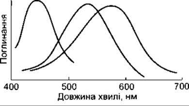
Fig. 8-26. Absorption spectra of the three types of cone pigments in the human retina. Pigment S, with an absorption peak at 440 nm, mediates blue perception, and pigment M, with an absorption peak at 535 nm, mediates green perception. Pigment L, with an absorption peak at 565 nm (yellow region of the spectrum), also absorbs long waves in the red part of the spectrum (reproduced with permission from Michael CR: Color vision. N Engl J Med 1973;288:724).
Neural Mechanisms
Color information is transmitted by ganglion Cells that subtract or add impulses received from Different types of cones. Processing within ganglion cells and Neurons of the lateral geniculate Nucleus generates impulses that travel via three distinct pathways to V1: the red-green pathway transmits signals reflecting the difference between L- and M-cone responses; the blue-yellow pathway transmits signals reflecting the difference between S-cone responses and the combined response of L- and M-cones; and the third pathway transmits the combined response of L- and M-cones. These three pathways project to the blobs and the deep layers of layer 4C in area V1. From the blobs and layer 4, color information is transmitted to area V8. The mechanism by which the signals arriving at V8 are further transformed into color sensation remains unclear to this day.
Color Blindness
There are numerous tests for detecting color blindness. The most common are yarn-matching tests and Ishihara plates (in Ukraine, Rabkin’s polychromatic plates are used). In the former test, the subject is given a set of variously colored yarns and asked to group similar colors. Ishihara plates and similar tests feature numbers composed of colored spots set against a Background of similarly shaped spots of a different color. The numbers are deliberately rendered in colors that blend into the background for individuals with color blindness.
Some individuals with color blindness are unable to distinguish certain colors (anopia), whereas others have only impaired color perception (anomaly). The prefixes prot-, deuter-, and tritat- refer to defects in the red, green, and blue cone systems, respectively. Individuals with normal color vision, as well as those with protanomaly, deuteranomaly, and tritanomaly, are called trichromats: they possess all three cone systems, although one may be deficient. Dichromats are individuals with only two cone systems, presenting as protanopia, deuteranopia, or tritanopia. Monochromats possess only a single cone system. Dichromats can broaden their color spectrum by combining two primary colors, whereas monochromats can only vary the intensity of a single color.
Color blindness is most commonly an inherited condition. Occasionally, it is observed in individuals with lesions in area V8 (see above), who develop achromatopsia. Additionally, men who use Viagra for the Treatment of erectile dysfunction experience temporary blue-green color vision impairment because this medication inhibits retinal phosphodiesterase activity (see Chapter 23).
Inheritance of Color Blindness
Population studies of individuals of Caucasian descent have revealed that color vision anomalies are inherited by 8% of males and 0.4% of females. Tritanomaly and tritanopia are relatively rare and show no sex bias. Approximately 2% of color-blind males are dichromats with protanopia or deuteranopia, and about 6% are anomalous trichromats with altered spectral sensitivity of the red- or green-sensitive pigments. These anomalies are inherited in a recessive manner and are X-linked, meaning they manifest when an abnormal gene is present on the X chromosome. Because all somatic cells in males contain one X and one Y chromosome In addition to 44 autosomes (see Chapter 23), color blindness occurs in men when a defective gene is present on their X chromosome. Female somatic cells contain two X Chromosomes—one from each parent. Given the recessive nature of this pathology, color blindness appears in women only when defective genes are present on both X chromosomes. Half of the daughters born to men with X-linked color blindness are carriers of latent color blindness and transmit the defect to half of their sons. Consequently, X-linked color blindness skips a generation and manifests in males every second generation. Hemophilia, Duchenne muscular dystrophy, and many other hereditary disorders are caused by Mutations in genes on the X chromosome.
The relatively high prevalence of deuteranomaly and protanomaly is likely related to the Molecular Organization of the genes encoding the green- and red-sensitive cone pigments. Located adjacent to each other (arranged HEAD-to-tail on the q-arm of the X chromosome), they are prone to unequal Homologous Recombination (Unequal Crossing over) during germ Cell development. This gives rise to hybrid pigments with altered spatial sensitivity; a significant number of such hybrids have already been studied and described.
Last update: 10/08/2026
Editorial and Educational Adaptation: This material has been compiled based on the primary/original source text. The project team performed an editorial review, corrected technical inaccuracies, structured sections, and adapted the content for an educational format.
What was processed:
- elimination of formatting defects (OCR errors, structural breaks, corrupted characters);
- editorial organization of content;
- standardization of terminology in accordance with academic sources;
- verification of factual statements against the original source text.
All mentions of the author, publication year, and origin of the primary text have been preserved in accordance with the source.