Review of Medical Physiology - William F. Ganong 2002
Functions of the Nervous System
Higher functions of the nervous system: conditioned reflexes, learning, and related phenomena
Functions of the Neocortex
Many Regions of the Brain are involved in memory and learning, yet the centers controlling various other higher nervous Functions—specifically, language-related mechanisms—are predominantly localized within the neocortex. Speech and other cognitive functions are especially well-developed in humans, largely because the protective mantle of the neocortex reaches its evolutionary peak in our species.
Only three modern animal species possess brains larger than those of humans (the bottlenose dolphin, elephant, and whale); however, in terms of brain-to-body mass ratio, humans proudly take first place. From a comparative perspective, the most distinctive hallmark of the human brain is the extraordinary expansion of three major association areas: the anterior region, situated rostral to the premotor cortex; the parieto-temporo-occipital region, intercalated between the somatosensory and visual cortical areas and extending into the posterior temporal lobe; and the temporal region, stretching from the inferior temporal cortex to the limbic system (Fig. 16-4). Although the proportions among various brain regions are generally similar between humans and apes, the overall size of these association areas is substantially greater in the human brain due to its larger absolute scale. Association areas constitute the Building Blocks of the six-layered neocortex. This mantle of Gray matter covers the lateral surfaces of the cerebral hemispheres, extending outward from the concentric allocortical and juxtallocortical rings surrounding the hilum (see Chapter 15). Neuronal connectivity within the neocortex forms an intricate, highly woven network (see Fig. 11-2). Descending axons originating from large pyramidal Cells in the pyramidal Cell layer emit collaterals that loop back—via interneurons—to synapse onto the dendrites of the parent cells, thereby establishing the foundation of a complex feedback regulatory mechanism. These recurrent collaterals also innervate adjacent cells. The large, complex dendrites of deep-layer Neurons receive specific and nonspecific thalamic afferents, reticular afferents, and association fibers originating from other cortical regions. Specific thalamic afferents terminate primarily within cortical layer IV. The plasticity of cortical connections and their capacity for adaptation are detailed in Chapter 7.
Class="center">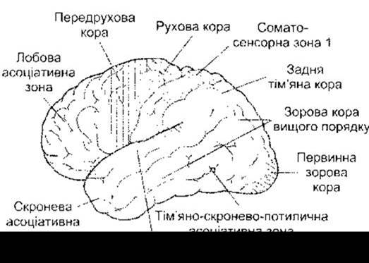
Fig. 16-4. LATERAL VIEW OF the human Cerebral Cortex, illustrating sensory, motor, and association areas.
Complementary Hemispheric Specialization versus "Brain Dominance"
One major functional domain mediated to varying degrees by the neocortex is language—encompassing the comprehension of spoken and written words, as well as the expression of ideas through speech or writing. It is now well established that human language functions depend much more heavily on one cerebral hemisphere than the other. This hemisphere specializes in categorization and symbolization and is frequently termed the "dominant hemisphere." Of course, the contralateral hemisphere is by no means less developed; rather, it reigns supreme in the realm of visuospatial relationships. It is this hemisphere that underlies, for instance, the identification of objects by their shape or the recognition of melodies, and it plays a primary role in face recognition. Consequently, the traditional concept of "brain dominance" and the dichotomy of dominant versus nondominant hemispheres has been superseded by METABOLISM/2.html">THE CONCEPT OF complementary hemispheric specialization. One hemisphere specializes in sequential-analytical Processing (the categorical hemisphere), whereas the other excels in visuospatial processing (the representational hemisphere). Language functions are governed by the categorical hemisphere. Hemispheric specialization is also observed in non-human primates, indicating that it predated the evolution of human language.
Damage to the categorical hemisphere results in language deficits that never occur following even severe lesions of the representational hemisphere. Conversely, lesions of the representational hemisphere typically lead to astereognosis (the inability to identify objects by Touch despite intact tactile and proprioceptive sensitivity) and Other forms of agnosia. Agnosia is a general term denoting the inability to recognize objects via a specific sensory modality. Such deficits typically stem from lesions within the parietal lobe. Damage to the inferior parietal lobule of the representational hemisphere, particularly near the parieto-occipital junction, precipitates unilateral neglect and inattention. Individuals with such lesions exhibit no overt primary visual, auditory, or somatosensory deficits; nevertheless, they completely ignore stimuli originating from the contralateral half of their body or the surrounding extrapersonal space. As a result, the patient neglects one entire side of their body. In extreme manifestations, an individual might shave only one side of the face, dress only one half of the body, or read only the right or left half of each page. This inability to mentally construct a complete map of visual space is partly driven by a pathological shift of visual attention toward the side of the brain lesion; it can be partially alleviated—though rarely fully corrected—by The Use of prismatic lenses.
It is worth noting that the specialization of each hemisphere is further modulated by other cortical regions. Patients suffering from damage to the categorical hemisphere often display deep concern over their condition and tend to
suffer from depression, whereas patients with lesions of the representational hemisphere are frequently nonchalant and occasionally euphoric. Furthermore, patients with right-hemisphere (representational) damage exhibit marked impairments in recognizing other people's emotions.
Hemispheric specialization is intimately linked to handedness, a genetically determined trait. In 96% of right-handed individuals—who comprise roughly 91% of the human population—the left, or categorical, hemisphere is dominant; the remaining 4% exhibit right-hemispheric dominance. Among left-handed individuals, approximately 15% have a categorical right hemisphere, another 15% show no clear lateralization, and the remaining 70% feature a categorical left hemisphere. Interestingly, learning disabilities such as dyslexia (a selective impairment in reading acquisition) occur roughly 12 times more frequently in left-handers than in right-handers. This may be explained by the possibility that certain early developmental anomalies in the left hemisphere prompt a shift in handedness during early childhood. On the positive side, the spatial abilities of left-handers are often significantly above average. A disproportionately large share of artists, musicians, and mathematicians are left-handed. For reasons not yet fully understood, the average life expectancy of left-handed individuals tends to be slightly shorter than that of right-handers.
Subtle anatomical asymmetries exist between the two hemispheres, which may underlie functional differences. As noted in Chapter 9, the planum temporale—a region of the superior temporal gyrus dedicated to language-related auditory processing—is consistently larger on the left side than on the right. This same anatomical Asymmetry is present in chimpanzees, even though language is an almost exclusively human trait. Neuroimaging studies have revealed that other cortical surface areas within the left temporal lobe are larger in right-handers, the right frontal lobe is typically more robustly developed (broader) than the left, and the left occipital lobe is wider and crosses the midline. Portions of the left temporal lobe surface are consistently larger in right-handed individuals. Biochemical differences between the two cerebral hemispheres have also been documented; for instance, the concentration of dopamine in the nigrostriatal pathway is higher on the left side in right-handers and on the right side in left-handers. The physiological significance of these asymmetries remains unknown.
Magnetic Resonance imaging (MRI) studies in schizophrenic patients have revealed a reduced volume of gray matter on the left side, specifically within the anterior hippocampus, amygdala, parahippocampal gyrus, and posterior superior temporal gyrus. The extent of gray matter reduction in the left superior temporal gyrus correlates directly with the severity of Thought Disorders in Schizophrenia. The pathology of this disorder is closely linked to dysfunctions of dopaminergic neurotransmitter systems (see Chapter 15) and alterations in cerebral Blood flow (see Chapter 32).
Physiological foundations of Language
Language is a foundational pillar of human intellect and a cornerstone of culture. The primary brain regions subserving language are clustered along and around the Sylvian fissure (lateral sulcus) of the categorical hemisphere. The posterior portion of the superior temporal gyrus, known as Wernicke’s area (Fig. 16-5), is responsible for processing both auditory and visual linguistic information. It projects via the arcuate fasciculus to Broca’s area (Area 44) in the frontal lobe, located immediately rostral to the lower end of the motor cortex. Within Broca’s area, information received from Wernicke’s region is analyzed, sequenced, and organized, after which signals are transmitted through the motor speech area of the insula to the primary motor cortex, thereby initiating the coordinated Movements of the Lips, Tongue, and Larynx required to produce speech. The putative sequence of neural events occurring when a person names a visually perceived object is illustrated in Fig. 16-6. In the angular gyrus, situated posterior to Wernicke’s area, visual inputs from written words are processed in such a way that they can be translated into their auditory word equivalents within Wernicke’s area.
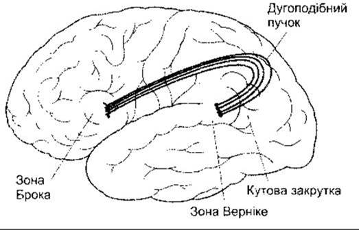
Fig. 16-5. Location of key language-related regions within the categorical hemisphere.

Fig. 16-6. Pathway traversed by neural impulses when naming a visual object, projected onto a horizontal slice of the human brain.
Functional MRI (fMRI) studies have demonstrated that in adults acquiring a second language later in life, the corresponding region within Broca’s area lies adjacent to, yet distinctly separate from, the area dedicated to their native language. In contrast, children who acquire two languages in early childhood utilize a single, shared neural zone for both. It is also well established that children master foreign languages with vastly greater ease than adults.
Speech Disorders
Impairments of language function not attributable to visual or auditory deficits or motor paralysis are collectively termed aphasia. Aphasias result from lesions within the categorical hemisphere, with cerebral embolism or thrombosis being the most common underlying causes. Although numerous Classification schemes exist, the most clinically practical division groups aphasias into fluent aphasia, nonfluent aphasia, and anomic aphasia. Nonfluent aphasia arises from damage to Broca’s area (Table 16-2); speech is halting, labored, and articulation is difficult. In severe cases involving extensive damage to this region, patients may be restricted to expressing a very narrow range of meanings and emotions using only two or three stereotyped words. Occasionally, the few words a patient can utter are precisely those they were speaking at the moment of trauma or vascular occlusion.
One form of fluent aphasia typically ensues following damage to Wernicke’s area. In this condition, the patient's speech output is effortless and often excessively profuse (logorrhea). However, their speech is riddled with paraphasias, jargon, and neologisms that frequently render it completely nonsensical. Furthermore, the patient cannot comprehend the meaning of spoken or written words, effectively blocking all other communicative uses of language.
In another variant of fluent aphasia, patients can speak relatively fluently and understand spoken language, yet they are unable to assemble word fragments into coherent wholes or mentally conjure specific words. This condition is termed conduction aphasia, as it was historically believed to stem from lesions of the arcuate fasciculus connecting Wernicke’s and Broca’s areas. Today, it is recognized that this syndrome typically results from damage localized to the auditory association cortex and its immediate surroundings (Areas 40, 41, and 42).
When a lesion affects the angular gyrus of the categorical hemisphere while sparing Broca’s and Wernicke’s areas, neither speech production nor auditory comprehension is disrupted; however, the patient experiences profound difficulty comprehending written words or pictures because visual information cannot be appropriately processed and relayed to Wernicke’s area. This specific disconnection syndrome is known as amnesic (or nominal) aphasia.
Table 16-2. Typical reactions of patients with lesions in various areas shown a drawing of a chair
Type of aphasia / lesion area |
Typical errors in object naming |
Non-fluent / Broca's area |
"Cha-chair" |
Fluent / Wernicke's area |
"Stool" or "chairlet" (neologism) |
Fluent / areas 40, 41, and 42; conduction aphasia |
"End... no, r-ring... chairlet" |
Anomic / angular gyrus |
"I know what it is... I have a lot of these" |
Numerous selective speech deficits have been described to date. For instance, lesions in the left temporal lobe (area 38) result in the inability to retrieve place names or personal proper nouns from memory, while leaving The ability to use common nouns, verbs, and adjectives unaffected.
Isolated lesions causing the selective deficits described above are relatively rare. Typically, brain damage is more widespread. Consequently, Multiple Forms of aphasia are more frequently observed. Most commonly, aphasia is global, impairing both receptive and expressive functions. In such cases, speech output is restricted. In all types of aphasia that affect speech functions, written language functions are also impaired, although the underlying neural processes remain to be fully elucidated. Interestingly, deaf patients suffering damage to the categorical hemisphere lose their ability to communicate in sign language.
As noted previously, aphasias result from lesions in the categorical hemisphere. Conversely, damage to the representational hemisphere produces distinct specific consequences. For example, it may impair the ability to tell a story or make a joke, as well as negatively affect an individual's capacity to comprehend the punchline of a joke or discern differences in vocal intonation. This provides further evidence supporting the concept of hemispheric specialization.
Stuttering is associated with dominance of the right CEREBRAL CORTEX AND widespread high activity in the Cerebellum and cerebral cortex, including elevated activity in the supplementary motor area. Stimulation of a portion of this area induces laughter, with its duration and intensity proportional to the strength of the stimulus.
Facial Recognition
In right-handed individuals, the processing of information required for facial analysis takes place within the fusiform gyrus on the ventral surface of the right temporal lobe. Facial recognition is enabled by this and adjacent areas (Fig. 16-7). Lesions in this region lead to prosopagnosia, the inability to recognize faces. Patients with this disorder can perceive and reproduce forms, as well as distinguish people by their voices; furthermore, many exhibit autonomic responses when viewing familiar faces. Predictably, such responses are absent when these patients view unfamiliar faces. Nonetheless, they remain unable consciously to identify familiar faces they see. Although the left hemisphere is also involved, The Role of the right hemisphere is primary. The presence of an autonomic response to a familiar face despite the inability to recognize it consciously is explained by the existence of a separate dorsal processing pathway for facial information that operates solely at a subconscious level.
Localization of Other Functions
Through the use of fMRI and PET, alongside the evaluation of patients with strokes and HEAD trauma, initial steps have been taken toward understanding how continuous sensory processing influences cognition, thought, comprehension, and language. Brain mapping of areas responsible for arithmetic calculations has revealed two distinct regions. The inferior region of the left frontal lobe houses the area responsible for numbers and exact calculations. Lesions in this area can result in acalculia—a selective inability to perform mathematical computations. Bilaterally, regions surrounding the intraparietal sulci of the parietal lobes are responsible for the visuospatial representation of numbers and, potentially, finger counting.

Fig. 16-7. Areas in the right cerebral hemisphere of right-handed individuals responsible for facial recognition (modified from Szpir M: Accustomed to your face. Am Sci 1992;80:539).
In humans, two subcortical structures in the right hemisphere are responsible for precise navigation. One of these is the right caudate Nucleus, which facilitates orientation and movement toward objects. Because male brains are larger than female brains, it has been suggested that men possess superior spatial orientation and navigational abilities. It has been proposed, partly in jest, that the larger brain mass in males is due to a higher concentration of neural components dedicated to wayfinding. Consequently, men are often reluctant to ask for directions when lost, whereas women do so without hesitation.
Other deficits observed in patients with localized cortical lesions include the inability to name animals while the ability to name other living creatures or objects remains intact. One patient with a left parietal lobe lesion experienced difficulty pronouncing the latter halves of words. Certain patients with parieto-occipital lesions write words consisting exclusively of consonants, omitting vowels. Such studies help elucidate the precise sequential information-processing pathways within respective brain regions. Further research in this direction aims to significantly broaden our understanding of neocortical functions.
Last update: 10/08/2026
Editorial and Educational Adaptation: This material has been compiled based on the primary/original source text. The project team performed an editorial review, corrected technical inaccuracies, structured sections, and adapted the content for an educational format.
What was processed:
- elimination of formatting defects (OCR errors, structural breaks, corrupted characters);
- editorial organization of content;
- standardization of terminology in accordance with academic sources;
- verification of factual statements against the original source text.
All mentions of the author, publication year, and origin of the primary text have been preserved in accordance with the source.