Human Anatomy Part 1 - K. A. Dyubenko, A. K. Kolomiytsev, Yu. B. Chaykovsky 2002
Special Part
Arthrology, artrologia [the study of bone joints] - Classification of joints and their general characteristics
Magnetic Resonance Imaging of the Spinal Column
MRI of the spine is performed in the sagittal and axial planes. It allows simultaneous visualization of the spine and Spinal Cord over a significant length without the Introduction of contrast agents into the subarachnoid space, and without The Use of ionizing radiation. This has made it widely applicable in the Diagnosis of spinal and spinal cord disorders.
MR scans provide the clearest picture of the Water/140.html">Anatomical Structure of the spine and spinal cord. On axial MR images of the cervical spine, the vertebral bodies appear more square-shaped (Fig. 80).
The vertebral arches with their processes extend posteriorly from the vertebral bodies. On axial MR images, the foramen transmitting the vertebral artery is clearly visible in each transverse process. The thoracic vertebral bodies increase in size in the caudal direction. Oval intervertebral foramina are well-defined in the sagittal plane; they are bounded by the posterior surfaces of the vertebral bodies and intervertebral discs, and superiorly and inferiorly by the vertebral notches of adjacent vertebrae. The articular processes lie nearly vertically. The lumbar vertebral bodies are massive, and their size increases from vertebra LI to LV (Fig. 81).
The arches of these vertebrae are large and form circular foramina with the vertebral bodies (Fig. 82).
MR images clearly delineate the soft Tissues of the spine, spinal cord, intervertebral discs, and ligaments. Furthermore, the contours of the spinal cord and its position within the spinal canal, which follow the physiological curvatures of the spine, are visualized with high clarity.
Class="center">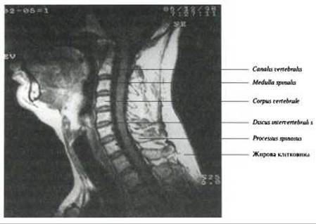
Fig. 80. Magnetic Resonance scan of the cervical spine in the sagittal plane (courtesy of O. P. Ivankov)
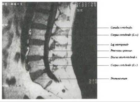
Fig. 81. Magnetic resonance scan of the lumbosacral spine in the sagittal plane (courtesy of O. P. Ivankov)
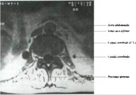
Fig. 82. Magnetic resonance scan of the lumbar spine in the axial plane at the level of LV (courtesy of O. P. Ivankov)
Last update: 08/08/2026
Editorial and Educational Adaptation: This material has been compiled based on the primary/original source text. The project team performed an editorial review, corrected technical inaccuracies, structured sections, and adapted the content for an educational format.
What was processed:
- elimination of formatting defects (OCR errors, structural breaks, corrupted characters);
- editorial organization of content;
- standardization of terminology in accordance with academic sources;
- verification of factual statements against the original source text.
All mentions of the author, publication year, and origin of the primary text have been preserved in accordance with the source.