Human Anatomy Part 1 - K. A. Dyubenko, A. K. Kolomiysev, Yu. B. Chaykovsky 2002
Special Part
Arthrology, artrologia [the study of bone joints] - Classification of joints and their general characteristics
X-ray anatomy of the vertebral column
Radiography of the spinal Column is performed in two projections: anteroposterior (AP) and lateral (Fig. 79). On the AP radiograph, the spinous processes are aligned along the midline of the spine, while the pedicles appear as shadow formations on either side of this line. The vertebral bodies, with the exception of the atlas and axis, are clearly outlined on the radiographs and assume a rectangular shape with slight lateral constrictions (the vertebral "waist") (Fig. 79A). The height of the vertebral bodies progressively increases toward the lumbar region. The massive shadows of the vertebral bodies, spinous and transverse processes, pedicles, and zygapophyseal joints are visible, with the radiolucent shadows of the intervertebral discs located between the vertebral bodies. Lateral radiographs clearly display the vertebral bodies, their upper and lower margins, articular processes, intervertebral spaces (housing the discs), and intervertebral foramina. Thus, plain radiographs allow for the Assessment of the shape, contours, and trabecular Structure OF THE vertebral bodies, the condition of the processes, ossification centers, the width and contours of the joint spaces between articular surfaces, as well as the condition of the spinal canal.
Class="center">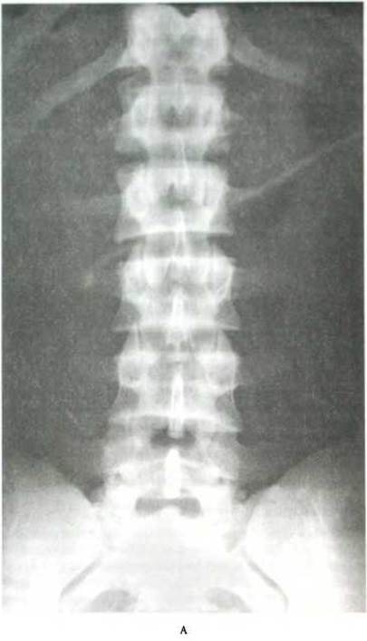
Fig. 79. Plain radiograph of the spinal column (after Ya. I. Kryzhanovsky):
A - lumbar spine (40-year-old male) - anteroposterior projection
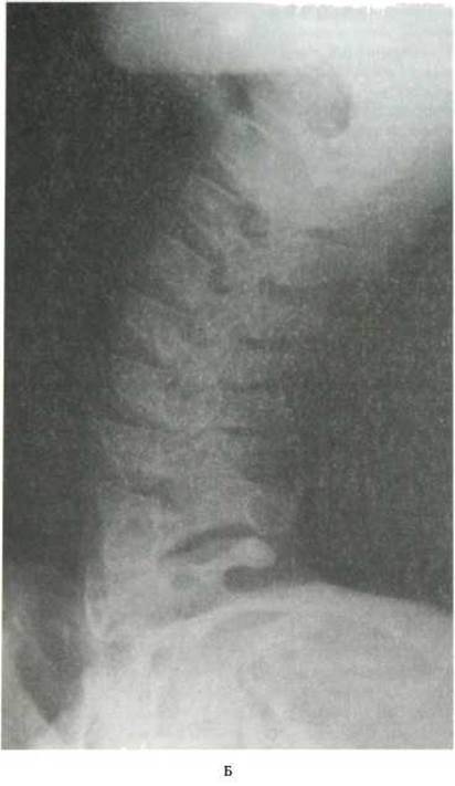
Fig. 79. Plain radiograph of the spinal column (after Ya. I. Kryzhanovsky) (Continued):
B - cervical spine (12-year-old boy) - lateral projection
In newborns, the vertebral bodies are oval-shaped, and the trabecular bone network is indistinctly visualized. The vertebral arches may exhibit slit-like gaps. This is an ossification anomaly known as clefting, or Spina bifida, which should be taken into account after three years of age. Occasionally, a meningocele may develop at the site of the cleft.
Plain radiographs occasionally reveal variations in the number of vertebrae. Of practical interest is an increase in the number of lumbar vertebrae, known as lumbarization. It occurs in 4% of cases and is considered a normal anatomical variant (M. K. Lysenkov). An increase in the number of sacral vertebrae, or when the 5th lumbar vertebra resembles a sacral vertebra and fuses partially or completely with the sacrum, is termed sacralization. Sacralization is relatively common on plain radiographs: 15% in men and 7% in women, with a tendency toward sacralization reaching 50% (M. K. Lysenkov).
Last update: 08/08/2026
Editorial and Educational Adaptation: This material has been compiled based on the primary/original source text. The project team performed an editorial review, corrected technical inaccuracies, structured sections, and adapted the content for an educational format.
What was processed:
- elimination of formatting defects (OCR errors, structural breaks, corrupted characters);
- editorial organization of content;
- standardization of terminology in accordance with academic sources;
- verification of factual statements against the original source text.
All mentions of the author, publication year, and origin of the primary text have been preserved in accordance with the source.