Review of Medical Physiology - William F. Ganong 2002
Functions of the Nervous System
Vision
Other Aspects of Visual Function
Dark Adaptation
The extraordinarily wide range of illumination to which the human eye can respond is illustrated in Fig. 8-27. When a person remains in bright light for a long period and suddenly moves into a dimly lit environment, the retina gradually becomes increasingly sensitive to light as it adapts to the dark. This reduction in the visual threshold is known as dark adaptation. Dark adaptation is almost entirely completed within 20 min, although a minor decrease in the light stimulus threshold can still be observed later. Conversely, when a person suddenly transitions from low light to a brightly lit environment, an uncomfortable sensation of excessively intense illumination occurs until the eyes adapt to the new conditions and the visual threshold rises. This process is called light adaptation and takes about 5 min, although it essentially represents the simple decay of dark adaptation.
In fact, two components can be distinguished in The Development of dark adaptation (Fig. 8-28). The initial drop in the visual threshold occurs rapidly and to a minor extent due to cone dark adaptation, which has been demonstrated by studying the foveal, rod-free area of the retina. The second drop in sensitivity threshold occurs in the peripheral Regions of the retina As a result of rod adaptation. The range of sensitivity threshold variation between a light-adapted eye and a fully dark-adapted eye is remarkably vast.
Radiologists, airplane pilots, and other individuals can, if necessary, shorten the 20-minute dark adaptation period by wearing red-tinted protective goggles when exposed to bright light. Red wavelengths provide relatively good cone function while only minimally stimulating the rods. Therefore, a person wearing red goggles can see well in bright light while dark adaptation of the rods takes place concurrently.
The duration of dark adaptation also depends on the time required to replenish rhodopsin stores. Under bright illumination, large amounts of this pigment are continuously broken down, so in low light conditions, it takes some time for sufficient pigment to accumulate for normal rod function. Cones also undergo dark adaptation, though additional factors are undoubtedly involved in this process.
Effects of Vitamin Deficiency on the Eye
Given the crucial role of vitamin A in the synthesis of retinal, it is not surprising that Vitamin A deficiency leads to visual impairments. Among other manifestations, night blindness (nyctalopia) is one of the earliest to develop. This phenomenon initially drew attention to Structure/19.html">The Importance of vitamin A in rod function; it was later discovered that vitamin A deficiency also leads to cone degeneration. Prolonged deficiency of this vitamin is accompanied by morphological changes in rods and cones, followed by the degeneration of retinal neural elements. Vitamin therapy helps restore retinal function if initiated before receptor destruction begins.
Other Vitamins, particularly those of the B complex, are also essential for the normal functioning of the retina and other neuronal Tissues.
Visual Acuity
Class="center">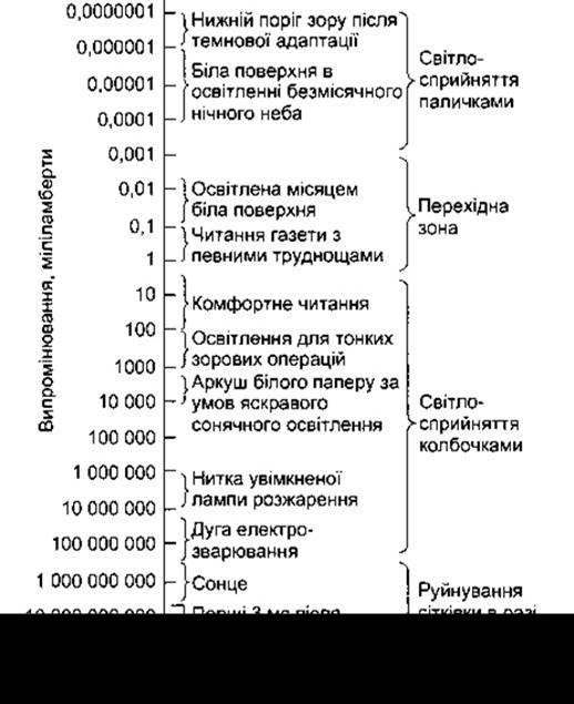
Fig. 8-27. Range of radiation to which the human eye responds, along with the characteristics of photoreceptor mechanisms (reproduced with permission from Campbell FW, in Bell GH, Emslie D, Paterson CR: Textbook of Physiology and Biochemistry, 9th ed. Churchill Livingstone, 1976).
Saccadic Eye Movements (see below) are among the many factors that contribute to visual acuity. This measure of visual function must not be confused with the visual threshold, which is the minimum amount of illumination required to produce a sensation of light. Visual acuity is the degree of perception of an object's details and contours. Although more precise Methods exist, visual acuity is conventionally defined as the minimum separable—the shortest distance at which two lines are perceived as distinct rather than merging. Clinically, visual acuity is measured using Snellen charts (rows of letters of varying sizes meant to be read from a distance of 6 m [20 ft]). The subject is asked to read aloud the smallest row of letters they can distinguish. Results are recorded as a fraction: the numerator is the distance to the chart, and the denominator is the maximum distance from the chart at which a person with normal vision can read the smallest line that the tested individual can read. Normal visual acuity is 20/20; an individual with 20/15 vision has better than normal acuity (though not farsightedness), whereas someone with 20/100 acuity has poor vision. On Snellen charts, the height of the letters in the bottom row that a healthy person can read from 20 feet subtends a visual angle of 5". All constituent lines of the letters are separated by an interval of 1' of arc. Thus, the minimum separable for a normal individual is approximately 1' of visual angle. Visual acuity is a complex metric influenced by numerous diverse factors, including optical factors related to the eye's image-forming mechanisms, retinal factors (the condition of rods and cones), and stimulus-related factors such as luminance, contrast between the stimulus and Background, and the duration of stimulus exposure to the observer.

Fig. 8-28. Dark adaptation. A graph showing Changes in the stimulus intensity required to stimulate the retina under dim illumination as a function of the observer's time spent in the dark.
Critical Fusion Frequency
The temporal resolving power of the Eyeball is determined by measuring the critical fusion frequency—the maximum rate at which stimuli can alternate and still be perceived as separate from one another. Stimuli alternating at a higher frequency are perceived as a continuous, steady stimulus. Moving objects in films appear smooth because frames change at a rate exceeding the critical fusion frequency; if a film projector slows down, the image begins to flicker.
Visual Fields and Binocular Vision
The visual field of each eye is the portion of the external world that the eye can see. Theoretically, the visual field should be circular, but in practice, it is bounded medially by the Nose and superiorly by the brow (Fig. 8-29). Mapping the visual field is essential in neurological Diagnosis. The peripheral PARTS OF THE visual fields are mapped using an instrument called a perimeter, and the process itself is termed perimetry. In this Procedure, one eye is closed while the other fixes on a central point. A small object is moved toward this point along selected meridians, and the point on each meridian where the object is first seen is recorded in degrees of arc from the central point (see Fig. 8-29). The central part of the visual field is mapped using a tangent screen—a black felt screen across which a white object is moved. By recording the locations where the moving object disappears and reappears, the blind spot and other objective scotomas (blind spots acquired due to disease) can be localized.
The central parts of the visual fields of both eyes overlap, so any object located in this region is viewed with binocular vision. Impulses generated by light rays in both retinas merge at the cortical level into a single image (fusion). Points on the retina where the image of an object must fall for that object to be perceived as a single entity during binocular vision are called corresponding points. If one eye is gently pushed out of alignment while observing an object in the center of the visual field, a double image called diplopia arises because the image on the retina of the deviated eye no longer falls on corresponding points.
Binocular vision plays a vital role in depth perception. In addition, monocular cues such as relative object size, shadows, and—for moving objects—motion parallax contribute to depth perception. Naturally, binocular vision enhances the perception of depth and proportions.
Effects of Lesions in the Visual Pathways
The Anatomy of the visual pathways from the eyes to the Brain is shown in Fig. 8-4. The level of a lesion in the visual pathways can be determined quite accurately from the resulting deficits.
Fibers from the nasal half of each retina decussate at the optic chiasm, so that each optic tract contains fibers from the temporal half of one retina and the nasal half of the other. A lesion of one Optic nerve causes blindness in the corresponding eye, whereas a lesion of one optic tract results in the loss of half of the visual field (see Fig. 8-4). This pathology is termed homonymous hemianopia (affecting corresponding halves of both visual fields). A lesion in the region of the optic chiasm (such as a pituitary tumor extending beyond the sella turcica) destroys fibers from both nasal hemiretinas, causing heteronymous hemianopia (affecting opposite halves of the visual fields). Because fibers originating from the maculas are located in the posterior part of the optic chiasm, hemianopic scotomas appear in pituitary tumors before complete loss of vision from both hemiretinas occurs. Partial visual field defects are further classified as bitemporal or binasal, right or left, etc.
Optic nerve fibers originating from the superior retinal quadrants, which supply the lower half of the visual field, terminate in the medial half of the lateral geniculate body, whereas fibers from the inferior quadrants terminate in its lateral half. Geniculocalcarine fibers from the medial half of the lateral geniculate body terminate on the upper lip of the calcarine sulcus, and fibers from the lateral half terminate on the lower lip. Furthermore, fibers from the lateral geniculate body subserving central vision are segregated from those subserving peripheral vision and terminate more dorsally on the Lips of the calcarine sulcus (see Fig. 8-5). Due to these Anatomical Features, occipital lobe lesions can cause quadrantanopia (loss of upper or lower quadrants in each half of the visual field). Macular sparing—the loss of peripheral vision with preservation of central light perception—is also a characteristic symptom of occipital lesions (see Fig. 8-4). This occurs because the cortical representation of the macula is segregated from that of the periphery and is considerably larger. Therefore, for complete elimination of both central and peripheral vision, the area of occipital damage must be very extensive. Bilateral destruction of the occipital cortex in humans leads to subjective blindness. At the same time, a phenomenon known as blindsight exists—residual responses to light stimuli even when they are not consciously perceived. For example, when a person with a damaged visual cortex is asked to locate a stimulus during perimetry, they can do so with greater accuracy than can be explained by chance alone. A certain capacity to detect motion, flicker, orientation, and even colors is also preserved. Similar responses may likewise be elicited by stimulating blind spots in patients with hemianopia resulting from visual cortex damage.

Fig. 8-29. Mono- and binocular visual fields. The dashed line outlines the visual field of the left eye, and the solid line outlines the visual field of the right eye. The overlapping area (the unshaded Heart-shaped central zone) is perceived via binocular vision. The shaded regions are visible through monocular vision.
The fibers passing from the pretectal areas, which mediate the pupillary light reflex, diverge from the optic tracts near the geniculate bodies. Therefore, blindness accompanied by a preserved pupillary light reflex occurs in bilateral lesions posterior to the optic tracts.
Last update: 10/08/2026
Editorial and Educational Adaptation: This material has been compiled based on the primary/original source text. The project team performed an editorial review, corrected technical inaccuracies, structured sections, and adapted the content for an educational format.
What was processed:
- elimination of formatting defects (OCR errors, structural breaks, corrupted characters);
- editorial organization of content;
- standardization of terminology in accordance with academic sources;
- verification of factual statements against the original source text.
All mentions of the author, publication year, and origin of the primary text have been preserved in accordance with the source.