BIOCHEMISTRY AND MOLECULAR BIOLOGY - W. ELLIOTT - 2002
CHAPTER 3. MOLECULAR ORGANIZATION OF CELL MEMBRANES
In Chapter 1, we briefly examined The Role of Three types of weak non-covalent bonds, as well as hydrophobic interactions driven by Water molecules excluding non-polar molecules that interfere with hydrogen bonding. The formation of weak bonds reduces the Free energy of a Structure; therefore, the more such bonds are formed, the more stable the structure becomes. This concept serves as an Introduction to the present chapter, which is dedicated to the Structure and function of Cell membranes.
We have yet to explore cellular METABOLISM. Recall that before nutrient molecules can be utilized, they must enter the bloodstream, be distributed throughout the body, and reach all its Tissues. Right at the outset of their journey, they must cross the membranes of the digestive tract Cells. This alone is reason enough to become familiar with membrane biochemistry before delving into The process of Digestion. Furthermore, cell membrane components are involved in The transport of fats within the body, so we will gain some insight into that process along the way.
Why does a cell need a membrane?
The answer is obvious: the Contents of the cell must be separated from the external environment. We do not yet know how life originated, although several hypotheses have been proposed to explain the potential mechanisms of this process. One of the fundamental Prerequisites for the origin of life is the presence of a primitive molecular self-Replication system. Such a system must function within a small, enclosed space; otherwise, its components would disperse via diffusion.
At first glance, this requirement seems impossible to fulfill because The Cell membrane is far too complex in structure for a primitive self-replication system to reproduce it. Of course, the conditions under which life originated on Earth differed significantly from modern ones; however, certain substances spontaneously form closed, membrane-like structures simply upon being shaken with water. If self-replicating systems were present in the water at the time, a fraction of them would become entrapped inside tiny droplets, or vesicles, bounded by membranes that bear a striking structural
similarity to the membranes of modern living cells. In other words, the assembly of the first membranes could have occurred spontaneously, without the involvement of specialized self-replication machinery.
What are these substances endowed with such unusual properties? One of them can be extracted from egg yolk using organic Solvents. Such an extract contains a mixture of polar Lipids. Polar lipids (Fig. 3.1) are termed amphipathic (i.e., possessing two opposing properties) because their molecules incorporate both polar (polar HEAD) and non-polar hydrophobic (hydrophobic tail) groups.
Class="center">Fig. 3.1. Structural Organization of amphipathic molecules that make up Biological Membranes

The molecules that could have formed a primitive membrane in the distant past were not necessarily identical to modern polar lipids. The crucial factor was that they possessed amphipathic properties.
In the Presence of water, such molecules form structures in which the polar heads face the water, while the contact between the hydrophobic tails and water is minimized. The interaction between hydrophobic tails is maximized by Van der Waals forces, which substantially decreases the free energy of the structure and, consequently, dramatically increases its stability.
When polar lipids are agitated in an aqueous medium, they form Liposomes — vesicles enclosed by a lipid bilayer. The bilayer consists of two rows of polar lipids whose hydrophobic ends are tucked inward, while the hydrophilic heads face outward, contacting water and each other, as illustrated in Fig. 3.2a.
Fig. 3.2. Structure of a liposome and a micelle: a — Cross-section of a synthetic liposome formed by a lipid bilayer; b — cross-section of a dense cylindrical micelle. Lipids with two hydrophobic chains are preferentially incorporated into bilayers, whereas those with a single chain favor micelles

If a liposome is sectioned and treated with a heavy metal that binds to the polar heads, The Lipid Bilayer appears under an Electron microscope as dark railroad tracks. The living cell membrane exhibits a similar appearance after a comparable Procedure, as the lipid bilayer serves as its primary structural framework.
Another potential type of structure formed by amphipathic molecules in water is a dense, cylindrical micelle (see Fig. 3.2b). However, the double hydrophobic tails of polar lipids favor the formation of a bilayer rather than a micellar structure.
The Lipid Bilayer
What are the Lipid Components of cell membranes?
There are numerous polar lipids whose structures, when written out as chemical formulas, vary widely. Nevertheless, schematically, all of them can be represented as shown in Fig. 3.1: with a polar head and two hydrophobic tails. Let us first examine The structure of non-polar lipids for comparison.
Fats are non-polar lipids that can be regarded as derivatives of Fatty acids. Free Fatty acids are never found in membranes. A fatty acid has the structure RCOOH, where R is a long hydrocarbon chain. Among fatty acids, the most common are hexadecanoic (C16) and octadecanoic (C18) acids, commonly known as palmitic and stearic acids, respectively. They are also designated as 16:0 or 18:0, indicating the number of carbon atoms and the number of unsaturated double bonds. Stearic acid (C18) has the structure CH3(CH2)16COOH, or

For simplicity, the hydrocarbon chain of fatty acids is depicted as a zig-zag line, without always ensuring that the number of zig-zags corresponds precisely to the number of carbon atoms.

Salts of fatty acids are called soaps. The hard soap we use for washing is a mixture of sodium salts of long-chain fatty acids.

In the human diet, fatty acids are ingested primarily as fats, which make up a significant portion of our nutritional intake. These are mostly neutral fats, which are derivatives of the trihydric alcohol glycerol, esterified with fatty acids at all three of its hydroxyl groups. Such glycerol esters are called triacylglycerols, or triglycerides.

When neutral fats are boiled in an aqueous alkali solution (NaOH or KOH), the ester bonds undergo Hydrolysis to yield glycerol and soaps. People learned how to make soap long before they understood what esters were. It is hardly surprising that the term saponification is often used to refer to the hydrolysis of any esters, not just neutral fats. Neutral fats are edible and are not detergents. They do not occur in biological membranes and are incapable of forming bilayer structures in water. Here, we discuss them solely for comparison with polar lipids.
The most common Membrane Lipids are derivatives of glycerol-3-phosphate, known as Glycerophospholipids (or phosphoglycerides).

The central carbon atom (C-2) in glycerol-3-phosphate is asymmetric, and therefore the molecule can exist in two isomeric forms. Naturally occurring phosphoglycerides feature exclusively the L-isomer; the prefix sn is used to indicate this stereochemistry. The other two hydroxyl groups are typically esterified with fatty acid residues. The simplest representative of phosphoglycerides is phosphatidic acid:

It is worth remembering this name, as it will make it easier to grasp the nomenclature of more complex compounds later on. Specifically, biological membranes frequently contain derivatives of phosphatidic acid in which various polar groups are attached to the phosphoryl residue. For instance, if we designate such a polar group as X, the corresponding compound is termed phosphatidyl-X:

The more polar the radical X, the more polar the "head" of the phospholipid molecule will be. The distribution of polarity within the molecule becomes much clearer when represented in the following way:

Presented this way, phosphoglycerides are readily recognized as amphipathic membrane lipids capable of forming bilayers, as they feature a polar head and two hydrophobic tails.
What polar groups are linked to the phosphatidic acid residue in Phospholipids?
Bilayers can be formed by a single type of polar lipid. However, to construct their membranes, living cells typically employ a variety of lipids—sometimes quite complex in structure, yet similar in molecular shape and amphipathicity. In phosphoglycerides, this is manifested in The Diversity of polar groups attached to phosphatidic acid.
One such group is the ethanolamine residue HOCH2CH2NH3+, whose phosphatidyl derivative is called phosphatidylethanolamine, or cephalin:

Ethanolamine triple-methylated at the nitrogen atom, HOCH2CH2N+(CH3)3, is called Choline, and the choline-containing phospholipid is known as phosphatidylcholine, or lecithin.
Carboxylated ethanolamine is none other than The amino acid Serine, HOCH2CHNH3+COO-. The corresponding phospholipid, a structural fragment of which is shown below, is called phosphatidylserine.
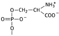
Another type of polar group is the residue of the six-carbon cyclic alcohol Inositol, which is present in phosphatidylinositol (PI).

Standing apart in this series is diphosphatidylglycerol, or cardiolipin, which consists of a glycerol molecule linked to two phosphatidic acid residues. Cardiolipin also exhibits amphipathic properties and is found in The inner mitochondrial membrane as well as in bacterial cell membranes.

All the polar lipids listed above are glycerol derivatives. However, evolution has crafted polar lipids with entirely different structures with unfathomable skill. Even so, their molecules share a similar shape and properties. We are referring to the derivatives of the polyhydric amino alcohol sphingosine, which differs quite significantly in structure from glycerol:

Unlike glycerol, sphingosine contains one of the two required hydrocarbon chains (typically consisting of 15 carbon atoms). The second hydrocarbon
chain in this type of polar lipid is linked to sphingosine via an amide bond, as illustrated by ceramide, whose molecular shape closely resembles that of diacylglycerol.
The phosphorylcholine derivative of ceramide is sphingomyelin, a naturally occurring phospholipid and a major component of the myelin sheath of Nerve Cells.

It is characteristic of evolution that, having discovered a successful solution, it applies it over and over again. For example, ceramide is used in various polar lipids in combination with different polar substituents at the free hydroxyl group. Such substituents are typically sugars.
The Monosaccharides glucose and galactose are typical of cerebrosides, which are present in Brain cell membranes. Recall that galactose is a C-4 stereoisomer of glucose.

Many MONOSACCHARIDES AND THEIR derivatives are known, including amino sugars. The structure of one of them, glucosamine, is shown below:

By combining even a small set of monosaccharides in various ways, nature produces numerous different Oligosaccharides (containing from 3 to 20 monosaccharide residues, whereas Polysaccharides contain hundreds or even thousands), including branched-chain structures. Oligosaccharide derivatives of ceramide are called gangliosides. In these lipids, the polar head groups are particularly large and structurally diverse.

While the structures of sugars are hard to memorize, the structures of N-acetylglucosamine and sialic acid must be known.

Both of these compounds are Components of the carbohydrate moieties of gangliosides and membrane Glycoproteins. Sialic acid is of particular interest because it is involved in cell infection by the Influenza virus (see p. 312). Gangliosides are sphingosine derivatives; they determine human Blood Groups (O, A, B). The carbohydrate parts of gangliosides exhibit antigenic properties and differ in their terminal residues within the oligosaccharide chains.
On the Nomenclature of Membrane Lipids
Alongside individual names for polar lipids, collective (group) names are frequently used. Thus, the terms membrane or polar lipids refer to lipids found in all biological membranes. Phospholipids include all phosphorus-containing lipids. These are subdivided into phosphoglycerides, whose structural backbone is glycerol, and sphingomyelins, which are sphingosine derivatives. Ceramide derivatives lacking phosphoryl groups and containing carbohydrate components are called Glycolipids or glycosphingolipids. A plasmalogen is a glycerophospholipid in which one of the hydrocarbon radicals is linked to the glycerol residue via an ether bond rather than an ester bond. Another essential membrane component is Cholesterol, whose structure has nothing in common with polar lipids.
Why Are There So Many Types of Membrane Lipids?
There is as yet no definitive answer to this question. Different membrane lipids determine various Surface Properties of membranes. For instance, lecithin molecules carry positively charged polar groups, phosphatidylserine molecules bear negatively charged ones, and cerebrosides are neutral. The membrane Lipid Composition can vary significantly among different cells. For example, brain cell membranes are rich in cerebrosides and gangliosides, whereas the membranes of nerve cell myelin sheaths are rich in glycosphingolipids. However, lipids differ in composition not only across different cell types. In all Plasma Membranes studied, the outer and inner surfaces of the bilayer differ markedly in lipid composition. For example, glycolipids are always located in the outer leaflet, with their carbohydrate portion exposed to the extracellular environment. Lipid molecules can hardly ever spontaneously flip from one leaflet to the other (a process known as a flip-flop), because this would require polar heads to pass through the Hydrophobic core of the membrane, which is thermodynamically unfavorable. Enzymes are known that catalyze this process; they likely participate in establishing and maintaining Asymmetry in biological membranes.
In contrast, lipids can move easily, freely, and rapidly within a single monolayer (lateral mobility) because they interact very weakly with one another.
One of the membrane lipids, phosphatidylinositol, serves as a carrier for two different chemical signals that regulate cellular functions (see Chapter 26). This is, however, a special case. There are still no rational explanations as to why membranes contain so many different polar lipids, what causes the unique Lipid Composition of various cells, or what purpose is served by the compositional asymmetry of the outer and inner monolayers. Certain membrane lipids, particularly gangliosides, are of medical interest. For instance, in childhood disorders belonging to the group of glycosphingolipids (such as Tay-Sachs and Gaucher diseases), glycosphingolipid metabolism is impaired, and the accumulation of their partial degradation products causes severe damage to The Nervous system.
In summary, cells utilize A wide variety of polar lipids to build their membranes, yet the reasons for this Variability remain unclear.
What is the Fatty Acid Composition of Membrane Lipids?
The fatty acid residues that form the hydrophobic tail of membrane lipids are structurally diverse. As a rule, they have unbranched chains and contain an even number of carbon atoms—from C14 to C24 (predominantly from C14 to C18). (The reasons for the even number of carbon atoms are discussed in Chapter 9.) The two fatty acid residues in a single phospholipid molecule may be identical or different, with the hydroxyl group at the first position in phosphoglycerides typically being esterified with a saturated-chain acid, and at the second position with an unsaturated one. The degree of unsaturation of the fatty acid tails plays a crucial role, since the central hydrophobic region of the membrane must be fluid-like in its properties rather than solid-like. The higher the unsaturation, the lower the corresponding phase transition Temperature of the membrane lipids.
Typically, double bonds in fatty acid tails have a cis configuration, which causes these tails (unlike saturated ones) to have
a bent rather than a straight shape. Such kinks prevent tight packing of the molecules.

The Effect of the degree of unsaturation on the phase state of a lipid is clearly seen when comparing mutton fat with olive oil. In both cases, we are dealing with triglycerides, but olive oil lipids are rich in unsaturated fatty acid tails. However, triglycerides are absent in membranes, and it is more appropriate to compare the fatty acid composition of membrane lipids in closely related warm-adapted and cold-adapted species: the former are dominated by fully saturated acids.
The Importance of maintaining membrane "fluidity" is illustrated by the fact that Bacteria regulate the fatty acid composition of their membranes so that the phase transition temperature of the membrane lipids is close to the growth temperature. In warm-blooded animals that undergo hibernation, the degree of saturation of membrane Lipid Fatty Acids also varies depending on seasonal changes in body temperature.
The most common monounsaturated acids are oleic (18:1) and palmitoleic (16:1) acids, while linoleic (18:2) and arachidonic (20:4) acids, with two and four double bonds respectively, are also quite frequent. The nomenclature of such acids will be discussed in detail in Chapter 10.
What role does cholesterol play in membranes?
Looking at the structural formula of cholesterol (Fig. 3.3, a), one might wonder why this substance is a component of biological membranes. However, everything falls into place when examining the conformation of its molecule (Fig. 3.3, b). Cholesterol is an elongated molecule with a rigid steroid nucleus and a flexible hydrocarbon chain. The cholesterol molecule is amphipathic, as one of its ends features a polar hydroxyl group.
Fig. 3.3. Cholesterol molecule: a — Planar view; b — Spatial view

By inserting itself between lipid molecules (with the hydroxyl group located in the region of the polar heads), cholesterol helps regulate membrane fluidity by preventing the tight packing of hydrocarbon chains, thereby lowering the lipid melting point. Externally, this manifests as a "broadening" of the phase transition range in the lipid bilayer. In other words, in the absence of cholesterol, this transition occurs over a narrower temperature interval. The presence of impurities in crystalline substances affects their melting point in a similar manner. The erythrocyte membrane may contain up to 25% cholesterol, whereas bacterial membranes lack it entirely, and animal mitochondrial membranes contain very little. In plant cells, the role of cholesterol is fulfilled by related compounds known as phytosterols.
Fusion of lipid bilayers
The lipid bilayer is somewhat analogous to a two-dimensional fluid in which individual lipid molecules move freely within their own monolayer. Lipid bilayers typically close in on themselves, eliminating free edges. For this reason, they are capable of spontaneously repairing damage by sealing the edges of torn areas; upon close contact, bilayers fuse. Such properties impart flexibility to cells and facilitate the resolution of constrictions, which is especially important during Cell Division (Fig. 3.4, a).
Similar processes occur during endocytosis—the process by which a cell engulfs particles (or large molecules) that cannot pass through the membrane bilayer. The engulfed particle is gradually surrounded by a small patch of The Plasma Membrane, which first invaginates and then pinches off to form an intracellular vesicle, or vacuole (Fig. 3.4, b).
The reverse phenomenon is also possible. For instance, the transport of protein molecules secreted by the cell is carried out by packaging them into membrane-bound vesicles, which then fuse with the plasma membrane, releasing their contents into the extracellular environment (Fig. 3.4, c).
This process is called exocytosis. It occurs, for example, during the secretion of digestive enzymes by pancreatic cells.
Fig. 3.4. Cell division (a), endocytosis (b), and exocytosis (c). Exocytosis is used for the secretion of digestive enzymes

Permeability of lipid bilayers
The ability of substances to diffuse across a lipid bilayer is mainly determined by their lipid solubility. Fat-soluble molecules easily cross the bilayer, whereas it is virtually impermeable to most polar molecules and ions. Large uncharged polar molecules such as glucose cross bilayers more poorly than smaller ones, such as ethanol and glycerol. The charged groups of polar molecules and inorganic ions in aqueous solution are surrounded by Hydration shells composed of specifically oriented water molecules. To cross the hydrophobic core of the membrane, they must shed these shells, which is energetically highly unfavorable. Therefore, the permeability of lipid bilayers to such molecules and ions is negligibly small.
Most molecules involved in biochemical processes are quite polar and therefore cannot diffuse across the lipid bilayer at a rate commensurate with the cell's needs. It follows that, firstly, the bilayer structure of biological membranes ideally fulfills the requirement of shielding the Cytoplasm from the external environment; secondly, the membrane must possess mechanisms that allow the cell to rapidly and selectively regulate the influx and efflux of molecules and ions according to metabolic demands.
Strikingly, water molecules, despite being polar, readily cross the lipid bilayer. Naturally, The small size of the water molecule and its lack of charge play a role, but this alone is insufficient; additional factors are likely at work. In the membranes of renal tubule cells and secretory epithelial cells, where water transport is particularly intensive, a specialized protein called aquaporin is present, which facilitates the free Movement of water across the membrane. The lipid bilayer is also permeable to dissolved gases, notably oxygen.
In summary, Cells must be surrounded by a barrier that prevents the leakage of their components, which are predominantly polar compounds. This goal is achieved through The Use of a lipid bilayer—a structure whose formation and stability are determined by weak interactions among various amphipathic polar lipids.
The monolayers that make up the bilayer membrane differ in their lipid composition. This asymmetry arises during Membrane Assembly and is maintained because lipid exchange between monolayers is energetically unfavorable. Lateral diffusion of lipids (within each monolayer) occurs quite freely. Different cell types vary in the lipid composition of their membranes. It remains unclear why nature required such an extensive array of lipids for Biomembranes, especially since only one of them—phosphatidylinositol—has a specific role as a chemical signal. The hydrocarbon core of membranes is in a "fluid" state. In animal cells, this is achieved by maintaining a balance between saturated and unsaturated fatty acid residues. The latter lower the phase transition temperature in bilayers: their chains, bent by cis double bonds, hinder the tight packing of lipid molecules. Cholesterol is used to fine-tune the phase transition temperature. In bacterial membranes, the same effect is achieved using Branched-Chain Fatty Acids. The lipid bilayer is practically impermeable to strongly polar molecules, yet small weakly polar molecules diffuse through it relatively easily. These properties enable the bilayer to perform its primary function—serving as a barrier that prevents the leakage of cytoplasmic components. The ability of bilayers to fuse ensures that processes such as cell division, endocytosis, and exocytosis can take place.
The lipid bilayer serves as the structural foundation of biological membranes, yet by itself it is incapable of carrying out all their functions, including selective transmembrane transport. Membrane Proteins are responsible for this.
Membrane Proteins and the Structure of Biological Membranes
Owing to their remarkable structure, biological membranes exhibit diverse properties. Different cells perform various functions, and each of these functions can be carried out by a specialized membrane protein synthesized for a specific task before being targeted to the membrane (for details on this process, see Chapter 22). Crucially, the protein must be capable of integrating into the membrane. Experiments have demonstrated that functional proteins isolated from biomembranes are able to insert into artificial lipid membranes (liposomes) and perform the same functions there as they do in the native membrane.
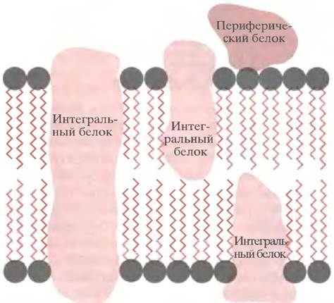
The structure of biomembranes is often referred to as fluid-mosaic (Fig. 3.5). It is likened to a two-dimensional sea of lipids populated by "icebergs"—integral
proteins that are either partially embedded in the membrane or span it completely. In addition, there are so-called peripheral proteins, whose molecules are not embedded in the membrane but are held on its surface via weak interactions.
Fig. 3.5. Fluid-mosaic model of Biological Membrane Structure
What holds integral proteins within the lipid bilayer?
Integral membrane proteins are retained in the lipid bilayer due to structural features that enable weak interactions between the proteins and their lipid environment.
As is well known, proteins are Polypeptides composed of covalently linked Amino Acids. If all amino acid residues in a polypeptide contain hydrophobic side chains, the polypeptide as a whole will be hydrophobic, and so forth. In integral membrane proteins, the terminal Regions of the polypeptide chain consist of hydrophilic amino acid residues, whereas the central region comprises hydrophobic residues (Fig. 3.6, a).
Fig. 3.6, b schematically illustrates the structure of Glycophorin, the major protein component of the erythrocyte membrane. The extracellular side of the membrane features the N-terminal region of this protein, which is rich in hydrophilic amino acids. The hydrophilicity of this fragment is enhanced by A large number of serine, Threonine, and asparagine residues covalently linked to carbohydrate moieties. The C-terminal region of the polypeptide chain, located on the cytoplasmic side of the membrane, is also rich in hydrophilic amino acids but lacks carbohydrate components (carbohydrate residues are invariably located on the cell surface). Both hydrophilic fragments of the glycophorin molecule are separated by a hydrophobic stretch of the polypeptide chain situated within the membrane (this segment lacks any strongly hydrophilic amino acids). It consists of 19 amino acid residues folded into an α-Helix.
The dimensions of the central hydrophobic region of the glycophorin molecule correspond to the thickness of the hydrophobic core of the lipid bilayer, while the outer hydrophilic regions are localized on opposite sides of the membrane and are capable of interacting with both water and the polar head groups of membrane lipids (see Fig. 3.6, b). Proteins whose molecules feature distinctly segregated hydrophilic and hydrophobic regions are termed amphipathic.
Fig. 3.6. Structural organization of integral proteins (a) and the STRUCTURE OF THE glycophorin molecule (b). The helical segment of glycophorin, comprising 19 amino acid residues, is approximately 3 nm long, which corresponds to the thickness of the hydrophobic region of the lipid bilayer

In certain membrane proteins, the polypeptide chain folds repeatedly so that it traverses the membrane not once, but multiple times (Fig. 3.7). Accordingly, hydrophilic regions alternate with hydrophobic ones; the latter are folded into helices consisting of 15–20 amino acid residues. Among such proteins, the most extensively studied is Bacteriorhodopsin, a pigment found in the purple bacterium Halobacterium halobium. It captures light energy and uses it to pump protons out of the bacterial cell into the environment, thereby generating a proton gradient whose energy is subsequently utilized by the cell to synthesize ATP (this mechanism is described in Chapter 7).
Fig. 3.7. Topography of the bacteriorhodopsin polypeptide chain in the membrane. Seven tightly packed α-helical segments span the lipid bilayer

The arrangement of membrane proteins ensures their most energetically favorable interaction with the lipid bilayer. Any attempt by a protein to leave the membrane would bring its hydrophobic regions into contact with Water and Its hydrophilic regions into contact with the hydrocarbon core of the membrane. Both outcomes are energetically unfavorable, making Transmembrane Movement of integral proteins virtually impossible. The situation is quite different for their lateral diffusion within the monolayer. The mechanisms by which proteins target biomembranes and their initial orientation will be discussed in Chapter 22.
Anchoring of peripheral proteins to the membrane
Peripheral, water-soluble membrane Proteins can be held on the membrane surface through hydrogen bonding and ionic interactions.
However, other mechanisms exist as well. For instance, a protein may contain a covalently linked long-chain fatty acid residue that inserts into the lipid bilayer, acting as a membrane anchor (Fig. 3.8). In some proteins, N-terminal Glycine residues are linked to fatty myristic acid:
Р-СO-NН-СН2-СO- polypeptide,
in others, C14, C16, and C18 fatty acids esterify the hydroxyl groups of Serine and threonine residues or the thiol group of Cysteine. Even more complex fatty acyl structures linked to proteins via ether bonds are also employed. An example of a more sophisticated mechanism for anchoring peripheral proteins to the membrane is the covalent attachment of phosphatidylinositol to the oligosaccharide moiety of a membrane glycoprotein. Such a phosphatidylinositol residue is incorporated into the membrane and behaves there as a free lipid molecule.
Fig. 3.8. A fatty acid anchor residue attached via an amide bond to a protein molecule. In some cases, fatty acid residues are attached to proteins through ester or thioester bonds

Membrane Glycoproteins
Many membrane glycoproteins, much like glycolipids such as cerebrosides and gangliosides, contain branched oligosaccharides exposed on the outer surface of the membrane. These oligosaccharides, covalently linked to the side chains of serine or asparagine residues, are composed of fucose, mannose, galactose, N-acetylgalactosamine,
N-acetylglucosamine, and sialic acid. In the erythrocyte membrane glycoprotein glycophorin, the carbohydrate portion accounts for half the mass of the molecule. The exact Biological Role of these oligosaccharides remains unclear, although in some cases they appear to act as markers for cell surface recognition.
Thus, the diversity of membrane proteins is dictated by the multifaceted functions of biological membranes. Integral proteins are embedded within the membrane, whereas peripheral proteins are held at the surface through non-covalent interactions with the polar head groups of lipids. Integral proteins are amphipathic: their structure clearly distinguishes a central hydrophobic region interacting with the hydrocarbon core of the lipid bilayer, and hydrophilic domains at the C- and N-termini exposed to the aqueous environment on opposite sides of the membrane. Membrane proteins are frequently glycosylated, meaning they contain covalently attached sugar residues, and these sugars are invariably located on the extracellular face of the plasma membrane. In principle, a wide variety of protein types can be incorporated into a lipid membrane, imparting functional flexibility.
Biological membranes perform a vast array of functions. We will restrict our Discussion here to the Functions of the plasma membrane surrounding the cell, leaving intracellular membranes for subsequent chapters. Aside from its primary role of containing the cellular contents, the plasma membrane serves numerous other purposes. This section will examine its involvement in the Transport of Molecules into and out of the cell, signal Transduction, maintenance of cell shape, and intercellular interactions.
Transport of substances Across the Cell Membrane
We have already noted that many, if not most, of the substances a cell must acquire from its environment cannot penetrate the plasma membrane at rates sufficient to meet its metabolic demands. (It should be noted that certain intracellular membranes, such as the outer membranes of Mitochondria and Chloroplasts, possess pores so large that any molecule with a mass of less than 600 Da passes through them freely.) Therefore, the Transmembrane Transport of polar molecules—such as sugars, amino acids, inorganic ions, and the like—requires specialized proteins. Only these proteins can provide the requisite selectivity and allow external control over cellular processes. This demand for selectivity explains the necessity for a multitude of specialized transport systems and, consequently, transport proteins.
Passive Transport, or Facilitated Diffusion
Transport systems are broadly categorized into passive and active. The former mediate the movement of substances across the membrane without requiring an external energy input, as transport is driven by a concentration gradient. For this reason, passive transport is also termed facilitated diffusion. A prime example is the transport protein responsible for the bidirectional movement of anions (Cl- and HCO-3) across the erythrocyte membrane (Fig. 3.9). The physiological significance of this process is discussed in Chapter 27. Another classic example of facilitated diffusion is the uptake of glucose across the membranes of animal cells—an essential and vital step in the assimilation of this nutrient delivered by the bloodstream.
Fig. 3.9. Anion channel in the erythrocyte membrane. Cl- and HCO3- ions can move through the channel in opposite directions in accordance with their concentration gradients. This counter-transport of anions ensures the maintenance of electroneutrality.

Important components of passive transport systems are gated pores, commonly referred to as channels. These include structures that facilitate transmembrane ion fluxes whose permeability is regulated by external signals. Some channels open in response to depolarization (see Chapter 26), while others are activated by specific chemical messengers, notably acetylcholine. The Structural elements of channels directly responsible for controlling their permeability are termed gates, whereas the structural elements that recognize the signaling molecule (chemical signal) are called receptors. These channels play a critical physiological role for Ca2+, Na+, and K+ ions.
This type of transmembrane transport is driven by an external energy source. In all cells (except bacteria), this source is ATP hydrolysis. Active transport involves the movement of a substance against its concentration gradient. To calculate The amount of energy required to move a substance into a cell against a concentration gradient, one can use the equation introduced in Chapter 1 for Chemical Reactions:

For substances whose structure remains unchanged during transport (∆G°′ = 0), this equation simplifies to:

where C1 is the extracellular concentration of the substance and C2 is its intracellular concentration. If C2/C1 = 10 and T = 298 K, then:

Thus, under these conditions, the transport of one mole of a substance requires 5706 J (at 25° C). It is hardly surprising that nerve cells, whose Membrane Potential is largely determined by the concentration gradients of sodium and potassium ions, expend a major fraction of their generated ATP to maintain these gradients.
A classic example of an active transport system is the so-called Na+/K+ pump in the plasma membranes of animal cells. These cells maintain a high intracellular concentration of potassium ions (140 mM) and a low concentration of sodium ions (12 mM), whereas in blood and extracellular fluid, these concentration ratios are reversed at approximately 4 and 145 mM, respectively. The ion permeability of the plasma membrane is such that the potassium and sodium concentration gradients correspond to an electrical potential difference of 50–70 mV (positive outside, negative inside). This maintenance comes at a metabolic cost: animal cells expend about one-third of their total energy consumption to sustain these ion gradients. However, this energy is not wasted. These gradients are essential for the GENERATION AND PROPAGATION of electrical signals in excitable membranes, and furthermore, the energy stored in these gradients drives the secondary transport of various other molecules across the membrane.
Architecture of the Na+/K+ Pump
This pump is also known as the Na+/K+-ATPase because the extrusion of sodium and the uptake of potassium are coupled to the hydrolysis of ATP to ADP + Pi.
The Na+/K+-ATPase is a protein composed of two pairs of subunits (αα and ββ). Certain proteins undergo conformational changes upon binding specific ligands (ligands are compounds that bind to other molecules at highly specific sites). These structural shifts occur either as conformational transitions within a single protein molecule or via the reorganization of subunits within Protein Complexes (as seen, for example, in Hemoglobin). In the Na+/K+-ATPase, such changes can be triggered by protein phosphorylation, which alters its binding affinity for sodium and potassium ions.
According to the model presented in Fig. 3.10, the Na+/K+-ATPase can exist in two Conformations. The binding of Na+ followed by the phosphorylation of the ATPase from the cytoplasmic side (see Fig. 3.10, a) induces a conformational change in the protein that drives Na+ across the membrane and releases it into the extracellular space. Concurrently, K+ binds to the extracellular surface of the membrane (see Fig. 3.10, b), and subsequent dephosphorylation returns the protein to its original state, releasing K+ into the cytoplasm as it crosses the membrane (see Fig. 3.10, c). Thus, phosphorylation of the enzyme results in the extrusion of a sodium ion from the cell and the binding of an extracellular potassium ion. If dephosphorylation resets the enzyme to its initial conformation, the process becomes cyclical, resulting in the hydrolysis of ATP coupled to the transmembrane counter-transport of sodium and potassium ions. Indeed, a single catalytic cycle of the Na+/K+-ATPase is accompanied by the reciprocal transport of three sodium ions and two potassium ions. All of these events can be described by the following equation:
3Na+(inside) + 2К+(outside) + ATP + Н2O —> 3Na+(outside) + 2К+(inside) + ADP + Рi + Н+.
The medical aspect here is quite fascinating. There is a group of compounds known as cardiac Glycosides, which are carbohydrate derivatives of Steroids (structurally similar to cholesterol). They are found in foxglove and closely related plant species.
These compounds inhibit Na+/K+-ATPase by preventing the removal of the phosphoryl group, thereby halting ion transport at stage b (see Fig. 3.10). In small doses, they have long been used in medicine to treat Heart Failure, while in large doses, they are lethal. The therapeutic effect of cardiac glycosides stems from the fact that by inhibiting Na+/K+-ATPase, they increase the concentration of sodium ions within heart Muscle cells, which in turn reduces the sodium gradient. This leads to an increase in cytoplasmic Ca2+ ion levels because there is another system that transports Na+ into the cell and Ca2+ out; that is, the extrusion of Ca2+ from muscle cells occurs via exchange for sodium and depends on the sodium gradient. The reduction in the Na+ gradient caused by cardiac glycosides helps decrease the outward transport of Ca2+ and increases its intracellular level. Ultimately, this stimulates heart Muscle contraction. A similar effect is produced by ouabain, a plant-derived substance used by Africans to poison their arrow tips.
Fig. 3.10. Hypothetical mechanism of the Na+/K+ pump: a–c — Successive states of Na+/K+-ATPase

Cotransport of molecules across membranes (symport)
The transmembrane Na+ gradient generated by Na+/K+-ATPase is essentially a form of stored energy. This potential energy would be dissipated as heat if sodium ions were simply allowed to diffuse back into the cell. Suppose, however, that they do so in conjunction with other molecules. Such coupled transport is called symport. For example, a protein is responsible for the symport of sodium ions and glucose, transferring them in strictly equimolar amounts. As a result, glucose can move into the cell against its own concentration gradient, driven by the sodium ion gradient. And since the latter is established via ATP hydrolysis, ATP indirectly acts as the driving force for glucose transport. A similar mechanism operates during the Absorption of Amino acids and glucose in the intestine (Fig. 3.11), with different transport proteins participating in The transfer of different substances.
Fig. 3.11. Symport — a system for the coupled transport of glucose and sodium ions

Antiport
In connection with the action of cardiac glycosides, we have already mentioned that The entry of sodium ions into heart muscle cells is coupled with the pumping out of Calcium Ions. Such tightly coupled counter-flow is termed antiport (Fig. 3.12). In this case as well, calcium can be transported against its own concentration gradient utilizing the energy of the sodium gradient, which, in turn, is generated through ATP hydrolysis. When we move on to examine muscle contraction, you will see that There are also Ca2+ pumps (Ca2+-ATPases) that directly utilize ATP hydrolysis as an energy source for the transmembrane transport of calcium ions.
Fig. 3.12. Antiport — a system of tightly coupled counter-flow of sodium and calcium ions

Uniport
The term uniport refers to the transfer of any solutes or ions from one side of the membrane to the other with the participation of specialized transport proteins, such as the Ca2+-ATPase from the sarcoplasmic reticulum.
Thus, most substances are transported across the plasma membrane with the help of a transport protein specific to each substance, which explains why there are so many such proteins. Transport is classified as passive when its driving force is solely a concentration gradient (such as the transfer of anions in erythrocytes or glucose in animal cells). However, this is not always the case, and substances can move across the membrane against their own gradient. Animal cells possess Na+/K+-ATPase, which establishes opposing concentration gradients of potassium and sodium ions across the plasma membrane. The inhibition of this enzyme by cardiac glycosides is utilized in medicine for the Treatment of heart failure. The sodium gradient generated across the plasma membrane can serve as an energy source for the transport of other molecules (the symport of sodium and amino acids or glucose in the intestine) or for exporting them into the extracellular environment (the antiport of sodium and calcium ions). In both sodium-coupled symport and antiport, ATP hydrolysis serves as the ultimate energy source for establishing all these gradients. Alongside Na+/K+-ATPase, other transport ATPases also exist.
Signal transduction
All animal cells must function in a coordinated manner, in accordance with the needs of the Organism as a whole, which they learn about through signals sent to them. Life is a chemical process, and therefore it is hardly surprising that these signals are transmitted in the form of chemical compounds, whether they are Hormones produced by glands or Neurotransmitters released by nerve endings.
Among signaling compounds, there are non-polar substances, such as Steroid Hormones, which enter cells by diffusing through the lipid bilayer, as well as polar substances, which cannot enter the cell and instead bind to specialized receptor proteins on its surface, which are responsible for signal transmission. The binding of a signaling molecule to a membrane receptor protein initiates a cascade of sequential events inside the cell. This is precisely what is meant when the plasma membrane is described as a signal transducer. The biochemical aspects of signal transduction will be discussed in Chapters 12 and 26.
The role of the plasma membrane in maintaining cell shape and motility
Eukaryotic cells possess a structural framework that, on the one hand, gives them a definite shape and, on the other hand, allows for its modification, enabling cells to move and relocate their Organelles from one place to another. This framework, known as the Cytoskeleton (see Chapter 29), unlike the Skeletal System of animals, is neither rigid nor of a fixed structure. It is formed by protein filaments that permeate the cytoplasm and are linked at numerous points to the Proteins of the plasma membrane. Naturally, these proteins are incapable of the lateral diffusion typical of ordinary membrane proteins.
The most striking example illustrating The connection between the cytoskeleton and membrane proteins is the erythrocyte (red blood cell). It has a biconcave disc shape, which maximizes the surface area for gas exchange between the cytoplasm and the external environment. When squeezing through the tiniest capillaries, an erythrocyte experiences significant mechanical stress and, owing to its flexibility and elasticity, undergoes substantial yet reversible changes in shape. The erythrocyte exhibits these properties due to a dense submembranous network of fibers formed by the protein spectrin. The name of this protein derives from the Latin word spectrum (ghost or phantom), which is synonymous with the English word ghost—a term used for erythrocytes that have retained their shape but lost their contents. Spectrin is linked to the protein ankyrin (from the Greek anchora, meaning anchor), which, in turn, is anchored to the inner surface of the membrane by binding to the anion exchanger protein. Other anchor proteins attach spectrin to glycophorin (Fig. 3.13).
Fig. 3.13. Interaction of the anion transport protein and glycophorin with the cytoskeleton

Certain patients are known to have a defective erythrocyte cytoskeleton resulting from structural abnormalities in spectrin or ankyrin. In such cases, THE RED BLOOD cells exhibit abnormal shapes and are destroyed in the Spleen. Diseases caused by these disorders are referred to as hereditary spherocytosis or hereditary elliptocytosis.
Intercellular interactions: tight junctions, Gap Junctions, and Cell Adhesion proteins
Epithelial cells lining the digestive tract mediate the transfer of digestion products into the bloodstream. These substances penetrate the epithelial cell through the plasma membrane facing the intestinal lumen, are transported across the cell, and then exit through the opposite membrane into blood capillaries. Absorption occurs precisely in this manner rather than through the intercellular spaces, which is physically impossible as the cells are tightly apposed. Tight junctions are impermeable protein structures that encircle adjacent cells and bind them together. Within a tight junction, even the lateral movement of membrane proteins is restricted. This plays a crucial functional role. The domains of the epithelial cell plasma membrane facing the intestinal lumen and the blood capillary contain different transport proteins which, in the absence of lateral diffusion, cannot migrate from one domain to another. At the same time, it is essential to facilitate the exchange of molecules between cells to synchronize their activity throughout the entire tissue. This is indeed achieved through specialized proteins that form intercellular tunnels, or gap junctions, which allow molecules such as ATP to pass while retaining larger molecules like proteins. The importance of such Intercellular Communication is unquestionable. For instance, it is vital for the coordinated contraction of heart muscle cells.
Specialized tissue-specific adhesion proteins drive the aggregation of similar cells into tissues. When embryonic cells of different types are mixed, they form aggregates of identical cells: Liver cells associate with liver cells, Kidney cells with kidney cells, and so on. The general term for proteins responsible for cell adhesion is cadherins. These include distinct families, such as NCAMs (Neural Cell Adhesion Molecules), which play a critical role in neural tissue formation.
Intracellular membranes
So far, this chapter has focused solely on the outer, or plasma, cell membrane. However, Eukaryotic cells contain a multitude of diverse membrane-bounded structures. All of them are based on a lipid bilayer, but they differ in lipid composition and the set of functional proteins embedded within them. These internal membrane systems partition the cell into distinct compartments. The principal components of an animal cell are illustrated in Fig. 3.14.
Fig. 3.14. Membrane-bound organelles of a typical animal cell

Let us outline the most prominent intracellular organelles and their functions.
1. The nucleus, which contains DNA and serves as the site of DNA and RNA Synthesis.
2. The Endoplasmic reticulum (ER), a network of membrane tubules and cisternae that accounts for half of the cell's volume. It is the site of synthesis for all membrane proteins, as well as proteins destined for secretion or targeted delivery to specific cellular locations. Part of the ER is studded with Ribosomes—structures responsible for Protein Synthesis. These regions are referred to as rough endoplasmic reticulum, in contrast to the smooth endoplasmic reticulum, which lacks ribosomes.
3. The Golgi apparatus, consisting of stacks of flattened cisternae; here, proteins synthesized in the ER are sorted and dispatched to their respective destinations. Both the ER and the Golgi apparatus are entirely closed structures, meaning that everything leaving them is enclosed within budding, membrane-enclosed vesicles.
4. Mitochondria, the sites of oxidative metabolism where the majority of ATP molecules (generated in the inner mitochondrial membrane) are produced to meet the various energy demands of the cell.
5. Lysosomes, which contain hydrolytic enzymes that break down unwanted cellular components.
6. Peroxisomes, vesicles containing enzymes that catalyze various oxidation reactions. Different cells rely on specific organelles to varying degrees. An electron micrograph of a Testis tissue section (Fig. 3.15, a, b) reveals regions of smooth endoplasmic reticulum enriched with enzymes involved in steroid synthesis. The ER is particularly well-developed in cells that secrete proteins, such as digestive enzymes or Antibodies. Cells with high energy demands, such as muscle cells, are rich in mitochondria. Cells completely devoid of organelles are extremely rare; mature erythrocytes serve as a prime example.
Fig. 3.15. Cytology/cytology/93.html">ELECTRON MICROGRAPHS OF testis sections: a — A steroid-secreting cell (note the smooth Endoplasmic reticulum and macrophage); b — the same cell at higher magnification, clearly showing the Golgi apparatus

Chapter 3 Questions
1. What Structural Features of amphipathic compounds determine their ability to form micelles or liposomes in water?
2. Are triglycerides components of biological membranes?
3. Can proteins cross the lipid bilayer?
4. What is the function of cholesterol in Eukaryotic Cell membranes?
5. Draw the structure of phosphatidic acid.
6. Name three phospholipids derived from phosphatidic acid and specify the head groups that distinguish them.
7. Describe the structure of sphingomyelin.
8. How do cerebrosides and gangliosides differ from sphingomyelin?
9. What is sialic acid? What is its structure?
10. What is the functional role of cis-unsaturated fatty acid tails in membrane phospholipids?
11. Why are lipid membranes poorly permeable to polar molecules?
12. How does facilitated diffusion across membranes differ from simple diffusion? Give an example.
13. Explain how to calculate the energy required to transport a substance into a cell. How much energy must be expended to transport one mole of a substance if its extracellular concentration is ten times lower than its intracellular concentration?
14. How do cells manage to maintain a high internal concentration of potassium ions and a sodium ion concentration much lower than that of the surrounding environment?
Last update: 06/08/2026
Editorial and Educational Adaptation: This material has been compiled based on the primary/original source text. The project team performed an editorial review, corrected technical inaccuracies, structured sections, and adapted the content for an educational format.
What was processed:
- elimination of formatting defects (OCR errors, structural breaks, corrupted characters);
- editorial organization of content;
- standardization of terminology in accordance with academic sources;
- verification of factual statements against the original source text.
All mentions of the author, publication year, and origin of the primary text have been preserved in accordance with the source.