BIOCHEMISTRY - L. Stryer - 1984
VOLUME 1
PART I. CONFORMATION AND DYNAMICS
CHAPTER 10. AN INTRODUCTION TO BIOLOGICAL MEMBRANES
10.11. The erythrocyte membrane is spanned by an anion channel and a complex protein, glycophorin
The erythrocyte membrane contains an anion channel that makes the membrane permeable to НСО3- and Сl-. The rapid exchange of these ions across the membrane is of great importance for The transport of СO2 by erythrocytes. As was shown relatively recently, the anion channel is a dimer of a protein represented in Electrophoresis as band 3; it constitutes one-quarter of the total protein in the erythrocyte membrane. The mass of the dimer is 95 kDa. To determine the localization and orientation of the band 3 protein, the following approach was used: intact erythrocytes, ruptured ghosts, and inside-out membrane vesicles were subjected to the proteolytic action of Chymotrypsin. The outcome of proteolysis depended on what was accessible to chymotrypsin: the outer or inner surface of the membranes, or both surfaces at once. These experiments showed that the band 3 protein is localized on both sides of the erythrocyte membrane and all protein molecules are oriented identically (Fig. 10.23). It also turned out that the carbohydrate component of the protein is located on the outer surface. The transmembrane localization of the band 3 protein is fully
consistent with its function as a channel across the membrane.
Class="center">Fig. 10.23. Schematic diagram of the localization of band 3 protein (anion channel) in the erythrocyte membrane
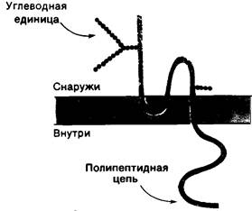
Another well-studied transmembrane protein of erythrocytes is Glycophorin A, which consists of 16 oligosaccharide units attached to a single polypeptide chain. CARBOHYDRATES account for 60% of the mass of this protein, so its name (derived from the Greek for "sugar-carrying") is well deserved. Due to its high carbohydrate content, glycophorin stains intensely with the PAS reagent.
This protein constitutes the PAS-1 band. Studies using proteolysis, chemical modification, and Electron Cell/15.html">Microscopy have shown that glycophorin A consists of three domains: 1) an N-terminal region containing all carbohydrate units and localized on the outer side of the membrane; 2) a hydrophobic middle region embedded in the hydrocarbon core of the membrane; and 3) a C-terminal region containing many polar and ionized side chains and located on the inner side of the erythrocyte membrane (Fig. 10.24). Despite a large body of data on The Structure of glycophorin, its function remains unknown.
10.12. Carbohydrate units are located on the outer side of the plasma membrane
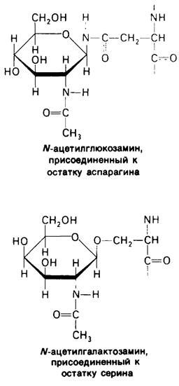
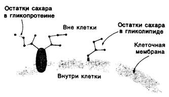
Introduction/5.html">Eukaryotic Cell membranes contain 2 to 10% carbohydrate in the form of Glycolipids and Glycoproteins. As mentioned above (Section 10.3), glycolipids of higher organisms are sphingosine derivatives with one or more sugar residues. In membrane glycoproteins, one or more carbohydrate chains are attached to the side chains of Serine, Threonine, or asparagine (usually via N-acetylglucosamine or N-acetylgalactosamine). The localization of these carbohydrate groups in the membrane is determined using specific labels. Lectins—plant Proteins with high affinity for specific carbohydrate residues—proved to be suitable labels. For example, concanavalin A binds to internal and nonreducing terminal α-mannose residues, whereas wheat germ agglutinin binds to terminal N-acetylglucosamine residues. These lectins are clearly visible under an Electron microscope when conjugated to ferritin, an iron-hydroxide-containing protein that is highly electron-dense. It turned out that the concanavalin A–ferritin conjugate binds specifically to the outer, but not the inner, surface of the erythrocyte membrane. A similar Asymmetry has been observed for the binding of many other lectins to the membranes of various Cells. Thus, the outer localization of the carbohydrate units of the anion channel and glycophorin (Figs. 10.23 and 10.24) reflects a general principle of Membrane Structure: sugar residues are located on the outer surface of The Plasma Membrane. In all mammalian cells studied to date, sugar residues in Plasma Membranes are located exclusively on the outer side of the membrane (Fig. 10.25).
It is possible that carbohydrate groups serve to orient glycoproteins in the membrane. Due to their highly hydrophilic nature, sugar residues in glycoproteins or glycolipids must be located on the membrane surface rather than in its hydrocarbon core. The free-energy cost of inserting an oligosaccharide chain into the hydrocarbon environment inside the membrane is extremely high. Consequently, There is a barrier that prevents the free rotation of a glycoprotein from one side of the membrane to the other. The carbohydrate components of membrane glycoproteins thus help maintain the asymmetry of Biological Membranes.
Fig. 10.24. Amino Acid Sequence and transmembrane localization of glycophorin A from erythrocytes. Fifteen carbohydrate units attached to Serine and threonine residues are shown in light green, and one carbohydrate unit attached to an asparagine side chain is shown in dark green. Hydrophobic amino acid residues embedded in the bilayer are highlighted in yellow. The C-terminal portion of the protein molecule, located inside The Cell, carries many negatively charged
(red) and positively charged (blue) amino acid residues. The carbohydrate units in the extracellular N-terminal portion of the protein molecule contain many negatively charged sialic acid groups
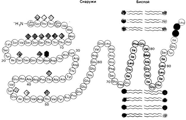
Fig. 10.25. Carbohydrate residues of glycolipids and glycoproteins are typically localized on the outer surface of mammalian cell plasma membranes
Cell-surface carbohydrates may also play an important role in intercellular recognition. Processes such as tissue formation through the interaction of different cells, or the recognition of foreign cells by The Immune System of higher organisms, depend on cell-to-Cell Recognition. Carbohydrates possess the potential for immense structural diversity. Thus, the repertoire of cell-surface carbohydrates has a colossal number of variations because: 1) Monosaccharides can link to one another through any of their hydroxyl groups; 2) the linkage at C-1 can be in either the α- or β-configuration; and 3) extensive chain branching is possible. Indeed, four sugars can form far more different Oligosaccharides than four Amino Acids can form different oligopeptides.
Last update: 06/08/2026
Editorial and Educational Adaptation: This material has been compiled based on the primary/original source text. The project team performed an editorial review, corrected technical inaccuracies, structured sections, and adapted the content for an educational format.
What was processed:
- elimination of formatting defects (OCR errors, structural breaks, corrupted characters);
- editorial organization of content;
- standardization of terminology in accordance with academic sources;
- verification of factual statements against the original source text.
All mentions of the author, publication year, and origin of the primary text have been preserved in accordance with the source.