Glycoproteins - Hughes R. 1985
Structure
Membrane Glycoproteins: General Concepts
Glycophorin
Above, we discussed The Structure of Glycoproteins isolated from Body Fluids, such as Blood, or from Cell secretions. However, glycoproteins are also vital components of all cellular membranes studied to date [18]. Plasma Membranes are particularly rich in glycoproteins, and researchers are currently focusing on elucidating the structure of these specific Proteins, their Organization within the membrane, and the Functions of membrane glycoproteins, particularly their carbohydrate components. Human erythrocytes have proven to be particularly convenient research subjects because they possess only a single membrane. Studies on these Cells have yielded a wealth of data that can presumably be extended to membrane systems of more complexly organized cells.
Human Erythrocyte membranes contain a relatively small number of major proteins. Of these, three are particularly rich in CARBOHYDRATES and can be isolated by selective extraction—for example, using a chloroform-methanol mixture, phenol, or lithium diiodosalicylate—followed by molecular weight Separation [19]. These glycoproteins are designated as glycophorins A, B, and C. Glycophorin A is the major glycoprotein component of The erythrocyte membrane. Its complete structure is shown in Fig. 2.19.
Glycophorin A, with a Molecular Weight of 29,000, consists of a single polypeptide chain composed of 131 amino acid residues. Analysis of its Primary Structure revealed two substitutions at positions 1 and 5 of the chain, which, as will be shown below, are of great biological significance. Landsteiner described another major blood group system, designated as MN. To identify the corresponding Antigens, researchers used an antiserum obtained by immunizing rabbits with erythrocytes from a single individual. Antibodies in this antiserum agglutinated erythrocytes only in a certain fraction of other individuals. When researchers used erythrocytes that were not agglutinated by the initial antiserum for immunization, the resulting antisera agglutinated erythrocytes only from those individuals who also failed to react with the initial antiserum. The erythrocyte surface antigens detected using both types of antisera proved to be products of two alleles: M and N. It is now well established that differences in MN blood group antigenicity are determined, at least in part, by the STRUCTURE OF THE peptide chain segment of glycophorin A between amino acid residues 1 and 5. Thus, in individuals possessing the M antigen, this sequence is as follows:
Class="center">
In individuals with the N antigen, two Amino Acid Substitutions are found:

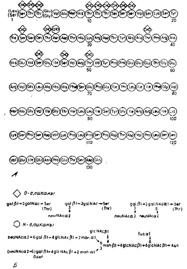
Fig. 2.19. A — Introduction/19.html">Primary structure of glycophorin A; glycosylated amino acid residues are indicated. B — general structural scheme of the carbohydrate chains.
O-glycans attached to The amino acid residues marked with an asterisk are essential for the expression of antigenic properties. These carbohydrates presumably help maintain the required conformation of the peptide chain recognized by specific antisera. Another important region of the glycophorin A chain is the segment between residues 73 and 95, which contains Amino Acids with neutral or nonpolar side chains. This domain spans the lipophilic bilayer of the membrane, with one part of the polypeptide chain located on one side of the membrane and the other part on the opposite side. The amino-terminal portion of the chain, exposed on the exterior, is very rich in two MAIN TYPES OF carbohydrate chains. It contains 15 O-glycans, which are di-, tri-, or tetrasaccharides linked via an N-acetylgalactosamine residue to Serine or Threonine. This structure is similar to the O-glycans discussed previously, as it also contains the galactoside β1→3N-acetylgalactosamine sequence. A sialic acid residue may be attached to one or both of these residues. Such Oligosaccharides are found not only in glycophorin but also in other Soluble Glycoproteins, such as fetuin, the major glycoprotein of fetal serum. More complex carbohydrate chains are attached to a single asparagine residue of the glycophorin peptide chain. They share A number of common features with the N-glycans of soluble glycoproteins. For instance, the core region β-mannose residue is linked to chitobiose [20]. Two α-mannose residues are attached to the β-mannose residue, which in turn are linked to N-acetyllactosamine sequences. The terminal residues of the latter are sialic acid and fucose. An N-acetylglucosamine residue is also attached to the central β-mannose residue via a (β1→4) linkage, similar to the hybrid chains of Ovalbumin glycopeptides (Fig. 2.7).
The primary structures of minor glycophorins B and C appear similar to that of glycophorin A, although their chains are shorter. They lack complex oligosaccharide chains, indicating a deletion of the peptide chain segment containing Asn-26 (Fig. 2.19). It remains unclear whether this deletion occurs after chain synthesis is complete or if separate genes encode The polypeptide chains of each of the three glycophorins.
Last update: 06/08/2026
Editorial and Educational Adaptation: This material has been compiled based on the primary/original source text. The project team performed an editorial review, corrected technical inaccuracies, structured sections, and adapted the content for an educational format.
What was processed:
- elimination of formatting defects (OCR errors, structural breaks, corrupted characters);
- editorial organization of content;
- standardization of terminology in accordance with academic sources;
- verification of factual statements against the original source text.
All mentions of the author, publication year, and origin of the primary text have been preserved in accordance with the source.