Glycoproteins - Hughes, R. 1985
Structure
Structure of O-glycans
Blood Group Antigens
Strictly speaking, the term "Blood group" refers to antigenic differences between THE RED BLOOD Cells of different individuals, which can be detected using specific Antibodies [2]. Early in this century, Karl Landsteiner described the major ABO(H) blood group system. This discovery generated immense interest, largely because it revealed a source of naturally occurring antibodies whose origin, however, remains unknown to this day. For instance, individuals carrying blood group A Antigens On the surface of their erythrocytes also possess high titers of antibodies directed against the B determinants, and vice versa. People with blood group O have antibodies against both A and B determinants. Naturally, the discovery of Blood Groups was of paramount importance for The Development of blood transfusion practices, as it is glaringly obvious what catastrophic consequences result from blood-typing errors. Intensive studies on the structural basis of ABO(H) antigenicity were carried out by Walter Morgan, Winifred Watkins, Elvin Kabat, and many others. Thanks to their work, we now know that blood group antigen properties depend on carbohydrate Structure, which, in turn, is entirely determined by the Genetic control of their assembly. Moreover, it has now become clear that Glycoproteins from various Tissues also possess antigenic determinants similar to those of erythrocyte antigens. Therefore, the BIOLOGICAL Functions OF carbohydrate chains—whatever they may be—as well as their strict genetic control in humans cannot be explained by the presence of these chains solely on The surface of red Blood Cells.
The carbohydrate secreted by individuals of the O genotype is referred to as the H antigen. As an essential component, it contains a fucose residue linked to C-2 of a galactose residue, which in turn is attached to C-3 or C-4 of an adjacent N-acetylglucosamine. Occasionally, the fucose residue may be absent, which results in the lack of A, B, or H antigens on the erythrocyte surface or in secreted glycoproteins (Fig. 2.16). Individuals with this rare anomaly were first discovered in India, and the corresponding phenotype was consequently named Bombay.
Through the attachment of N-acetylgalactosamine or galactose residues via an (α1→3)-glycosidic bond, the H determinant can be converted into either the A or the B determinant, respectively.
It is quite evident that the A, B, and H determinants reside at the terminals of O-glycans in secreted glycoproteins. But What is the core region sequence of these O-glycans? As we have already seen, they exhibit considerable diversity. For example, in glycoproteins from porcine submandibular gland secretions, the antigenic determinants are attached directly to the N-acetylgalactosamine residue, which is linked to the polypeptide (Fig. 2.14). A significantly longer sequence serving as the attachment site for antigenic determinants is found in rat colonic glycoproteins (Fig. 2.15). The core sequence of human secreted glycoproteins with A, B, and H activity has not yet been fully elucidated. The putative structure of substances secreted by an ovarian cyst is illustrated in Fig. 2.16.
Class="center">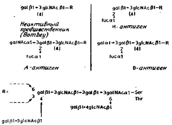
Fig. 2.16. Secreted human glycoproteins with ABO(H) system antigenic determinants.
It should be emphasized that the CARBOHYDRATES discussed here pertain to secreted glycoproteins. It is therefore natural to ask: What are the active blood group substances located within Erythrocyte membranes, which were first discovered by Landsteiner? Although this issue is not yet fully resolved, there is reason to believe that the majority of carbohydrate chains with antigenic properties are largely absent from glycoproteins and occur instead in glycolphids [17]. The basic structure of Glycolipids is shown in Fig. 2.17; these compounds contain exclusively galβ1→4gIcNAc chains. The structurally simplest glycolipids exhibiting blood group activity are substances of the A–G type, although more complex structures, such as D and E, have also been identified.

Fig. 2.17. Human erythrocyte membrane glycolipids possessing blood group antigenic activity. Bold arrows indicate bonds sensitive to endo-β-galactosidase.
A distinctive feature of all these chains is the presence of repeating N-acetylglucosaminyl (β1→3) galactose sequences linked by (β1→4) bonds. The A, B, and H antigenic determinants are formed by The addition of Monosaccharides such as fucose and N-acetylgalactosamine or galactose to the terminal galactose residue attached to the repeating sequence via a (β1→4) bond. In the absence of these residues, antigens of another human blood group system, designated as Ii antigens, are expressed. Much like Landsteiner's initial work on the human ABO(H) blood groups, antibodies found in the blood of certain individuals are used to identify Ii antigens. It is highly likely that specific antibodies against the I and i determinants are capable of recognizing branched (E) and unbranched (D) Variants of the carbohydrate chains, respectively (Fig. 2.17).
Accumulating evidence indicates that a significant number of blood group antigenic determinants are also present in membrane glycoproteins of erythrocytes and other cells. These determinants are located on long chains similar to those found in the complex glycolipids D and E, and they are sensitive to endo-β-galactosidase from E. freundii. Treatment of erythrocytes with this enzyme results in the complete loss of Ii antigenic determinants, while the glycoprotein designated as "band 3" protein (Section 2.6.2) is thereby converted into a low-molecular-weight component with a low carbohydrate content. According to other data, this same glycoprotein possesses only a single such carbohydrate chain [17]. The long carbohydrate sequences under Discussion, termed polyglycosyl units and constituting part of erythrocyte band 3 glycoproteins, are also minor components of membrane glycoproteins in certain other Cell types, such as fibroblasts. The polyglycosyl chains of glycoproteins are attached to the core region of N-glycans, as illustrated in Fig. 2.18. In polyglycosyl chains exhibiting I activity, branches consisting of several galactose residues extend from the main repeating backbone. Such branched polyglycosyl chains predominate in mature erythrocytes, whereas fetal erythrocytes contain the unbranched chain forms characteristic of the i antigen. Evidently, the Enzymes responsible for branching emerge during organismal development, though the exact significance of this for the normal maturation of the erythroid cell Lineage remains unclear.

Fig. 2.18. Structure of glycoprotein "polyglycosyl"-N-glycans. R — glcNAcβ1→4glcNAc, Asn.
Last update: 06/08/2026
Editorial and Educational Adaptation: This material has been compiled based on the primary/original source text. The project team performed an editorial review, corrected technical inaccuracies, structured sections, and adapted the content for an educational format.
What was processed:
- elimination of formatting defects (OCR errors, structural breaks, corrupted characters);
- editorial organization of content;
- standardization of terminology in accordance with academic sources;
- verification of factual statements against the original source text.
All mentions of the author, publication year, and origin of the primary text have been preserved in accordance with the source.