Glycoproteins - Hughes R. 1985
Structure
Structure of O-glycans
Secrets of the glycoprotein nature (mucins)
In addition to the aforementioned points regarding Collagen O-glycans, several Characteristic Properties of glycans attached via glycosidic bonds to amino acid hydroxyl groups merit further Structure/133.html">Discussion.
Anyone who has ever noticed the slimy trail left by a snail crawling across a dry path, or has tasted bird's nest soup in a Chinese restaurant, is already familiar with Glycoproteins containing large amounts of O-linked glycans. These and similar glycoproteins are often called mucins or mucoids due to their exceptionally high viscosity and jelly-like appearance. The German chemist Hoppe-Seyler used the unmelodious term Schleimstoff, meaning mucus, to designate them. These substances are quite unpleasant in appearance and awkward to work with, particularly when attempting to perform chemical and physical analyses. Nevertheless, It is important to remember that mucins perform vital Functions. It is crucial for higher organisms (both vertebrates and invertebrates) that the cavities of the respiratory, digestive, and urogenital tracts are lined with a layer of Cells that secrete viscous glycoproteins. Examples of such secretions include saliva (which, incidentally, is the main ingredient in bird's nest soup), bronchial and intestinal secretions, seminal fluid, and cervical mucus. All of these can act as lubricants, for instance, within the digestive tract. Furthermore, this thick, viscous layer serves to protect the underlying Tissues from both mechanical and chemical damage (such as from digestive juices).
Secretory glycoproteins have been studied intensively for many years. This is due both to their crucial biological role and to their association with certain severe diseases, such as cystic fibrosis. The carbohydrate composition is best understood in salivary secretions, particularly the glycoproteins of the submandibular gland. Twenty years ago, Alfred Gottschalk, working first in Australia and later in West Germany, studied in great detail The structure of the simplest substance found in sheep glands. In this glycoprotein, a disaccharide consisting of N-acetylneuraminic acid and N-acetylgalactosamine residues is attached to numerous Serine and Threonine residues of the polypeptide chain (Fig. 2.14). It turned out that one in every six amino acid residues is a glycosylated serine or threonine, with about 200 such residues present per polypeptide chain. The third most frequent amino acid in submandibular gland glycoproteins is Proline, making the glycosylated regions extremely rich in it. The protein itself resembles a comb in structure: short carbohydrate chains protrude like Teeth from a rigid, proline-rich polypeptide backbone. Through disulfide bridges between protein globules, these comb-like structures form large glycoprotein molecules with unique viscous properties [4]. In the submandibular gland glycoproteins of other animal species, not only the Disaccharides described above (Fig. 2.14) are attached to the Polypeptides, but also many other oligosaccharide chains. For example, the well-characterized pig submandibular gland secretion contains the neutral disaccharide galß1→3gaINAc, alongside its sialylated forms. Of particular interest are the fucose-containing chains of porcine glycoprotein. Such chains are very easy to detect because they also occur in human secretions, such as the Blood group H antigen. The attachment of an N-acetylgalactosamine residue converts the H antigenic determinant into the blood group A antigen, a structure that is likewise present in pig submandibular gland glycoproteins (Fig. 2.14).
Class="center">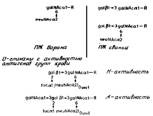
Fig. 2.14. Structure of O-glycans from submandibular gland (SMG) glycoproteins. The N-acetylgalactosamine residue is linked to serine (or threonine); R represents a serine or threonine residue of the polypeptide chain.
Very little is known about the mucous secretions of The Stomach and intestine, despite the relative ease of obtaining them. The reason for this is that such secretions represent complex mixtures of numerous glycoproteins that are exceedingly difficult to separate. Figure 2.15 illustrates the structure of some O-glycans recently identified [16] in the rat colon. Several Characteristic Features of these glycans deserve mention, most notably a novel type of linkage: N-acetylglucosaminyl (ß1→3) N-acetylgalactosamine. The first of these residues serves as an elongation point, converting the chain into a highly complex sequence. The longest chain terminates in a region characteristic of the A determinant (compare with the chain responsible for A-activity found in pig submandibular secretions; Fig. 2.14).
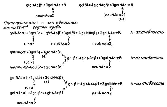
Fig. 2.15. Structure of O-glycans from rat colonic glycoproteins, where R is a serine or threonine residue of the polypeptide chain [16].
Last update: 06/08/2026
Editorial and Educational Adaptation: This material has been compiled based on the primary/original source text. The project team performed an editorial review, corrected technical inaccuracies, structured sections, and adapted the content for an educational format.
What was processed:
- elimination of formatting defects (OCR errors, structural breaks, corrupted characters);
- editorial organization of content;
- standardization of terminology in accordance with academic sources;
- verification of factual statements against the original source text.
All mentions of the author, publication year, and origin of the primary text have been preserved in accordance with the source.