BIOCHEMISTRY AND MOLECULAR BIOLOGY - W. ELLIOTT - 2002
CHAPTER 27. THE RED BLOOD CELL AND HEMOGLOBIN FUNCTION
The most crucial molecule for oxygen delivery from the Lungs to the Tissues is Hemoglobin. Rather than acting merely as a passive oxygen-binding protein, it Functions as a sophisticated molecular machine designed to load the maximum amount of oxygen in the lungs, transport it to peripheral tissues, pick up CO2, and carry it back to the lungs.
Hemoglobin is a globin protein featuring heme as its prosthetic group. In this chapter, we will explore several aspects of gas transport, beginning with the synthesis and regulation of heme. Following this, we will examine The regulation of globin synthesis and conclude with a Structure/133.html">Discussion of The Physiological Role of hemoglobin in the body.
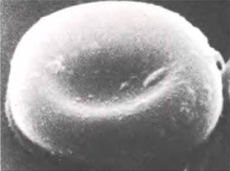
The Human Body produces 160 million red blood cells, or erythrocytes, every minute. They circulate in the bloodstream for about 110 days before being destroyed. Mature mammalian erythrocytes are anucleate and appear as flattened, biconcave discs (Fig. 27.1). This shape, maintained by the Cytoskeleton, provides a much larger surface area than that of spherical cells. Combined with a shorter diffusion distance, this optimizes gas exchange between The Cell and the extracellular environment. The total surface area of all erythrocytes combined is approximately 4000 m2.
Class="center">Fig. 27.1. Scanning electron micrograph of an erythrocyte
In the embryo, erythrocytes are produced in The Liver and Spleen. In adults, however, this process takes place in the Bone Marrow of flat bones, where hematopoietic stem cells continuously divide to generate precursors for all blood cell lineages, as illustrated in Fig. 25.1 (stem cells continuously self-renew, while their progeny are capable of differentiation). Individual cells differentiate into specialized blood cell types (erythrocytes, various leukocytes, and platelets). The formation of different blood cell types is a tightly regulated process, and various regulatory Proteins have been identified that stimulate the proliferation of specific progenitor cells. The protein Erythropoietin, produced in the renal medulla, stimulates erythrocyte production. Because The rate of erythropoietin production is inversely proportional to the partial pressure of oxygen in the tissues, red blood cell levels are regulated automatically: oxygen deprivation triggers increased hormone synthesis, which in turn leads to a higher production of red blood cells.
In the early stages, erythrocyte precursors contain a Nucleus; however, this is later lost in mammals to form an immature red blood cell known as a reticulocyte, which is then released into the Circulation. Reticulocytes contain mRNA, and Protein Synthesis continues within them until the cell fully matures into an erythrocyte. When stained, the Ribosomes and mRNA appear as a dark, reticular network—hence the name reticulocyte. Mature cells lack Mitochondria and rely on Glycolysis coupled with lactate production to generate energy. The Pentose Phosphate Pathway is also active in mature erythrocytes, serving to regenerate NADPH.
Heme and Its Synthesis
The complete structure of heme (Fig. 27.3)—which few biochemists commit entirely to memory—is a ferrous iron complex with protoporphyrin (Fig. 27.2).
Fig. 27.2. Outline STRUCTURE OF THE heme molecule
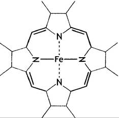
Fig. 27.3. Structure of heme. This specific form is found in hemoglobin; other Hemes with varying side chains occur in Cytochromes. One side of the molecule features two nucleophilic propionic acid residues (CH2-CH2-COO-), while the other side chains are hydrophobic. In Myoglobin, heme sits within a hydrophobic pocket of the protein, with its hydrophilic edge oriented toward the surface and hydrophobic groups facing the interior of the protein molecule

The latter consists of four pyrrole rings; these four substituted pyrroles are linked by methenyl bridges (=CH-), creating a system of conjugated double bonds (meaning that single and double bonds alternate continuously around the ring). This imparts a deep red color to both porphyrin and heme.
In heme, the four pyrrole nitrogen atoms coordinate with Fe2+ (Fig. 27.4), leaving two of the six coordination bonds of Fe2+ free and available to bind other ligands.
Fig. 27.4. Coordination of Fe2+ in heme. The iron atom in heme can bind up to six ligands: four bonds are occupied by the pyrrole nitrogen atoms, while the remaining two are positioned perpendicularly above and below the plane of the ring, respectively

Heme Synthesis
Erythrocytes account for the vast majority of heme in the human body. Therefore, discussing its synthesis here is entirely appropriate, although the heme in the prosthetic groups of cytochromes and other Hemoproteins is synthesized in other cell types as well.
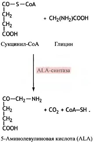
Heme synthesis is a remarkable pathway because, surprisingly, animals require only two starting precursors to build it: Glycine and succinyl-CoA. You have already encountered the latter in The Citric Acid Cycle (see p. 120). These substrates are condensed to form aminolevulinic acid (ALA), a reaction catalyzed by the enzyme aminolevulinate synthase (ALA synthase) (Fig. 27.5).
Fig. 27.5. The initial step of heme synthesis, catalyzed by 5-aminolevulinate synthase (ALA synthase). Structures are shown in the un-ionized form
5-Aminolevulinic acid is the sole precursor for porphyrin synthesis. Its two molecules, which are used to form the pyrrole - porphobilinogen (PBG), undergo dehydration catalyzed by ALA dehydratase (Fig. 27.6). The remaining stages of heme Biosynthesis involve the joining of 4 PBG molecules into a single structure, modification of side-chain groups, and the formation of chelate complexes with a divalent iron atom. The intermediate tetrapyrroles in the steps between PBG formation and heme formation are colorless uro- and coproporphyrinogens (containing methylene bridges) and red protoporphyrin (containing methenyl bridges). The heme biosynthetic pathway is shown in Fig. 27.7. The metabolic pathway of heme synthesis has several fascinating features.
Fig. 27.6. Synthesis of porphobilinogen (PBG) - the dehydration stage catalyzed by ALA dehydratase. Deionized structures are shown. Porphobilinogen is a monopyrrole; hemoglobin is a tetrapyrrole
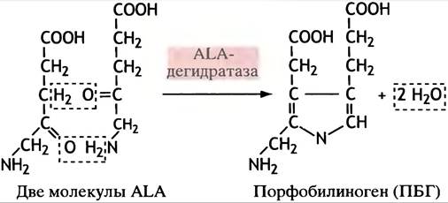
Fig. 27.7. Heme biosynthesis

The first reaction—the synthesis of ALA—takes place inside the mitochondria, after which ALA is transported into the Cytoplasm. However, the final three steps occur in the mitochondria once again. Why this happens remains unclear.
Regulation of Heme Biosynthesis and Iron Delivery to the Erythrocyte
The regulation of heme biosynthesis is of particular interest because it is directly linked to cellular iron uptake and storage.
This process occurs as follows.
✵ The activity of ALA synthase limits the entire heme synthesis process.
✵ The synthesis of ALA synthase, and consequently its level, is regulated by The amount of iron.
✵ Iron levels depend on the amount of transferrin receptor protein.
✵ Iron levels regulate transferrin synthesis via a feedback mechanism.
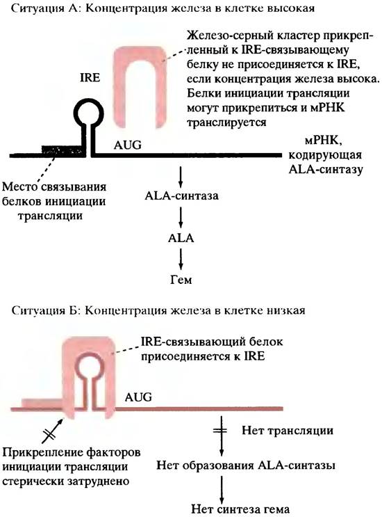
During heme biosynthesis, the rate-limiting step is the synthesis of ALA. The production of the ALA synthase enzyme itself is regulated in red blood cells at the translational level. At the 5' untranslated region (i.e., at the Translation initiation site), the enzyme's mRNA features a stem-loop structure called the iron-responsive element (IRE). When iron levels are low, the IRE-binding protein attaches to the IRE and prevents translation (Fig. 27.8). When iron levels are high, the protein is released As a result of its interaction with an iron-sulfur cluster (a small complex consisting of iron, Cysteine,
and inorganic sulfur), after which translation begins. In this way, a balance is maintained between the rate of heme synthesis and iron stores.
Fig. 27.8. Regulation of heme synthesis by varying intracellular iron concentrations in erythrocytes. This regulatory mechanism apparently relies on ALA synthase having a short half-life: as soon as enzyme synthesis ceases, heme synthesis stops immediately as well. The enzyme is known to be unstable in reticulocyte lysates in vitro and presumably in erythrocytes themselves. The question of how cellular iron levels are controlled is discussed separately. IRE is an iron-responsive element representing an mRNA stem-loop. When the IRE-binding protein attaches to the IRE, it likely sterically hinders the binding of translation initiation proteins. The binding of iron to the IRE-binding protein reduces its affinity for the IRE
Heme synthesis begins long before reticulocyte formation, starting in the nucleated proerythroblast, meaning that mRNA Translation occurs in these cells and continues into the reticulocyte stage.
This raises the question: How is the iron level regulated within the cell? Iron is transported in Blood Plasma as a complex with the protein transferrin, which is produced in the liver. This complex is taken up by cells via receptor-mediated endocytosis. In non-erythroid cells, this uptake is determined by the level of transferrin receptor protein, the synthesis of which is regulated by cellular iron levels. Iron exerts control over the Stability of the transferrin receptor mRNA, which contains an iron-responsive element at its 3' end. When cellular iron levels are low, the IRE-binding protein binds to the receptor mRNA, protecting it from degradation. When cellular iron levels are high, the IRE-binding protein dissociates, leading to decreased stability of the transferrin receptor mRNA. Thus, iron causes down-regulation, or a reduction in the number of transferrin receptors, which in turn decreases cellular iron uptake. When iron levels are low, the entire process is reversed. It has not yet been established whether this mechanism applies to erythroid cells, but it is known that erythrocytes possess transferrin receptors whose numbers increase as the cell matures.
Iron stores in erythrocytes are maintained in the form of ferritin, a complex of the protein apoferritin and inorganic iron. The liver is also a major storage site for iron. Interestingly, apoferritin synthesis is regulated by essentially the same mechanism as the Synthesis of the enzyme ALA synthase: iron induces the binding of the IRE-binding protein to apoferritin mRNA, thereby promoting protein synthesis.
All human cells synthesize heme via the same pathway as blood cells (with minor differences). Heme is required for cytochromes in the Electron Transport Chain (see p. 123) as well as for Other Enzymes. Erythrocytes have the greatest need to fill mature cells with the heme-containing protein hemoglobin. Following red blood cells, the liver has the next highest heme content due to its high concentration of cytochrome P450 (see p. 210). The regulation of ALA synthase differs somewhat across different cell types. In the liver, heme destabilizes ALA synthase mRNA and also inhibits The transport of enzyme molecules to their destination—the mitochondria. The enzyme's mRNA lacks the iron-responsive element found in erythroid cells. Overproduction of ALA synthase and high blood levels of ALA in patients with acute intermittent porphyria correlate with the neurological symptoms characteristic of this disease (the Molecular Basis of which is still unknown). In such patients, the heme biosynthetic pathway is partially blocked. Chlorophyll is also a tetrapyrrole, but interestingly, ALA in plants is synthesized via a different reaction.
Returning to red blood cells, however:
Heme Degradation
Erythrocytes are degraded primarily by the reticuloendothelial Cells of the spleen, Lymph Nodes, bone marrow, and liver. The removal of sialic acids from erythrocyte membrane Glycoproteins serves as a signal for erythrocyte Aging. Carbohydrate moieties lacking sialic acids bind to receptors on these cells, leading to the endocytosis of aged erythrocytes.
The enzyme heme oxygenase cleaves the tetrapyrrole ring, releasing iron for reuse and producing the linear tetrapyrrole biliverdin (Fig. 27.9). Biliverdin is reduced to bilirubin. The latter is insoluble in Water. Bound to serum albumin, it is transported by the blood to the liver, where, following The addition of two glucuronic acid residues, it becomes much more polar and is subsequently excreted via the Bile into the intestine. Modification and partial reabsorption of certain bile components impart a yellow color to urine (bilirubin is thought to function as an antioxidant, see p. 213).
Fig. 27.9. The reaction catalyzed by heme oxygenase and the subsequent reduction of biliverdin. Double bonds in the ring structures are not shown. As a result of the reaction, one methylene group is converted into carbon monoxide, thereby opening the ring structure. Biliverdin is reduced to bilirubin, binds to 2 molecules of glucuronic acid, and is excreted from the body. However, there is some ambiguity in the Stoichiometry of the heme oxygenase reaction; it is believed that 3 molecules of O2 are involved in the reaction. By analogy with cytochrome P450, it can be assumed that 3 molecules of NАDРН are required to convert 3 oxygen atoms into H2O

How Globin Synthesis Is Regulated
Hemoglobin consists of heme bound to the protein globin. The two components—globin and heme—must be produced in approximately equal amounts, which is why a coordinating regulatory mechanism exists. Experiments with reticulocyte lysates have shown that in the absence of heme, protein kinase phosphorylates one of the initiation factors (еIF2) of protein synthesis. It has The ability to halt translation initiation in red blood cells and thus prevent globin synthesis. In the presence of heme, the kinase is inactivated, and phosphatase restores the activity of the initiation factor. Thus, globin synthesis occurs only when heme is present. This mechanism is specific to erythroid cells.
Gas Transport into the Blood
The solubility of oxygen in ordinary solutions is insufficient to supply the body's tissues. Oxygen carriers in the blood are therefore necessary. Similarly, CO2 cannot be transported at a sufficient rate from the tissues to the lungs in a simple solution.
What is the chemical basis for The Role of the hemoprotein complex as an oxygen carrier?
The inorganic ion Fe2+, but not Fe3+, can bind oxygen. However, Fe2+ ions spontaneously oxidize to Fe3+, meaning that inorganic iron by itself is not a good oxygen carrier. Fe2+ as part of heme can also bind oxygen, but it too is rapidly oxidized to Fe3+, forming hematin. Therefore, free heme is likewise a poor oxygen carrier. For heme to oxidize, 2 of its molecules must interact with 1 oxygen molecule. Binding heme within a protein pocket prevents this interaction; consequently, the Fe2+ ions within the heme of hemoglobin are much more resistant to oxidation than free heme. It is important to clarify why heme in cytochromes during electron transport (see p. 123) alternates between the Fe2+ and Fe3+ states, whereas heme in hemoglobin functions solely in the Fe2+ state, which is unaffected by interaction with oxygen. The reason is that the iron atom in hemoglobin is bound to 4 nitrogen atoms of the pyrrole rings, while the fifth and sixth coordination bonds are located directly above and below the plane of the heme structure; one of these bonds is linked to a Histidine residue, while the other is available for oxygen binding.
Structure of Myoglobin and Its Role in Oxygen Binding
Myoglobin is the red pigment of Muscle tissue. A preliminary look at its structure will help in understanding the more complex Organization of hemoglobin.
Myoglobin simply supplies the muscle cell with oxygen. It captures oxygen from the blood and transports it into the cell, where it is used by mitochondria to generate energy. Myoglobin is a complex protein consisting of a single polypeptide chain and an associated heme molecule. The protein has a globular shape and contains a series of α-helices (Fig. 27.10), with the heme molecule located in a pocket between two of them.
Fig. 27.10. Models of Myoglobin and hemoglobin molecules. a - A simple computer model demonstrating the folding of the polypeptide chain and THE POSITION OF the heme in the myoglobin molecule; b - a more detailed model of myoglobin; c - a model showing the arrangement of subunits in the hemoglobin molecule. 2,3-Bisphosphoglycerate fits into the central cavity of the molecule in the deoxygenated state (marked with X). In Sickle cell anemia, two glutamic acid residues at position 6 in the β-chains are replaced by valines, creating hydrophobic patches on the molecule

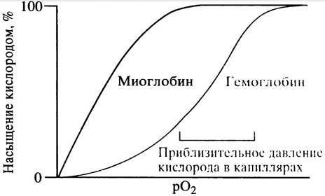
The oxygen saturation of myoglobin with increasing partial pressure of oxygen is characterized by a hyperbolic curve (Fig. 27.11) of the same type as the curve of enzyme activity versus Substrate Concentration described earlier for a "classical" enzyme (see p. 157). Myoglobin has a high affinity for oxygen and therefore readily extracts it from blood oxyhemoglobin (see Fig. 27.11, which illustrates the relative oxygen affinities of the two carriers). In Muscles, where oxygen concentration drops during Electron Transport and H2O formation, myoglobin releases its bound oxygen. Thus, it functions as a very simple oxygen carrier.
Myoglobin was the first protein whose three-dimensional structure was determined, an achievement considered a turning point in biochemistry.
Structure of Hemoglobin
Hemoglobin consists of 4 protein subunits (see Fig. 27.10), each resembling the folded polypeptide structure of myoglobin and containing an oxygen-binding heme. The adult human hemoglobin molecule is composed of 2 identical α- and 2 identical β-subunits.
Oxygen Binding to Hemoglobin
The hemoglobin molecule binds 4 oxygen molecules, one per subunit. Hemoglobin oxygen saturation is described not by a hyperbolic curve, as in the case of myoglobin, but by a sigmoidal curve, which is shifted to the right relative to the myoglobin curve (see Fig. 27.11). A higher oxygen concentration is required to achieve 50% saturation of hemoglobin compared to myoglobin, indicating that myoglobin has a higher oxygen affinity. Hemoglobin needs to extract as much oxygen as possible from the lungs and release it to the capillaries in the tissues. The steepest part of the sigmoidal curve (indicating the greatest oxygen release) corresponds to the O2 pressure found in the capillaries (see Fig. 27.11). Hemoglobin becomes saturated with oxygen at the higher oxygen pressures found in the lungs.
Fig. 27.11. Hemoglobin oxygen-saturation curve. The higher oxygen affinity of myoglobin compared to hemoglobin means that myoglobin in muscles more readily extracts oxygen from the blood
How the Sigmoidal Oxygen-Saturation Curve Arises
You encountered kinetics described by a sigmoidal curve when discussing Metabolic Regulation (Chapter 12). It was mentioned there that Allosteric enzymes are typically multisubunit proteins that undergo conformational changes upon binding a substrate molecule. This leads to A change in the enzyme's affinity for subsequent substrate molecules. The same principle applies to oxygen binding by hemoglobin.
Although the heme molecules in hemoglobin are quite far apart, the initial binding of oxygen to one subunit accelerates the binding of oxygen molecules to the remaining subunits. This phenomenon is known as homotropic positive cooperativity (homotropic because only oxygen is involved). This is precisely what causes the sigmoidal shape of the curve. The effect is that the initial oxygen affinity of deoxyhemoglobin is 200 times lower than the affinity at The final stage of binding. Oxygen saturation corresponding to the final segment of the curve is limited by the number of available binding sites.
Mechanism of the Allosteric Change in the Hemoglobin Molecule
As already mentioned, the binding of one oxygen molecule to a hemoglobin molecule facilitates the binding of oxygen by the remaining subunits of the
hemoglobin molecule. According to the concerted model (see p. 159), hemoglobin exists in two conformational states: the "tense", or T-state, which has a low affinity for oxygen, and the "relaxed", or R-state, which has a high affinity for oxygen. Both states are in a free equilibrium. In the absence of oxygen, the T-state predominates. Upon oxygen binding, the probability increases that all four subunits of the hemoglobin molecule will be in the R-state (high affinity); consequently, the T <-> R equilibrium shifts to the right.
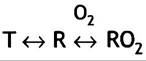
Since The Study of hemoglobin oxygen saturation is carried out on A large number of molecules, it is possible that the more oxygen is bound, the greater the number of hemoglobin molecules that are in the R-state, and, therefore, the higher the oxygen affinity in the solution. A similar situation is characteristic of allosteric enzymes (see pp. 159-160). X-ray crystallography has been used to determine the conformation of the tetrameric structure of hemoglobin in both the oxygenated and deoxygenated states. As previously mentioned, hemoglobin is a tetramer consisting of 2 identical α- and 2 identical β-subunits. In the tetramer, they are designated as α1, α2, β1, and β2. According to X-Ray Diffraction data, the contacts between the α- and β-subunits differ slightly from one another.
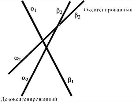
To gain a clearer picture of hemoglobin, it is useful to view its structure in terms of two heterodimers formed by the α- and β-subunits: α1β1 and α2β2. It is precisely the interaction between the two heterodimers within the tetramer that drives the T <-> R rearrangement. The relative movement of the dimers during the transition from the T- to the R-state is driven by oxygen binding (Fig. 27.12).
Fig. 27.12. Schematic representation of the relative arrangement of subunits in deoxygenated and oxygenated hemoglobin molecules, whose axes are represented by straight lines. The subunit pairs α1, β1 and α2, β2 should be considered as a single subunit dimer. Upon oxygenation, these dimers rotate at an angle of approximately 15° relative to each other. The black and gray lines show the relative arrangement of the dimers in the T-state. In the oxygenated (R) state, the red line shows the Rotation of the α2/β2 dimer relative to the α1/β1 dimer, which is depicted as a fixed line. This displacement alters the α1β2/α2β1 contacts between the dimers
What triggers this allosteric change in the entire hemoglobin molecule upon binding to one or more oxygen molecules? The change is initiated by a slight Displacement of the iron atom when Oxygen binds to the heme, which drives the T <-> R transition. Although heme, as already mentioned, is a planar molecule, in the deoxygenated state the iron atom lies out of the plane of the molecule because the atom is too large to fit entirely within the tetrapyrrole ring structure. In deoxygenated hemoglobin, the tetrapyrrole structure is not completely planar (Fig. 27.13).
Fig. 27.13. Structural Changes in the heme of the hemoglobin molecule upon oxygenation (schematic representation). a — Heme molecule in deoxyhemoglobin with a tetrapyrrole structure pulled into a slightly bell-like shape; b — heme binding in oxyhemoglobin

The Fe2+ atom is linked to a histidine residue of one of the α-helices that make up the subunit. This α-Helix is designated as F, and the histidine group as F8 (Fig. 27.13, a). Due to the electronic changes that occur upon oxygen binding, the iron atom in the heme significantly decreases in diameter and moves into the plane of the tetrapyrrole ring, making the entire molecule more planar. This is accompanied by a rearrangement of the protein molecule itself (see Fig. 27.13).
This minor shift of the iron atom leads to major structural changes elsewhere in the protein molecule via a "lever-like" effect that influences The structure of the polypeptide chain. This "elsewhere" is the point where the α-subunit of one dimer interacts with the β-subunit of the other (at the α1β2/α2β1 interface). This interaction forms a network of weak bonds between The amino acid residues of the two subunits. We have one set of bonds in the T-state and another in the R-state. These bonds hold the dimers together. Thus, the iron-initiated displacement leads to the replacement of one set of interdimer bonds with another, causing a relative rotation of the dimers (see Fig. 27.12).
It should be noted that the existence of structures corresponding to the T- and R-states of hemoglobin has been conclusively proven by X-ray crystal diffraction analysis of hemoglobin and oxyhemoglobin crystals, respectively.
However, it is impossible to obtain crystals of partially oxygenated hemoglobin, and therefore the structure (or structures) of the latter remains unknown. Consequently, the intermediate stages (if any exist) during which the T- and R-forms mutually interconvert have not been established.
The Important Role of 2,3-Bisphosphoglycerate (BPG) in Hemoglobin Function
You are already familiar with the glycolysis intermediate 1,3-bisphosphoglycerate (see Fig. 8.6), but until now we have not discussed the fact that 2,3-bisphosphoglycerate (BPG) is also synthesized in the cell.
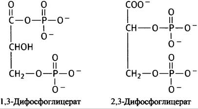
BPG plays a vital physiological role in Oxygen transport by decreasing the oxygen affinity of hemoglobin and thereby enhancing the release of oxygen to tissues. BPG shifts the oxyhemoglobin dissociation curve to the right. A cleft runs through the entire tetrameric hemoglobin molecule (see Fig. 27.10). The side chains of positively charged Amino Acids project into this cleft. The BPG molecule, which carries 4 negative charges at blood pH, matches the size and configuration of the cleft and forms ionic bonds with the positively charged Regions of the protein. In doing so, cross-linking of the β-subunits helps hold hemoglobin in the deoxygenated state. In the deoxygenated (T) state, hemoglobin can accommodate a BPG molecule. However, upon oxygenation into the R-state, Conformational Changes in the protein make the cleft smaller, so it can no longer accommodate the BPG molecule. The ability of BPG to bind tightly and stabilize the deoxygenated state favors the release of oxygen in the capillaries. As a result, the following process takes place.
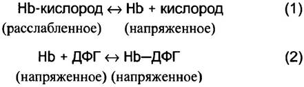
Reaction (2) with BPG helps drive the equilibrium of reaction (1) toward the release of oxygen.
If the blood has depleted its reserve of BPG, hemoglobin remains virtually saturated with oxygen even at concentrations lower than those in tissue capillaries; consequently, it becomes incapable of delivering oxygen to tissues efficiently enough.
The Effect of BPG on oxygen binding to hemoglobin is shown in Fig. 27.14.
Fig. 27.14. Hemoglobin oxygen saturation curves illustrating the effect of 2,3-bisphosphoglycerate (BPG)
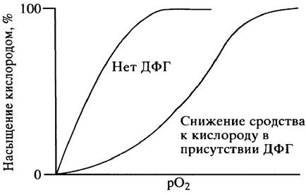
The higher the concentration of BPG, the more pronounced the predominance of the deoxygenated form of hemoglobin. A regulatory system is thus established: when oxygen pressure in tissues is low, red blood cells synthesize a greater amount of BPG (via a regulatory mechanism that promotes increased oxygen delivery, the details of which we will not discuss). Upon acclimatization to high-altitude conditions, the BPG content in red blood cells increases. It should be noted that although BPG induces greater oxygen delivery to tissues, the reduced oxygen affinity of hemoglobin has practically no effect on the degree of hemoglobin oxygenation in the lungs. The normal molar concentration of BPG in the blood is roughly equivalent to the concentration of tetrameric hemoglobin.
There is another subtle aspect of the DPG regulatory system that demonstrates how minor changes in proteins can produce significant physiological effects. When supplying the fetus with oxygen, it is essential that fetal hemoglobin extracts oxygen from the mother's oxyhemoglobin across the Placenta. To achieve this, fetal hemoglobin must possess a higher oxygen affinity than the maternal carrier. This is accomplished by replacing the adult hemoglobin β-subunits with fetal γ-subunits, each of which lacks one of the positive charges found on the β-subunit. Specifically, these are the exact charges that, in adult hemoglobin, reside in the cleft where DPG binds. Having two fewer ionic groups, fetal hemoglobin binds DPG less tightly. Consequently, DPG is less effective at lowering hemoglobin's oxygen affinity, thereby ensuring that fetal hemoglobin maintains a higher affinity for oxygen compared to maternal hemoglobin, which readily releases its oxygen.
Effect of pH on Oxygen Binding to Hemoglobin
Deoxygenated hemoglobin exhibits a higher affinity for protons than oxyhemoglobin. In other words, the R-form is a stronger acid than the T-form (deoxygenated); as a result, oxygen binding triggers the dissociation of protons from the hemoglobin molecule. This phenomenon is known as the Bohr effect:
Нb + 4O2 <-> Нb(O2)4 + (Н+)n (1)
where n is a value of approximately 2; this number depends on a complex set of parameters.
Protons are released, for instance, from histidine residues within the protein. This release is driven by conformational changes occurring during the transition from the T to the R state, which affect the ionization (pKа) of such groups (on histidine dissociation, see p. 40).
Role of pH Changes in Oxygen and CO2 Transport
The Bohr effect described above carries significant physiological importance. CO2 generated in tissues must be transported to the lungs. It enters erythrocytes, where the enzyme Carbonic anhydrase converts it into H2CO3, which then dissociates into a bicarbonate ion and a proton:
СO2 + Н2O <-> Н2СO3 + НСО3- (2)
The latter shifts the equilibrium in equation (1) to the left, compelling HbO2 to release its oxygen—an effect that aligns with physiological demands.
HCO3- moves passively through the anion channel (see p. 63) down its concentration gradient into the serum. This movement of HCO3- is not accompanied by the translocation of H+, as there is no channel allowing protons to cross The erythrocyte membrane. To maintain electrical balance (equilibrium) as HCO3- exits the cell, Cl- moves inward through the same anion channel. This reciprocal transport is known as the chloride shift (Fig. 27.15).
Fig. 27.15. CO2 transport in the blood. a - Reactions in tissue capillaries; b - reactions in the lungs. Transport of CO2 in the form of hemoglobin carbamino groups is not shown in the figure

Dissolved HCO3- travels with venous blood back to the lungs. Here, shifts in proton concentration also help achieve the desired physiological outcomes. The release of a proton from hemoglobin upon oxygenation leads to two main results. H2CO3 is formed from HCO3- through a simple equilibrium process:
НСО3- + Н+ <-> НСO3
This enables carbonic anhydrase to generate CO2. It should be noted that the substrate for carbonic anhydrase is H2CO3 (rather than HCO3-):
НСO3 <-> Н2O + СO2
The breakdown of H2CO3 inside the red blood cell drives the influx of HCO3- from the serum and the efflux of Cl- down its concentration gradient, so that a reverse chloride shift occurs in the lungs, leading to the elimination of CO2 via exhaled air.
Small amounts of CO2 are carried in the blood in dissolved form, but the major portion (about 75%) is transported as HCO3-. Approximately 10–15% of CO2 is carried by hemoglobin. CO2 interacts spontaneously with uncharged uncharged NH2 groups of globin to form carbamino groups:
RNН2 + СO2 <-> RNНСООН <-> RNНСОO- + Н+
This reaction involves Lysine and Arginine residues, whose side-chain amino groups possess high pKa values and therefore remain predominantly uncharged. The available RNH2 groups are primarily terminal amino groups (the side chains of lysine and arginine have pKa values that are far too high to remain uncharged).
Maintenance of Blood pH
From the foregoing, it is clear that substantial fluctuations in blood hydrogen ion concentration are associated with CO2 and oxygen transport. When acid is produced in tissues, the pH within red blood cells must drop. The buffering capacities of HCO3-, phosphates, and hemoglobin itself play a vital role in maintaining physiological pH levels. The Bohr effect described above also acts as a buffer. Upon releasing oxygen, hemoglobin takes up protons (see equation 1), which accounts for roughly half of the H+ ions generated by CO2 in the tissues, thereby maintaining red blood cell pH within the physiological range.
Sickle Cell Anemia
This condition is commonly cited in textbooks to illustrate how the substitution of a single amino acid in a protein can lead to severe consequences. Normally, in the β-subunits of the tetrameric hemoglobin structure, the amino acid at the sixth position is glutamic acid, whose side chain is negatively charged and highly hydrophilic (see Fig. 27.10, c). In patients with sickle cell anemia, glutamic acid is replaced by a hydrophobic valine residue. The codons for glutamic acid in mRNA are the triplets GAA and GAG. A mutational change of the central base A to U is all that is required to substitute the amino acid, since the triplets GUA and GUG serve as valine codons. The valine-induced appearance of an abnormal hydrophobic patch on the globin subunit causes the deoxygenated hemoglobin molecule to bind to a hydrophobic pocket on another molecule, resulting in the formation of long, intertwined, rigid structures. Oxygenated hemoglobin, owing to a different conformational structure, prevents this. The long deoxygenated hemoglobin polymers distort the normal biconcave disc shape of erythrocytes into a sickle shape, which can block capillaries. Such red blood cells break down prematurely, contributing to The Development of anemia. If both homologous Chromosomes are affected, the disease can be fatal, particularly under low oxygen pressure at high altitudes, where hemoglobin deoxygenation is exceptionally high. The disease is widespread in geographic areas where the malignant form of malaria is most prevalent. The high incidence rate can be explained by positive Selection of The Genome in carriers of the abnormal genes. Sickled red blood cells are "unfavorable" for the Development of the malarial parasite. Since mortality rates among healthy individuals during malarial infection are significantly higher than among those suffering from heterozygous sickle cell anemia, it can be concluded that the latter enhances the survival of individuals who contract malaria.
Questions for Chapter 27
1. Describe the first two stages of heme biosynthesis in animals.
2. What is the Clinical significance of regulating ALA synthase synthesis in the liver?
3. Explain how iron levels regulate the activity of ALA synthase in red blood cells.
4. Compare the oxygen dissociation curves of myoglobin and hemoglobin.
5. What is the plausible mechanism for oxygen binding to hemoglobin described by the sigmoidal curve?
6. The binding of an oxygen molecule to hemoglobin induces conformational changes in the protein. Describe The Mechanism of these changes.
7. Explain how fetal hemoglobin is able to bind oxygen delivered from the maternal carrier.
8. Explain The Significance of the chloride shift in red blood cells.
Last update: 06/08/2026
Editorial and Educational Adaptation: This material has been compiled based on the primary/original source text. The project team performed an editorial review, corrected technical inaccuracies, structured sections, and adapted the content for an educational format.
What was processed:
- elimination of formatting defects (OCR errors, structural breaks, corrupted characters);
- editorial organization of content;
- standardization of terminology in accordance with academic sources;
- verification of factual statements against the original source text.
All mentions of the author, publication year, and origin of the primary text have been preserved in accordance with the source.