BIOCHEMISTRY - L. Stryer - 1984
VOLUME 1
PART I. CONFORMATION AND DYNAMICS
CHAPTER 3. OXYGEN CARRIERS—MYOGLOBIN AND HEMOGLOBIN
The transition from anaerobic to aerobic life was a pivotal milestone in evolution, as it unlocked vast sources of energy. In the presence of oxygen, glucose yields up to 18 times more energy than in its absence. Over the course of evolution, vertebrates developed two primary mechanisms to ensure a continuous and adequate supply of oxygen to Cells. The first is the Circulatory system, which actively delivers oxygen to Tissues. Without a circulatory system, the size of aerobic organisms could not exceed a millimeter, because oxygen diffusion over longer distances would be far too slow to meet cellular demands. The second crucial adaptation for supplying cells with oxygen was the evolutionary emergence of specialized oxygen-carrier molecules, which overcame the limitations imposed by the low solubility of oxygen in Water. In vertebrates, this role is fulfilled by the Proteins Hemoglobin and Myoglobin. Hemoglobin, contained within erythrocytes, serves as the Blood's oxygen carrier. Its presence dramatically increases the oxygen-carrying capacity of blood—from 5 to 250 mL of O2 per liter of blood. Hemoglobin also plays a vital role in the Transport of Carbon dioxide and hydrogen ions. Myoglobin, located in Muscle tissue, acts as an oxygen reserve and facilitates intracellular Oxygen transport.
3.1. Oxygen Binds to the Heme Prosthetic Group
The capacity of Myoglobin and hemoglobin to bind oxygen stems from the presence of a non-polypeptide component known as heme. Heme is also responsible for the red color of these proteins. In fact, many proteins contain tightly bound, specific non-polypeptide components that are essential for their biological activity. Such components are referred to as prosthetic groups. A protein stripped of its prosthetic group is called an apoprotein.
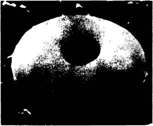
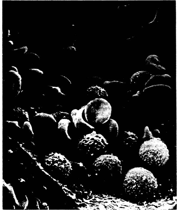
Class="center">Fig. 3.1. Scanning electron micrograph of an erythrocyte
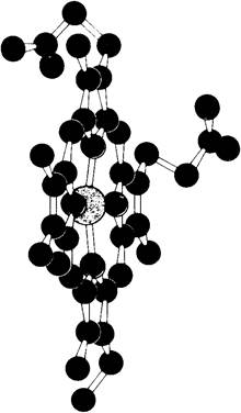
Heme consists of an organic moiety and an iron atom. The organic portion—protoporphyrin—is built from four pyrrole rings. These four pyrroles are linked by methenyl bridges to form a tetrapyrrole ring system. Attached to this ring are 4 methyl, 2 vinyl, and 2 propionate side chains. There are 15 possible spatial arrangements for these substituents. Biological systems utilize only a single isomer, known as protoporphyrin IX.
Scanning electron micrograph showing erythrocytes (biconcave discs) and leukocytes (spherical) within a small blood vessel
The iron atom in heme is bonded to 4 nitrogen atoms at the center of the protoporphyrin ring (Figs. 3.2 and 3.3). Iron can form two additional bonds on either side of the heme plane. These bonding directions are designated as the fifth and sixth coordination positions. The iron atom in heme can exist in the ferrous (+2) or ferric (+3) oxidation state. The corresponding forms of hemoglobin are termed ferrohemoglobin and ferrihemoglobin, respectively. Ferrihemoglobin is also known as methemoglobin. Only ferrohemoglobin (+2) is capable of binding oxygen. A similar nomenclature applies to myoglobin.
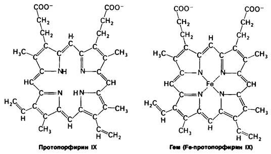
Fig. 3.2. The iron atom in heme is capable of forming six bonds. Four of these lie in the plane of the heme ring. The fifth is located on one side of this plane, and the sixth on the other. The arrangement of the atoms bound to iron is also referred to as coordination positions
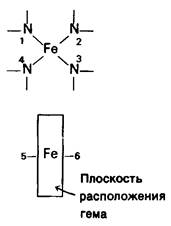
Fig. 3.3. Heme of hemoglobin (Fe shown in yellow, N in blue, O in red, C in black)
Last update: 06/08/2026
Editorial and Educational Adaptation: This material has been compiled based on the primary/original source text. The project team performed an editorial review, corrected technical inaccuracies, structured sections, and adapted the content for an educational format.
What was processed:
- elimination of formatting defects (OCR errors, structural breaks, corrupted characters);
- editorial organization of content;
- standardization of terminology in accordance with academic sources;
- verification of factual statements against the original source text.
All mentions of the author, publication year, and origin of the primary text have been preserved in accordance with the source.