FUNDAMENTALS OF MEDICAL BIOLOGY - 2012
Molecular Foundations of Heredity. Characteristics of Nucleic Acids. Transformation and Transduction
Transformation (from Lat. transformatio – metamorphosis) is The transfer of Genetic information from one bacterium to another via isolated DNA. The phenomenon of transformation was first discovered in 1928 by the English microbiologist F. Griffith while working with two strains of pneumococci (Streptococcus pneumoniae).
One strain (S) possesses a polysaccharide capsule and exhibits high virulence (causing Pneumonia), whereas the other (R) is non-capsulated and avirulent (does not cause disease). The ability of S-pneumococci to induce disease is attributed to the presence of a capsule that protects them from phagocytosis within the Organism. In a series of experiments on white mice, Griffith obtained the following results:
✵ administration of the live R-strain resulted in the mice surviving;
✵ administration of the live S-strain resulted in the mice dying from pneumonia;
✵ administration of the heat-killed S-strain resulted in the mice surviving;
✵ administration of the heat-killed S-strain combined with the live R-strain resulted in the death of most mice, and live S-strain Bacteria were isolated from their Blood.
It was concluded that the traits of the killed bacteria—capsule formation and virulence—were transmitted to the live bacteria. Consequently, a transformation of the avirulent R-strain into the virulent S-strain occurred (R - S).
The Chemical Nature of the transforming factor was established in 1944 by American geneticists O. Avery, C. MacLeod, and M. McCarty, who demonstrated that it was DNA.
Transformation does not occur in all Cells, but only in those capable of taking up DNA. Such cells are referred to as competent. The Mechanism of transformation involves the following stages: 1) adsorption of the double-stranded donor DNA molecule onto The surface of the recipient Cell; 2) uptake of the donor DNA by the recipient cell; 3) Conversion of the double-stranded donor DNA into single-stranded fragments by cellular Nucleases; 4) Integration of the single-stranded DNA fragment into the recipient chromosome; 5) expression of the integrated donor Gene within The Cell, enabling it to acquire new, heritable traits.
Transduction (from Lat. transductio – transfer) is the transfer of DNA fragments from a donor bacterium to a recipient bacterium mediated by temperate Bacteriophages. Upon release from the bacterium in which they parasitized, temperate bacteriophages package a portion of the host DNA and transfer it to a new bacterium, thereby conferring the traits of the previous bacterial host. Transduction is extremely widespread among bacteria. It not only corroborates the genetic role of DNA but is also utilized to investigate chromosome and gene Structure in Introduction/32.html">Genetic Engineering.
Class="center">Nucleic Acids: DNA and RNA
There are two Types of Nucleic acids (NAs)—deoxyribonucleic acid (DNA) and ribonucleic acid (RNA). Prokaryotic and Eukaryotic cells contain both types of nucleic acids (DNA and RNA), whereas Viruses contain only one of them (either DNA or RNA). Nucleic acids are present in All living organisms and perform unique biological Functions associated with the storage, expression, and transmission of hereditary information across generations.
Deoxyribonucleic acid (DNA) is a high-molecular-weight organic compound, a biopolymer whose monomers are NUCLEOTIDES linked together into a long polynucleotide chain. Each DNA nucleotide (deoxyribonucleotide) consists of three components: 1) a pentose sugar, deoxyribose (hence the name deoxyribonucleic acid); 2) one of four nitrogenous bases belonging to different classes: adenine (A) and guanine (G) belonging to Purines, and cytosine (C) and thymine (T) belonging to Pyrimidines; 3) a phosphoric acid residue.
A DNA macromolecule consists of two long polymer chains. Within a DNA chain, nucleotides are linked via phosphodiester bonds formed between the 3’- and 5’-hydroxyl groups of adjacent deoxyribose sugars. Two distinct ends are distinguished in a DNA polynucleotide chain: one end bears a hydroxyl group (-OH) attached to the 3’-carbon of the deoxyribose sugar, while the other end features a phosphoric acid residue at the 5’-position of the sugar. Within the nucleotide structure, deoxyribose occupies a central position between the phosphoric acid and the nitrogenous base. The relative molecular mass of DNA can reach 1,500,000 to 2,000,000 or more.
DNA is characterized by specific regularities established in 1950 by the American biochemist E. Chargaff (born in Chernivtsi). Chargaff's rules state that:
1) the sum of purine bases equals the sum of pyrimidine bases: A+G=T+C;
2) the combined amount of guanine and thymine equals The amount of adenine and cytosine: G+T=A+C, or (G+T)/(A+C)= 1;
3) the content of adenine equals that of thymine, and the content of guanine equals that of cytosine (the rule of equivalence): A=T, G=C.
4) the base composition of DNA varies among different organisms only in terms of the (G+C)/(A+T) ratio, yet this ratio remains constant for organisms of a given species: in humans it is 1.54, and in the bacterium Escherichia coli it is 1.0.
5) There are Two Types of DNA: the AT-type, where A+T>G+C, and the GC-type, where G+C>A+T. The AT-type is characteristic of higher plants, animals, and humans, whereas the GC-type is typical of Fungi, bacteria, and viruses.
Nitrogenous Bases and Their Corresponding Nucleotides
Name of nitrogenous base |
Designation |
Name of nucleotide |
||
Adenine |
A |
dAp |
Deoxyadenosine-*5’-phosphate* |
Adenosine-3’-phosphate |
Thymine |
T |
dTp |
Thymidine-3’-phosphate |
- |
Uracil |
U |
Ur |
- |
Uridine-3’-phosphate |
Guanine |
G |
dGp |
Deoxyguanosine-3’-phosphate |
Guanosine-3’-phosphate |
Cytosine |
C |
dCp |
Deoxycytidine-3’-phosphate |
Cytidine-3’-phosphate |
In 1953, American biochemist J. Watson and English physicist F. Crick (Cambridge University), drawing upon chemical and X-Ray Diffraction data, proposed a model of the three-dimensional structure of DNA AS A double helix (Nobel Prize, 1962). According to the Watson-Crick model, a DNA molecule consists of two polynucleotide chains coiled right-handedly around a common axis into a double helix. The two strands of the DNA molecule are antiparallel: one strand runs in the 5’>3’ direction, while the other runs 3’>5’. The Double Helix of the DNA molecule is stabilized by hydrogen bonds between the bases of opposite strands following THE PRINCIPLE OF complementarity: adenine of one strand always pairs with thymine of the other (A-T), and guanine pairs with cytosine (G-C). Adenine and thymine are linked by a double Hydrogen bond, whereas guanine and cytosine are linked by a triple bond. The paired bases lie perpendicular to the central axis between the two strands. The entire structure resembles a spiral staircase, where the handrails are formed by the sugar-phosphate backbone, and the steps consist of the paired bases. The diameter of the helix is 2 nm, the distance between adjacent nitrogenous bases along the helical axis is 0.34 nm, and ten nucleotide pairs comprise one complete turn of the double helix, measuring 3.4 nm in length.
Several forms of DNA are known: 1) the B-form serves as the primary structural type under normal physiological conditions (Watson-Crick model structure); 2) the A-form is found in a dehydrated environment (the DNA is wider (2 nm) and a single helical turn contains 11 nucleotide pairs); 3) the C-form has fewer bases per turn; 4) the Z-form is left-handed.
Ribonucleic Acids (RNA) are high-molecular-weight Organic compounds consisting of a single polynucleotide chain. RNA is a biopolymer whose monomers are ribonucleotides. An RNA nucleotide, much like a DNA nucleotide, comprises three components: 1) a pentose sugar, ribose (hence the name of this acid — ribonucleic); 2) one of four nitrogenous bases: adenine (A) and guanine (G) belong to the purine class, while cytosine (C) and uracil (U) belong to the pyrimidine class; 3) a phosphoric acid residue.
Single-stranded RNA can sometimes form a double-stranded structure if different PARTS OF THE chain contain antiparallel complementary fragments that bind via Hydrogen Bonds (A-U, G-C). In some viruses, RNA acts as the carrier of hereditary information instead of DNA. Three MAIN TYPES OF RNA are found in cells, performing diverse functions: messenger or mRNA (m-RNA), Transfer RNA (t-RNA), and ribosomal RNA (r-RNA). All Three types of RNA are synthesized on DNA templates with the help of RNA polymerase Enzymes according to The principle of complementarity. All types of RNA are synthesized as precursor RNAs, which undergo specific modifications after METABOLISM/31.html">Transcription, turning into biologically active RNAs. This process is called maturation, or Processing.
Ribosomal RNA (rRNA) is a large, single-stranded, branched molecule that is the longest in length, contains up to 3–5 thousand nucleotides, and makes up to 90% of all cellular RNA. rRNA is formed in the Cell Nucleus within the nucleolus on the nucleolar organizers of Chromosomes. Since Ribosomes are abundant in both prokaryotic and eukaryotic cells, the genes encoding rRNA synthesis are represented by multiple copies. In the karyoplasm, rRNA combines with ribosomal Proteins to form the small and large ribosomal subunits. rRNA forms the structural framework of ribosomes to support the cell's protein-synthesizing machinery.
Transfer RNA (tRNA) is the shortest in size, single-stranded, cloverleaf-shaped, contains 70–90 nucleotides, accounts for 10–20% of total cellular RNA, and, unlike other RNAs, is soluble. Transfer RNAs transport activated Amino Acids to the site of Protein Biosynthesis (ribosomes) for incorporation into the polypeptide chain being synthesized there. A specific tRNA exists for each amino acid. Since Most amino acids are coded by multiple codons, the number of Different types of tRNA exceeds the number of amino acids (20). A tRNA molecule has four distinct regions: the acceptor stem, the anticodon, the enzyme attachment site, and the ribosome-binding site.
A common property of all types of tRNA is the presence of the CCA nucleotide triplet at the 3'-end (the acceptor end). The terminal adenosine of the anticodon accepts (attaches) the activated amino acid via the 3'-OH group of ribose during Translation. tRNAs serve as intermediate molecules between mRNA and the polypeptide.
Messenger, or template, RNA (mRNA) makes up 0.5–3% of the total cellular RNA. Its length can vary—from 300 to 3000 nucleotides—depending on the length of the gene. mRNA molecules are formed on specific regions of DNA known as structural genes. They carry genetic information regarding the Primary Structure of a protein from the DNA in The Nucleus to the ribosomes in the Cytoplasm. Messenger RNA is called template RNA (mRNA) because it acts as a template on which a polypeptide is built according to the information it carries. An mRNA that carries information for the synthesis of a single protein molecule is called monocistronic. If an mRNA molecule contains several cistrons for different proteins, it is polycistronic.
Messenger RNA is initially formed as immature pre-mRNA, which contains non-coding regions called introns—removed during processing (mRNA maturation)—and coding regions called exons. Mature mRNA enters the cytoplasm, binds to a ribosome, and translates its information. However, mRNA sometimes accumulates in the cell, binds to specific proteins that "conserve" it, and forms informosomes. Physiological changes that trigger the activation of Protein Synthesis serve as the signal for utilizing the RNA stored in informosomes.
DNA performs the following functions: it serves as the chemical basis of chromosomal genetic material (the gene); participates in the synthesis of RNA and DNA; and carries information regarding Protein Structure. The Biological Role of DNA lies in storing and executing genetic information throughout the life of the cell, and transmitting hereditary information from mother to daughter cells, and from parents to offspring.
RNAs perform various functions:
- mRNA carries information about protein structure from DNA to the site of protein synthesis at the ribosomes;
- tRNA delivers amino acids to the site of protein synthesis at the ribosomes;
- rRNA is a component of ribosomes, forming their functional Skeleton.
DNA Replication (self-reproduction at THE MOLECULAR LEVEL) is a complex biological process of template-directed synthesis that ensures the duplication of the DNA molecule before Cell Division. This process takes place in the cell nucleus during the S phase of interphase. The capacity for self-duplication is a unique property inherent solely to DNA and no other chemical substance. DNA replication is the most critical molecular process underlying all types of reproduction, and consequently, the sustained existence of individual organisms, populations, and all living species (maintaining the constancy of genotypes and phenotypes). It occurs via a semi-conservative mechanism and according to the principle of complementarity. Replication is an enzymatic, energy-dependent process.
The DNA model proposed by Watson and Crick made it possible to understand the principle of DNA molecule duplication (DNA replication). The double helix of the parent DNA molecule separates into two strands, and each of them serves as a template for synthesizing a new strand based on the principle of complementarity. As a result, two daughter molecules are formed from a single parent DNA molecule, acting as exact copies of the parent. Because each newly formed daughter molecule retains one old (parental) strand and one new strand, this mechanism of replication was named semi-conservative.
DNA Synthesis is catalyzed by the enzyme DNA polymerase. The DNA polymerase enzyme was first isolated from the bacterium Escherichia coli by A. Kornberg in 1957. There are several types of DNA polymerases. The DNA replication process is driven by the coordinated action of a series of Proteins and Enzymes. These proteins identify the corresponding DNA genes, meaning that DNA itself dictates its own replication. Initiator proteins form the Replication fork. DNA topoisomerases unwind the strands, DNA helicase and single-strand binding proteins split DNA into two separate strands, DNA polymerase and DNA primase catalyze the polymerization of nucleotide triphosphates and The formation of the new strand, while DNA ligases remove RNA primers and join short segments of the newly synthesized polynucleotide chain into a single continuous strand.
Fig. 7. DNA replication according to the model of J. Watson and F. Crick:
1, 2 - parental DNA strands; 1a, 2a - daughter DNA strands
Replication includes the following stages:
I - Initiation (Lat. initialis — initial).
II - Elongation (extension).
III - Termination (Lat. terminalis — final).
Initiation begins with enzymes identifying the replication origin—the ori site (from English origin), where the DNA double helix unwinds, its strands locally separate, single-stranded DNAs are stabilized, and a Y-shaped structure (the replication fork) emerges from the two separated strands. In prokaryotes and viruses, There is a single replication origin, and the DNA constitutes a single unit of replication known as a replicon. In eukaryotes, DNA molecules are divided into multiple replicons, meaning they possess multiple replication origins.
Deoxyribonucleotides, which serve as the "raw material" for DNA synthesis, undergo activation. Deoxyribonucleotide monophosphates (dAMP, dGMP, dCMP, dTMP) are activated upon interaction with ATP to form triphosphates (dATP, dGTP, dCTP, dTTP). This reaction is called phosphorylation and is catalyzed by the enzyme phosphorylase.
Next, DNA helicase enzymes break the hydrogen bonds between complementary nitrogenous bases, forming a replication bubble. The region where the replication bubble forms is called the replication origin. It contains about 300 nucleotides and is recognized by specific proteins.
DNA topoisomerases (gyrases) stabilize the DNA molecule ahead of the replication fork, preventing supercoiling by cleaving One DNA strand, which rotates around the other. Single-strand DNA-binding proteins (SSBs) attach to the free single strands, preventing the two strands from spontaneously re-associating—thus forming a replication fork.
Elongation. Free deoxyribonucleoside triphosphates attach via their nitrogenous bases through hydrogen bonds to the nitrogenous bases of both DNA strands according to the complementary base-pairing rules: A-T, G-C.
The enzyme DNA polymerase begins adding complementary activated nucleotides (deoxyribonucleoside triphosphates: dATP, dGTP, dCTP, dTTP) to one of the strands with a free 3' end. Thus, complementary Synthesis of the daughter strand proceeds gradually and continuously along one DNA strand, which has a 5' 3' directionality (from the carbon 5'-end to the carbon 3'-end of the DNA molecules). The other strand is antiparallel, with a 3' 5' directionality. The action of DNA polymerase in this direction is impossible. Consequently, the duplication process at the replication fork is asymmetric. On one strand, it occurs sequentially, continuously, and rapidly. This strand is called the leading strand. Synthesis on the second strand proceeds slowly, which is why this strand is called the lagging strand.

Fig. 8. DNA replication according to R. Okazaki's model.
On the lagging strand, synthesis of the daughter strand occurs discontinuously and with a delay. First, short RNA chains are synthesized using the DNA template—these are RNA primers (10-60 nucleotides). Primer formation is catalyzed by RNA primase. The primers are subsequently removed, and the resulting gaps are filled with DNA deoxyribonucleotides. DNA polymerase synthesizes fragments of the daughter DNA strand in the 5' to 3' direction. In other words, short DNA fragments, known as Okazaki fragments, are formed at the primer site. In prokaryotes, such fragments contain 1000...2000 nucleotides, while in eukaryotes, they contain 100...200 nucleotides. Next, these fragments are joined (spliced) by DNA ligases, the primers are degraded, and DNA polymerase fills in the complementary DNA nucleotides.
The result of synthesis is the formation of 2 identical DNA molecules, each consisting of one parental strand and one newly synthesized daughter strand.
Precise complementarity of Base Pairs ensures accurate DNA replication. However, errors in base pairing sometimes occur. These are corrected by DNA polymerase, which re-associates with the DNA molecules for this purpose (repair).
Termination is the completion of the replication process. The newly formed DNA molecules coil up, forming a chromosome consisting of two chromatids.
The rate of replication is extremely high (1000 nucleotides/sec in prokaryotes; 100 nucleotides/sec in eukaryotes). A region of DNA controlled by a single origin is called a replication unit (replicon). Prokaryotic DNA has a single origin, and the entire molecule replicates as a single replicon. Eukaryotic DNA has multiple origins and, accordingly, multiple replicons, which is a necessary condition for giant eukaryotic DNA molecules to complete duplication within a single Cell Cycle.
Throughout evolution, cells have developed specific mechanisms that ensure the stability of hereditary material (DNA) and protect it against Mutations. Without these mechanisms, organisms would perish from the effects of natural mutagens due to The high frequency of genetic DNA damage. Most DNA damage, which serves as a potential source of mutations, is repaired through several mechanisms:
- high chemical Stability of the DNA molecule itself
- the presence of specialized self-correction and repair mechanisms for emerging alterations.
Repair (Latin reparatio - restoration) is The process of restoring the primary structure of DNA
after damage by mutagens. Based on their developmental mechanisms, light (non-excision), dark (excision), and recombinative DNA repair are distinguished. All of them are enzymatic in nature, repair only single-strand DNA Lesions, and are controlled by specialized genes.
Light repair (photoreactivation) utilizes visible light energy and repairs damage caused exclusively by ultraviolet rays. Such damage consists of dimers—adjacent pyrimidine bases on the same DNA strand linked together. Dimers form between two thymines, a thymine and a cytosine, two cytosines, a thymine and a uracil, a cytosine and a uracil, or two uracils. The mechanism of light repair is as follows: the enzyme DNA photolyase specifically binds to the UV-damaged region of DNA, is activated by daylight quanta, and subsequently dissociates; upon dissociation, the enzyme simultaneously unlinks the dimers, restoring the normal DNA Structure. Light in the blue region of the spectrum is the most effective for light repair.
Dark (excision) repair, unlike photoreactivation: does not require visible light energy; repairs damage caused not only by ultraviolet rays but also by other Mutagenic Factors; is carried out by excising the damaged fragment of the DNA strand (excision - to cut out). The following stages of dark repair are distinguished: 1) recognition of the damaged DNA region by endonuclease; 2) incision of the DNA strand near the damage by endonuclease; 3) excision of the damaged DNA region accompanied by gap enlargement in both directions by exonuclease; 4) template-directed synthesis of a DNA fragment ("patch") at the gap site (repair replication); 5) joining of the newly synthesized segment to the main DNA strand using the enzyme ligase.
Post-replication repair occurs via recombination between two newly formed DNA Double helices. An example of such repair is the restoration of DNA structure upon the formation of thymine dimers (T-T) when they are not resolved by light (light repair) or during pre-replicative Excision Repair.
Light and dark repairs eliminate DNA damage prior to its replication (pre-replicative repair). If these mechanisms fail, the damage is resolved after replication in the daughter DNA strands (post-replicative repair). Replicatively coupled repair refers to the set of DNA restoration processes occurring during replication.
Impairment of various DNA repair pathways leads to hereditary disorders: xeroderma pigmentosum, Bloom syndrome, Fanconi anemia, and juvenile progeria (premature Aging).
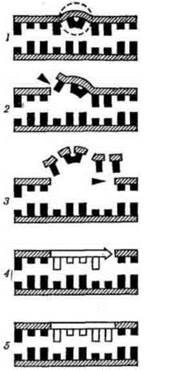
Fig. 9. Scheme of DNA excision repair:
1 - UV light-induced damage to the DNA molecule; 2 - incision of one DNA strand by endonucleases; 3 - excision of the damaged region by exonucleases; 4 - synthesis of a new strand via polymerase action; 5 - ligation of the newly synthesized segment using ligases.
Xeroderma pigmentosum is a hereditary disorder associated with impaired dark DNA repair. It manifests as photophobia even in newborns.
Patients exhibit a characteristic hypersensitivity of the Skin to ultraviolet rays. This leads to atrophy of exposed skin areas, hyperpigmentation, and can potentially develop into skin Cancer. Several forms of xeroderma pigmentosum are distinguished. The mode of inheritance is autosomal dominant and autosomal recessive.
The principle of complementarity entails the formation of hydrogen bonds between strictly defined nitrogenous bases of two strands: A=T; G=C. The nucleotide sequence in one strand of the DNA molecule corresponds to the nucleotide sequence In the second strand.
The principle of antiparallelism implies that the two polynucleotide strands are joined in such a way that the 5' end of one connects with the 3' end of the other, and vice versa. One strand runs from top to bottom from the 5' to the 3' carbon atom, while the other runs from bottom to top.

Last update: 08/08/2026
Editorial and Educational Adaptation: This material has been compiled based on the primary/original source text. The project team performed an editorial review, corrected technical inaccuracies, structured sections, and adapted the content for an educational format.
What was processed:
- elimination of formatting defects (OCR errors, structural breaks, corrupted characters);
- editorial organization of content;
- standardization of terminology in accordance with academic sources;
- verification of factual statements against the original source text.
All mentions of the author, publication year, and origin of the primary text have been preserved in accordance with the source.