BIOLOGY Volume 3 - A Guide to General Biology - 2004
25. APPLIED GENETICS
25.7. Human Genetics
25.7.2. Sickle-Cell Anemia
This disorder is monogenic in nature, meaning it is caused by a single Gene mutation. Sickle-Cell Anemia is a striking example of The Role of natural Selection in regulating gene frequency within a population.
In 1904, a young Chicago physician named James Herrick examined a 20-year-old African American man who was admitted to the hospital complaining of fever, headache, weakness, dizziness, and a cough. The patient's Lymph Nodes were enlarged, his Heart was abnormally large, and urinalysis indicated Kidney disease. However, the most striking findings came from the Blood analysis. Under the Microscope, the patient's red Blood Cells were sickle-shaped rather than round (Fig. 25.20). His Hemoglobin level was roughly half of normal; in other words, the young man was suffering from anemia. Although the patient was discharged four weeks later, Herrick did not publish a Description of the case until 6 years afterward. Shortly thereafter, numerous reports of patients with similar symptoms appeared in medical literature, prompting research into the underlying cause of their illness.
Class="center">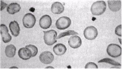
Fig. 25.20. Sickled red blood cells.
It was discovered that the condition—named sickle-cell anemia—is more prevalent among African Americans, has a genetic basis, and the gene responsible for it appears to be inherited recessively. A person develops the disease only if they carry two copies of the mutant gene, one inherited from the mother and the other from the father; in other words, affected individuals are always homozygous for this gene. If only a single copy of the sickle-cell gene is present, the individual is referred to as a heterozygous carrier. It is estimated that approximately 100,000 people die worldwide from sickle-cell anemia each year. The disease is particularly widespread in Africa, Pakistan, and India.
Symptoms
The primary clinical manifestations of the disease are anemia and the alteration of red blood cell shape (sickling) under low oxygen concentrations. Sickled red blood cells accumulate in capillaries and small vessels, impeding normal blood flow and consequently failing to adequately supply cells with oxygen. As a result, a wide range of secondary symptoms may occur (Fig. 25.21). The Kidneys and joints are particularly affected. Blood vessel occlusion causes severe pain in the arms, legs, back, and abdomen. Joints become stiff and painful, and the extremities swell. Affected individuals are exceptionally susceptible to infections and frequently succumb to them.
Children with sickle-cell anemia generally feel well and lead a relatively normal lifestyle, although they may occasionally experience severe pain and anemia-related fatigue.
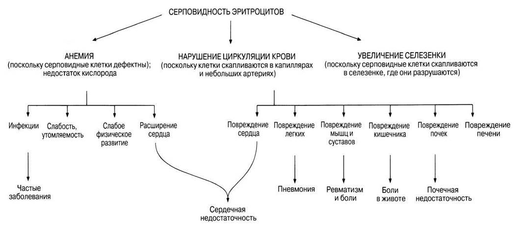
Fig. 25.21. Some of the potential effects of sickle-cell anaemia.
Cause
In 1949, a research group led by Linus Pauling discovered that the haemoglobin of patients with sickle-cell anaemia (HbS) differs from that of normal healthy individuals (HbA). Using Electrophoresis—a technique that separates Proteins based on their net charge—it was established that at pH 6.9, the net charge of HbS is positive, whereas for HbA it is negative. Thus, it was demonstrated for the first time that the disease is caused by a defective haemoglobin molecule. In 1956, Ingram showed that the difference between normal and abnormal haemoglobin is determined by a single amino acid, after which the complete Amino acid sequences of HbA and HbS were determined. Haemoglobin consists of four polypeptide chains (Fig. 3.36): two α-chains and two β-chains, consisting of 141 and 146 amino acid residues respectively. The defect occurs at the sixth amino acid of the β-chain. In normal haemoglobin, this position is occupied by glutamic acid. However, in HbS, it is replaced by valine. Using The amino acid abbreviation code (Table 23.4), this can be represented as follows:
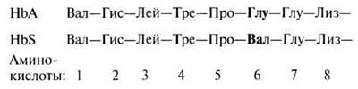
Glutamic acid carries a negative charge and is polar, whereas valine is non-polar and hydrophobic. The presence of valine makes deoxygenated haemoglobin less soluble. Therefore, when HbS loses its oxygen, the molecule comes out of solution and crystallises into rigid, rod-like fibres. This alters the shape of red blood cells, which normally appear as biconcave discs. The underlying cause of this amino acid substitution is a mutation in the DNA encoding that amino acid. Referring to Table 23.4, you can see how this change comes about. In mRNA, the possible codons for these Two Amino Acids are:

Consequently, the complementary triplet codons in the DNA are:

For the sequence to change from Glu to Val, T (thymine) must be replaced by A (adenine) In the second position of the triplet. Such a mutation is called a base substitution. We now know that in the β-globin gene, the CTC codon is replaced by CAC, and that this gene is located on chromosome 11.
In heterozygotes, roughly half of the molecules are HbS and half are HbA, meaning that the HbA and HbS alleles are co-dominant. Such individuals generally do not exhibit symptoms of the disease, except in situations where oxygen concentration is severely reduced, such as during high-altitude ascent.
As can be seen from Fig. 25.22, if both parents are heterozygous carriers of the sickle-cell trait, the probability of their children being affected is 1/4. The phenotype is determined via a blood test. If There is a METABOLISM/13.html">History of the disease in the family, prospective parents are advised to undergo a blood test before deciding to have children. Prenatal Diagnosis is also currently available. It is performed either by hybridisation using the HbS gene as a probe or by restriction analysis. Foetal cells are obtained via amniocentesis or chorionic villus sampling (Section 25.7.9).

Fig. 25.22. Genetic diagram showing the possible genotypes and phenotypes of children from two parents heterozygous for the sickle-cell gene.
The story of sickle-cell anaemia research takes an unexpected turn. Geneticists found it puzzling that a mutation causing such clear harm to its bearer could be so widespread in the population. To explain this phenomenon, a hypothesis was put forward suggesting that under certain conditions, the mutation might confer a distinct advantage upon the carrier. And such an advantage was indeed discovered. Fig. 25.23 presents distribution maps for sickle-cell anaemia and malaria. The geographic ranges overlap quite closely; wherever malaria is more prevalent, the mutant gene is also more common. In some regions of Africa, its frequency reaches 40% (40% HbS, 60% HbA in the population). In areas where malaria is endemic, it is a leading cause of mortality; however, carriers of the defective gene are far less susceptible to malaria (as the malaria parasite replicates only within normal red blood cells). Although homozygous individuals with sickle-cell anaemia often die before reaching reproductive age, heterozygous carriers possess a selective advantage over non-carriers and thus have a higher chance of survival and of passing on their genes to the next generation. The resulting gene frequency in the population varies depending on the incidence of malaria. This phenomenon is known as balanced polymorphism (Section 25.7.5).

Fig. 25.23. Distribution of the sickle-cell anaemia gene (A) and malaria in Africa, the Middle East, India, and southern Europe (B).
Last update: 06/08/2026
Editorial and Educational Adaptation: This material has been compiled based on the primary/original source text. The project team performed an editorial review, corrected technical inaccuracies, structured sections, and adapted the content for an educational format.
What was processed:
- elimination of formatting defects (OCR errors, structural breaks, corrupted characters);
- editorial organization of content;
- standardization of terminology in accordance with academic sources;
- verification of factual statements against the original source text.
All mentions of the author, publication year, and origin of the primary text have been preserved in accordance with the source.