Medical Genetics - V. M. Zaporozhan 2005
Classification, general characteristics, and symptomatology of hereditary diseases. General clinical diagnostics
General principles of clinical diagnostics of hereditary diseases
Description of the phenotype of a patient with hereditary pathology. Main micro-anomalies and developmental malformations
Examination of a patient with a hereditary condition is performed “from HEAD to toe,” as signs of a genetic disorder can manifest as abnormalities in any morphological structures of The Human Body.
Clinical examination of the patient begins with measuring height and body weight. The obtained data are compared with normal age-specific population standards. Anthropometric parameters in individuals with Genetic Disorders typically fall outside the range of normal variation.
Body Weight and Height
Hereditary diseases often manifest as early as the Embryonic Stage of development, leading to intrauterine growth restriction, hypotrophy, or hypoplasia in the newborn. This is observed, for example, in Chromosomal Disorders. In Postnatal development, symptoms of hypotrophy are common signs of inherited Metabolic Disorders, congenital defects of the digestive tract, etc.
Short stature in children at birth and during the postnatal period may be a symptom of genetic dwarfism. Body proportions may be altered or remain normal. Less commonly, an increase in birth weight and length—macrosomia—is observed. For instance, this is a leading feature of Beckwith-Wiedemann syndrome.
Obesity may also occur in hereditary diseases (Prader-Willi, Bardet-Biedl syndromes, etc.).
Body Symmetry
Attention must be paid to body symmetry (Fig. 3.1), as a specific sign of Certain genetic disorders is partial Asymmetry (hemifacial hypertrophy, Klippel-Trenaunay syndrome, etc.) or complete asymmetry of the right and left sides of the body (Russell-Silver syndrome).
Head, Face, and Neck
1. Head size deviation by more than 10 % from the age norm: microcephaly (Fig. 3.2) or macrocephaly. In newborns, a clinically significant deviation from the norm is 5 cm.
Hydrocephalus (Water on the Brain) differs phenotypically from macrocephaly by a disproportion between the facial and cerebral cranium—the face is relatively small, the forehead is prominent and overhanging, the cranial vault is enlarged, subcutaneous Veins are dilated, cranial sutures may be separated, and the fontanelles are bulging.
Class="center">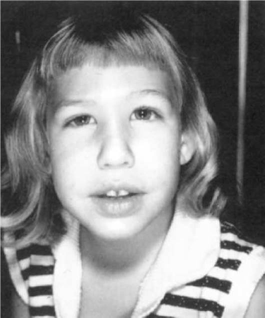
Fig. 3.2. Microcephaly
2. The shape of the Skull can be normal or abnormal—asymmetrical. Premature fusion of the cranial sutures (craniosynostosis) restricts skull growth in a particular direction, leading to its deformation. Brachycephaly (relative increase in transverse diameter, flattened face); dolichocephaly (increase in the longitudinal diameter of the skull) (see Fig. 5.8); scaphocephaly (narrow, elongated skull with a prominent forehead and occiput); trigonocephaly (skull widened in the occipital region and narrowed in the frontal area due to underdevelopment of the frontal eminences); acrocephaly, or oxycephaly (high “turret” skull — “sugar loaf”) (see Fig. 6.4) may be observed.
3. Low hairline on the forehead and neck.
4. Face: bird-like face (Marfan Syndrome) (see Fig. 6.6), doll-like face (Glycogen Storage Diseases), coarse facies with enlarged supraorbital ridges and thick Lips (mucopolysaccharidosis) (see Fig. 6.16); triangular face (Russell-Silver syndrome), etc.
5. Forehead: low, very high, or with prominent frontal eminences.
6. Malformations and micro-Anomalies of the eyes
The palpebral fissure may be horizontal (normal for European populations); mongoloid (the outer canthus is higher than the inner) (see Fig. 5.5); antimongoloid (the outer canthus is lower than the inner) (see Fig. 5.12, 5.15).
Hypotelorism—closely spaced eyes (see Fig. 5.9); hypertelorism—increased distance between the inner canthi (normally, the distance between the inner canthi is on average equal to the length of the palpebral fissure) (see Fig. 5.12, 9.14).
Anophthalmia—absence of one or both eyes (see Fig. 9.3); cryptophthalmia—absence of the palpebral fissure and eyelids, with underdevelopment of the Eyeball; buphthalmos—enlarged “ox-eye”; microphthalmia—small eye size (see Fig. 5.8); enophthalmos—posterior Displacement of the eyeball (deep-set eyes in the Orbit); exophthalmos—protrusion of the eyes forward.
Synophrys—meeting of the eyebrows at the bridge of the Nose; distichiasis and trichiasis—double and triple rows of eyelashes; coloboma of the eyelid—a slit-like defect of the eyelid margin; epicanthus—a Skin fold at the inner corner of the eye covering the lacrimal caruncle (see Fig. 5.5, 5.6); microblepharon—reduction in the vertical dimension of the eyelids leading to impaired closure; blepharophimosis—shortening of the eyelids and narrowing of the palpebral fissure; ptosis—drooping of the upper eyelid (see Fig. 9.2).
Blue sclerae due to their thinning are observed in Connective Tissue disorders (Marfan syndrome, Osteogenesis Imperfecta, etc.).
Microcornea and macrocornea—decrease or increase in corneal size; corneal leukoma—corneal opacity (“white spot”); iris coloboma—a slit-like defect of the iris (Fig. 3.3); aniridia—almost complete absence of the iris (inherited as a dominant trait); heterochromia—uneven pigment distribution within a single eye or different eye colors; aphakia—absence of the lens; cataract—opacity of the lens.

Fig. 3.3. Iris coloboma
Hereditary diseases may present with glaucoma, strabismus, myopia, and blindness.
If a hereditary disorder is suspected, an ophthalmologist consultation is mandatory, as ocular malformations and microanomalies are part of the symptom complex of approximately 280 hereditary diseases.
7. Bridge of the Nose and Nasal Anomalies
Nasal bridge: depressed, broad, flat, protruding forward with parallel edges ("Greek warrior helmet").
Nose: saddle nose (see Fig. 6.4), beak-shaped (pseudorhinine) nose (see Fig. 5.20), pyriform nose, short nose with nostrils turned forward (see Fig. 9.2), hypoplasia of one nasal half, hypoplasia of the alae nasi, nasal septum deviation, coloboma of the alae nasi, etc.
8. Anomalies and Malformations of the Nasolabial Region and Jaws
The upper jaw may be underdeveloped (micrognathia) or protrude forward (prognathia). Underdevelopment of the lower jaw is micrognathia/microgenia (see Fig. 9.2, 9.9), while its excessive development with a massive chin is progenia (mandibular prognathia) (see Fig. 5.20). Facial clefts also occur (Fig. 3.4).
Shortening or lengthening of the philtrum (see Fig. 9.2) (the philtrum is the distance from the subnasal point to the vermilion border of the upper lip).
9. Mouth
Macrostomia (see Fig. 5.20) and microstomia—enlargement or reduction of the mouth size; extremely thick or thin lips.
Cheiloschisis (cleft lip)—complete or partial, unilateral or bilateral, median (see Fig. 3.4).
Dental anomalies: changes in tooth number (adontia—absence of Teeth, oligodontia—reduced number of teeth), supernumerary teeth, altered shape (cone-shaped teeth) (see Fig. 6.23). Macro- and microdontia—enlargement or reduction of tooth size, fused teeth, diastema—a gap between the central incisors. Tooth discoloration—amelogenesis. Multiple caries.
High ("gothic") palate. Palatoschisis (cleft palate)—complete, incomplete, unilateral and bilateral, total (transverse/complete) or submucosal.
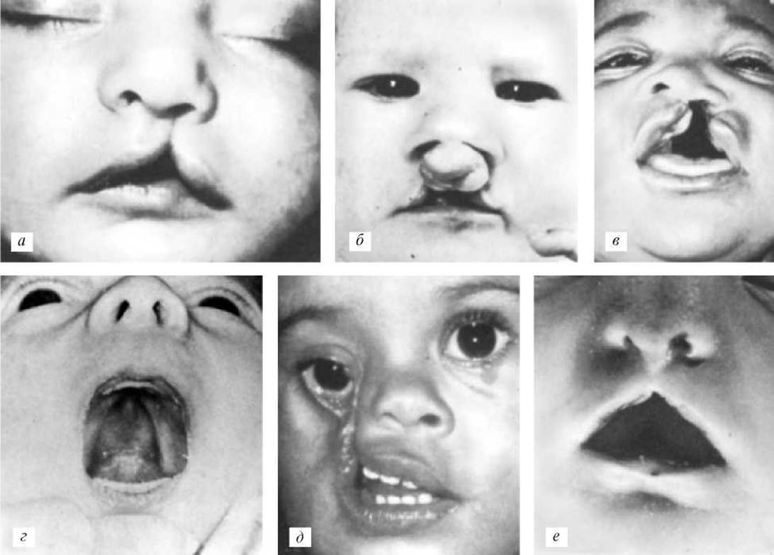
Fig. 3.4. Cleft lip and palate:
a — unilateral cleft lip; b — bilateral cleft lip; c — cleft lip and palate; d — isolated cleft palate; e — oblique facial cleft; f — median cleft lip
Macroglossia—enlargement of the Tongue (see Fig. 5.5, 6.16, 6.18), microglossia—small tongue.
11. Auricles (Pinnae)
The embryonic Formation of the auricle is highly sensitive to genotype alterations or teratogenic factors. In Hereditary and Congenital pathologies, enlargement of the auricles (macrotia) (see Fig. 9.2) or their reduction (microtia), abnormally high or low positioning (see Fig. 5.8), and structural deformation are frequently observed. Normally, the lower wall of the external auditory meatus in an adult lies at the level of the line connecting the free edge of the nasal ala base with the Base of the mastoid process of the Temporal bone.
Common findings include: attached earlobe, fleshy earlobe, prominent (protruding) ears (see Fig. 9.3), preauricular sinuses (blind-ending tract openings), preauricular tags/Papillomas (Fig. 3.5), atresia or stenosis of the external auditory meatus, and hypo- or hyperplasia of individual structures of the auricle.
Hearing impairment or deafness may also be observed.
12. Neck: shortened; webbed neck (pterygium colli) (see Fig. 5.15); median and lateral cysts; muscular Torticollis—shortening of the sternocleidomastoid Muscle, resulting in the child's head tilting toward the affected side.
Trunk
1. Chest and Spinal Deformities. Lateral curvature of the spine with rotation—Scoliosis; backward curvature of the spine—Kyphosis (typically in the thoracic region); kyphoscoliosis; forward curvature of the spine—lordosis (typically in the lumbar region). Flat back—absence of physiological curves. Sacral sinus—a skin depression along the midline in the lumbosacral region.
"Funnel chest" (Pectus excavatum) — a concave depression of the Sternum and costal cartilages; Flat chest; Pigeon chest (pectus carinatum) — a forward protrusion of the sternum and Ribs ("chicken breast").
2. Alterations of the nipples and Mammary glands: absence of nipples — athelia; supernumerary nipples — polythelia; widely spaced nipples — nipple hypertelorism (see Fig. 5.15). Excessive development of mammary glands in males — gynecomastia.
3. Hernias of the linea alba, umbilical hernias, umbilical cord hernias (omphalocele), inguinal hernias, and inguinal-scrotal hernias.

Fig. 3.5. Auricular anomalies:
a — microtia with a preauricular pit; b — preauricular pit and fistula; c — preauricular tags
4. Malformations of the reproductive Organs. In males: epispadias — a dorsal urethral fissure with displacement of the urethral opening upward; hypospadias — a ventral urethral fissure with displacement of the opening downward, extending as far as the Perineum (see Fig. 9.7). Macrophallus and microphallus — enlargement or reduction of the Penis. Cryptorchidism — absence of one or both Testes in the Scrotum. In females: clitoral hypertrophy (see Fig. 6.19), hypoplasia or hyperplasia of the labia, vaginal atresia, etc.
Extremities
Amelia — absence of a limb (see Fig. 9.21). Phocomelia — complete or partial absence of the proximal segments of the limbs (seal-like limbs). Brachymelia — shortening of a limb. Arachnodactyly — long, spider-like fingers (see Fig. 6.7). Brachydactyly — shortening of the fingers. Isodactyly — all fingers of equal length (see Fig. 6.2). Camptodactyly — flexion contracture of the fingers at the proximal interphalangeal joint. Clinodactyly — lateral curvature of the fingers. Oligodactyly — decreased number of fingers. Polydactyly — increased number of fingers (Fig. 3.6). Syndactyly — fusion of the fingers (cutaneous, osseous, partial, or complete) (see Fig. 6.4). Symphalangism — fusion of the finger Phalanges. Ectrodactyly — Aplasia of the central Components of the hand or FOOT, resulting in a cleft hand or foot ("lobster-claw" hand/foot) (see Fig. 6.8). Sandal gap — increased distance between the First and Second toes (see Fig. 9.2). Flatfoot (pes planus) — flattening of the arch of the foot. Hollow foot (pes cavus) — abnormally high arch of the foot. Rocker-bottom foot — flat foot with a posteriorly protruding heel (see Fig. 11.2). Congenital Clubfoot (talipes equinovarus) — persistent adduction-flexion contracture of the foot.

Fig. 3.6. Polydactyly
Cytology/cytology/66.html">Skin and its Appendages (hair, nails, glands)
Hyperkeratosis — excessive thickening of the cornified layer. Ichthyosis — marked hyperkeratosis with scale formation and horny encrustations (Fig. 3.7). Albinism — absence or marked reduction of pigment in the skin, hair, and iris. Pigmented spots and nevi (birthmarks) on the skin. Depigmented skin areas (leukoderma). Anhidrosis — absence of Sweat Glands. Hypohidrosis — reduced function of the sweat glands. Hyperhidrosis — excessive function of the sweat glands. Increased hair growth — hypertrichosis; excessive male-pattern hair growth in girls — hirsutism. Hypotrichosis — diminished hair growth (see Fig. 6.23). Alopecia — complete or partial absence of scalp hair. Anonychia — absence of nails. Nail hypoplasia — underdevelopment of the nail plates.

Fig. 3.7. Ichthyosis (harlequin-type ichthyosis)
Last update: 11/08/2026
Editorial and Educational Adaptation: This material has been compiled based on the primary/original source text. The project team performed an editorial review, corrected technical inaccuracies, structured sections, and adapted the content for an educational format.
What was processed:
- elimination of formatting defects (OCR errors, structural breaks, corrupted characters);
- editorial organization of content;
- standardization of terminology in accordance with academic sources;
- verification of factual statements against the original source text.
All mentions of the author, publication year, and origin of the primary text have been preserved in accordance with the source.