Orthopedics - Oleksa A.P. 2006
Congenital malformations of the upper extremity
Congenital hand deformities
Syndactyly
Syndactyly, the fusion of adjacent fingers or toes, is a congenital malformation resulting from the failure of the digits to separate during a specific stage of intrauterine development (Fig. 123). This developmental anomaly occurs twice as frequently in boys as in girls (Barsky A.J., 1951).
Syndactyly has an incidence of one in every 2,000 to 3,000 newborns (Donald, 1940; McCollum, Bunnel, 1944) and accounts for nearly half of all congenital Anomalies of the upper extremities (Paryin B.V., 1960).
Syndactyly can be unilateral or bilateral, and it occurs more frequently in the hands than in the feet. In most cases, the fused digits are the third and fourth or second and third fingers, and the second and third toes.
While simple forms of syndactyly exist—where the digits have normal anatomical structures and are simply joined to an adjacent digit—syndactyly is also frequently combined with other developmental anomalies (such as digital hypoplasia, phalangeal defects, polydactyly, and amniotic bands).
Accordingly, based on its etiopathogenesis, syndactyly is classified into embryonic and amniotic forms. The former result from impaired Embryogenesis (occurring up to 4–6 weeks of development), tend to be bilateral, and may be hereditary. The latter, amniotic syndactyly, are rare and caused by amniotic constriction bands.
Class="center">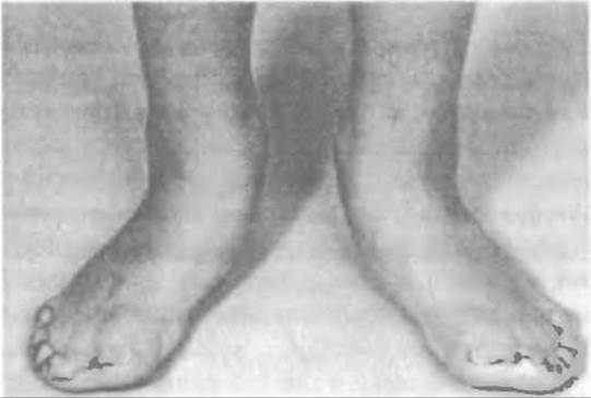
Fig. 123. Syndactyly with valgus deviation of the halluces (first toes) of both feet in Apert syndrome.
Regarding the Classification of syndactyly, Novachenko M.P. (1968) considers the classification proposed by Balabanits G.F. (1965) to be the most appropriate. Balabanits divides syndactyly into simple and complex forms, further subdividing the simple form into cutaneous, membranous, and osseous types, which can be either basal or distal. According to Balabanits, the complex form comprises four groups: cutaneous, membranous, osseous, and combined. Each of these groups is further categorized into amniotic, syndactylic polyphalangy, and syndactylic polydactyly, and each can be either complete or incomplete.
While Balabanits's classification encompasses all potential variants of syndactyly, it remains quite complex.
From a practical standpoint, alongside the aforementioned variants, the specific Nature of the digital fusion must also be considered. It may present as a webbed form (featuring a broad Skin fold, akin to webbed feet in aquatic birds), a cutaneous form (where the digits are directly apposed and connected solely by skin), or an osseous form (where the bone Phalanges, most commonly the distal ones, are fused together). Hand radiography is performed to determine the exact nature of the syndactyly.
Parents of a child with a hand malformation typically seek medical consultation and assistance immediately. Distressed by the cosmetic defect—particularly on the hands—they often demand prompt Surgical Treatment. The physician, in turn, must determine the optimal age for surgery based on the form and nature of syndactyly, selecting the best surgical approach for each individual case to eliminate the cosmetic defect and improve hand function.
In many cases, especially with basal cutaneous syndactyly or the webbed form, hand function is nearly unimpaired. Furthermore, children adapt remarkably well and quickly to more complex forms of syndactyly, remaining capable of performing a fairly wide range of tasks.
Nevertheless, it should be borne in mind that without treatment, the deformity progresses as the child grows; Tissues become denser, worsening both the aesthetic and functional condition of the hand. Discrepancies in the length of the fused digits and contractures may develop, particularly in cases of osseous fusion of adjacent distal phalanges, where the longer third digit remains persistently flexed at the proximal interphalangeal joint.
When syndactyly affects the feet, the toes develop normally and generally cause no discomfort or concern to the children or their parents.
Due to the diverse presentations of syndactyly, particularly complex forms, the surgical plan and timing for hand Procedures must be tailored individually.
Evidently, in webbed syndactyly where There is a sufficient skin reserve, surgery can be performed in early childhood. This allows for the restoration of normal anatomical shape and function of the digits.
The Procedure involves releasing the skin fold and suturing the wound margins on each digit without tension, with any excess skin excised. Such surgery typically leaves no prominent scars that could impair digital function.
Surgery is more challenging in cutaneous syndactyly, where the digits are closely apposed and there is no skin surplus. In the past, Dèֹdo's operation (Fig. 124) combined with Zeller's interdigital commissure reconstruction was widely used. The rationale was sound: to ensure that skin sutures and subsequent scars did not overlap. However, clinical experience confirmed that even this technique failed to provide sufficient skin to close the wound margins without tension, leading to suture failure, wound dehiscence, skin necrosis, and other complications.
This prompted a search for new surgical techniques to simplify the procedure and prevent complications. There is now a consensus that linear incisions along the interdigital space should be avoided, as the resulting scar eventually tightens, restricting the uniform, normal longitudinal growth of the fingers. We re-operated on a 12-year-old boy whose postoperative scars had caused tissue contracture, stunted unilateral digital growth, and resulted in a claw-like lateral deviation of the fingers.
Additionally, cases occur where a poorly reconstructed interdigital commissure leads to the fusion of linear scars in the region of the proximal phalanges, resulting in proximal desmogenic symphalangism.
Procedures involving skin incisions in the interdigital space combined with the creation of triangular flaps along the digits and the Base of the web space have gained widespread popularity. However, even with these techniques, primary wound closure is not always achievable, necessitating supplemental split-skin grafting (Beacfield, Hause, 1955; Erezy, Zoltan, 1958; Paryin B.V., 1964; Cronin et al.).
Many surgeons have since abandoned this approach in favor of free skin grafting, which allows wounds on the digits and the base of the interdigital web to be closed without tension.
Janelidze Yu. utilized Thiersch skin grafts, Paryin B. employed full-thickness perforated grafts, while Godunov G., Andruson M., Zoltan, Gurdin, and others utilized split-thickness skin grafts harvested from the abdomen, thigh, or arm.
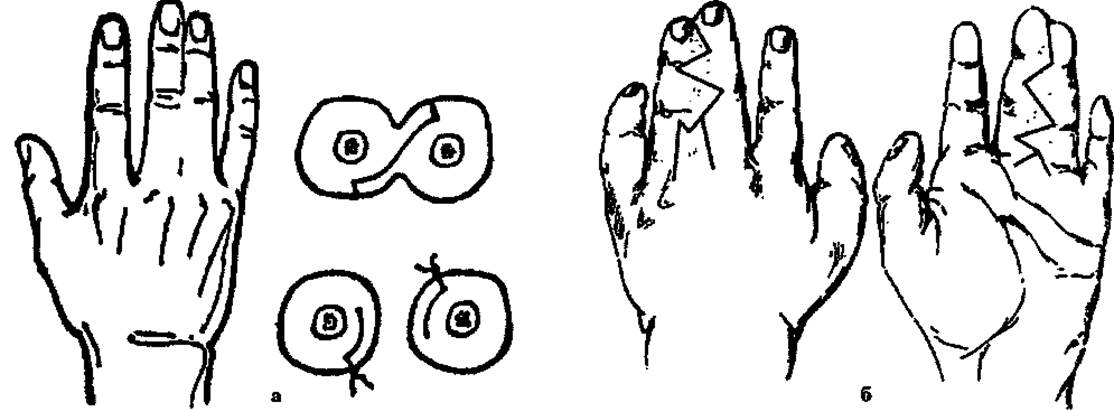
Fig. 124. Schematic of surgical techniques for syndactyly: a – Dèdo incision, b – zig-zag incision.
Thus, as we can see, the core issue lies in the skin deficiency at the site of syndactyly.
When parents sought medical help, we suggested having them gently stretch the skin of the web space by manually spreading the fingers apart as a preoperative preparation. In some cases, this helped gain a certain skin reserve, facilitating subsequent wound closure.
An original method of preoperative preparation was proposed by O. N. Gudushauri and L. O. Tvaliashvili—gradual Separation of the fused digits using a distraction device. This technique not only gradually stretches the skin but also maintains the fingers at the achieved distance. Only after obtaining a sufficient skin excess are the fingers separated using Dide's method.
It is crucial in all such procedures to reconstruct the base of the interdigital commissure. This is most commonly achieved via Zeller's method, which involves transplanting two opposing triangular skin flaps or a single flap. Naturally, any resulting skin defect must be covered with a full-thickness skin autograft, and a meshed graft if necessary.
To reliably reconstruct the base of the web space in syndactyly with a single hypoplastic digit, V. V. Demchenko and E. A. Padabed (1986) harvest a shaped pedicle musculocutaneous (or fasciocutaneous) flap from the dorsal aspect of the hand. This flap covers the base of the web space while simultaneously releasing the fingers and enveloping the underdeveloped digit.
Patients with bony symphalangism associated with syndactyly undergo single- or two-stage surgical correction. Previously, single-stage operations were performed, during which the fused distal phalanges were separated, and linear incisions were made along the volar and dorsal aspects of the interdigital region over the entire length of the fingers, as well as between the two nail plates. If there is a single broad nail, it is split in half. Afterward, a resection knife or osteotome is used to divide the osseous fusion of the phalanges. The wound surface and exposed bone are then covered with a full-thickness skin autograft. A plaster cast is applied to immobilize the hand until the wound heals and the graft engrafts.
A two-stage procedure involves separating the bony fusion of the distal phalanges in The First stage, effectively converting bony syndactyly into cutaneous syndactyly. For this purpose, a small incision is made on the dorsal aspect of the distal phalanx only, splitting the nail and the bony bridge. A distraction device is immediately applied to gradually spread the fingers apart, stretching the skin and preventing the phalanges from fusing again. Following progressive stretching and The formation of a sufficient skin reserve, the Second Stage of the surgery is performed—finger separation, as described above.
Rehabilitation treatment is administered during the postoperative period. Complex syndactyly requires thoughtful surgical planning. Additional digits and amniotic bands must be addressed and removed first. Subsequently, the surgical strategy most appropriate for each individual patient is applied. A technically well-executed operation yields excellent functional and cosmetic outcomes.
At what age is it most appropriate to operate on patients with syndactyly? Potel, Ombredanne, and other authors believed that surgery should not be rushed and did not recommend operating on children under 5–6 years of age. Having encountered unsatisfactory results following early surgeries, Janelidze suggested postponing operations until the patient reached 18–20 years of age, whereas Schwartz and Zatsepin believed that surgery should be performed during adolescence. According to E. Osten-Sacken, the optimal timing depends on the form of syndactyly. Amniotic hand deformities must be operated on during the child's first or second year of life. Minor surgical interventions at this stage successfully eliminate obstacles that impair the normal development of hypoplastic and deformed fingers. In cases of simple cutaneous syndactyly, where two or more digits are enclosed within a common skin sheath, Osten-Sacken recommended waiting until adulthood before performing surgery.
It is now widely accepted that the earlier a child undergoes surgery to separate the fused digits, the better the functional and cosmetic outcomes will be, as fingers growing under normal functional conditions will develop normally.
Many surgeons perform operations on children between the ages of 6 months and 1.5 years (Demchenko V.V., Padabed E.A., 1986; Godunova G.S. et al., 1986, etc.).
We believe that the timing of surgery depends on The complexity of the syndactyly. Simple membranous syndactyly should be operated on During the first year of life, whereas cutaneous syndactyly is best treated in the 2nd–3rd year. If the syndactyly involves three or more digits, the procedure is performed in two or more stages with an interval of 3–6 months between surgeries.
Last update: 10/08/2026
Editorial and Educational Adaptation: This material has been compiled based on the primary/original source text. The project team performed an editorial review, corrected technical inaccuracies, structured sections, and adapted the content for an educational format.
What was processed:
- elimination of formatting defects (OCR errors, structural breaks, corrupted characters);
- editorial organization of content;
- standardization of terminology in accordance with academic sources;
- verification of factual statements against the original source text.
All mentions of the author, publication year, and origin of the primary text have been preserved in accordance with the source.