Cytology, General Histology and Embryology - V. K. Napkhanyuk 2002
Fundamentals of General Embryology
Embryogenesis
Embryogenesis is the period of INTRAUTERINE DEVELOPMENT OF the Human and Animal embryo, which begins at the moment of Fertilization, is accompanied by the formation and development of all Tissues, Organs, Organ Systems, and a viable fetus, and ends with the birth of a fully formed individual.
Embryonic development occurs in stages, involving gradual Qualitative and quantitative changes; therefore, several successive stages are distinguished in The process of embryogenesis:
— fertilization;
— Cleavage and blastocyst formation;
— Gastrulation and germ layer differentiation;
— Formation of tissue primordia (histogenesis);
— formation of organs (Organogenesis);
— Formation of the fetal organ systems (systemogenesis).
Fertilization (fertilisatio) is the process of fusion of male and female Gametes, resulting in the restoration of the diploid chromosome set characteristic of each animal species and The formation of a single-celled embryo — the zygote.
Fertilization is preceded by The entry of seminal fluid into the reproductive tract (in internal fertilization) or into the medium where the egg Cell is located (in external fertilization).
Three phases are distinguished in the fertilization process:
— distant interaction between the spermatozoon and the egg cell, ensured by a combination of non-specific factors that increase the probability of gamete encounter;
— contact interaction and penetration of the spermatozoon into the egg cell, mediated by the acrosome (acrosomal reaction). During this process, the Enzymes hyaluronidase and proteases dissolve the contacts between the follicular Cells of the zona pellucida;
— penetration of the spermatozoon into the egg cell, followed by the formation of male and female pronuclei and the subsequent formation of the zygote.
Cleavage (fissio) is the successive mitotic division of the zygote into blastomeres, transforming it into a multicellular embryo — the blastula.
During cleavage, cell size progressively decreases. Each animal Class exhibits a specific type of cleavage, determined by the amount and distribution pattern of yolk in the egg cell. Yolk inhibits the cleavage process; therefore, the portion of the zygote containing yolk divides more slowly or does not divide at all.
As a result of cleavage, a blastula is formed. A cavity — the blastocoel, or primary body cavity of the embryo — forms in the center of the blastula. Depending on the type of cleavage, Various Forms of blastulae are produced (Figs. 23, 24).

Fig. 23. Types of cleavage and blastulae (after N. A. Yurina)

Fig. 24. Various Histology/18.html">Types of Blastulae:
1 — coeloblastula; 2 — amphiblastula; 3 — discoblastula; 4 — periblastula; 5 — blastocyst
Gastrulation is the period of embryogenesis during which, as a result of complex biochemical and morphogenetic changes accompanied by cell proliferation, growth, directed migration, differentiation, and interaction, the germ layers are formed: the outer (ectoderm), middle (mesoderm), and inner (endoderm), giving the embryo a triploblastic Structure.
The germ layers serve as the source for The Development of Tissue and organ primordia.
The redistribution of cellular material during gastrulation can occur via several mechanisms, which depend on preceding developmental stages and the degree of yolk accumulation in the egg cell. Accordingly, several types of gastrulation are distinguished (Fig. 25):
— immigration (immigratio) — migration of a group of blastomeres into the interior of the embryo, resulting in the formation of an additional layer of blastomeres, known as the germ layer;
— invagination (invaginatio) — the process by which a portion of the wall (floor) folds inward into the interior of the blastula;
— epiboly (epibolia) — the process whereby rapidly dividing cells of one region of the blastula wall overgrow other regions where Cell Division proceeds more slowly. This type of gastrulation typically occurs when the blastomeres of the vegetal pole are heavily laden with yolk, divide slowly, and are unable to invaginate;
— delamination (delaminatio) — a splitting process characterized by the tangential division of embryonic material, leading to the formation of two cell layers—the two primary germ layers.
In vertebrates, gastrulation generally involves a combination of two or three of the aforementioned types.
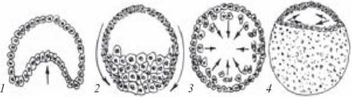
Fig. 25. Various types of gastrulation:
1 — invagination; 2 — epiboly; 3 — immigration; 4 — delamination
Gastrulation in birds, which exhibit meroblastic cleavage and a discoidal blastula, relies on two primary mechanisms: delamination and immigration. Delamination gives rise to the primary outer layer (epiblast) and the primary inner layer (hypoblast).
As a result of immigration, the material of the primary outer layer at the margins of the embryonic disc shifts in a caudal direction. Where the two streams of cells meet, an elongated cellular aggregation forms—the primitive streak, which terminates at its anterior end in a dense primitive (Hensen's) node. A cellular cord, the notochord, grows inward from the primitive node between the inner and outer germ layers, while two mesodermal layers develop from the material of the primitive streak on either side.
Gastrulation in mammals and humans also proceeds via delamination and immigration.
Slides for Study
Slide 7. Cross-section of a chick embryo at 26–38 hours of incubation (Fig. 26).
Low power. Examine the slide. In the center of the embryo, a dense, rounded cord—the notochord—is visible. The neural tube lies dorsal to the notochord. Externally, the embryo is covered by the secondary or cutaneous ectoderm. It is separated from the yolk by the inner germ layer, the entoderm (endoderm). Located between the ectoderm and the entoderm is the mesoderm, which is differentiated into somites (segmented dorsal mesoderm), nephrotomes or intermediate cell masses (intermediate mesoderm), and splanchnotomes (ventral mesoderm), between whose layers (parietal and visceral) lies the coelom (secondary body cavity).
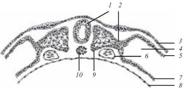
Fig. 26. Cross-section of a chick embryo at 26–38 hours of incubation. Stained with iron hematoxylin. Objective ×8, eyepiece ×5:
1 — neural tube; 2 — nephrotome (segmental stalk); 3 — ectoderm; 4 — coelomic cavity; 5 — parietal layer of the splanchnotome; 6 — Heart tube; 7 — visceral layer of the splanchnotome; 8 — entoderm (endoderm); 9 — somite; 10 — notochord
Last update: 10/08/2026
Editorial and Educational Adaptation: This material has been compiled based on the primary/original source text. The project team performed an editorial review, corrected technical inaccuracies, structured sections, and adapted the content for an educational format.
What was processed:
- elimination of formatting defects (OCR errors, structural breaks, corrupted characters);
- editorial organization of content;
- standardization of terminology in accordance with academic sources;
- verification of factual statements against the original source text.
All mentions of the author, publication year, and origin of the primary text have been preserved in accordance with the source.