Molecular Biology of the Cell - Volume 3 - Alberts B., Bray D., Lewis J., Raff M., Roberts K., Watson J. 1994
From Cells to Multicellular Organisms
Cellular Mechanisms of Development
Organogenesis: The Coordinated Assembly of Complex Tissues
Up to this point, we have considered the establishment of spatial Organization primarily in terms of changes in Cell properties while paying relatively little attention to cell position; we traced the accumulation of these changes within individual Tissues, specifically examining the epidermis in *Drosophila* and the skeletal Connective Tissue in vertebrate limbs. In reality, however, due to complex morphogenetic movements during Embryogenesis, all Organs are intricate composites of diverse tissues made up of Cells of different origins that follow distinct rules. Cell movements bring all these components together. In this final section, we will examine how positional information directs cell movements and coordinates the construction of complex organs such as limbs.
Class="center">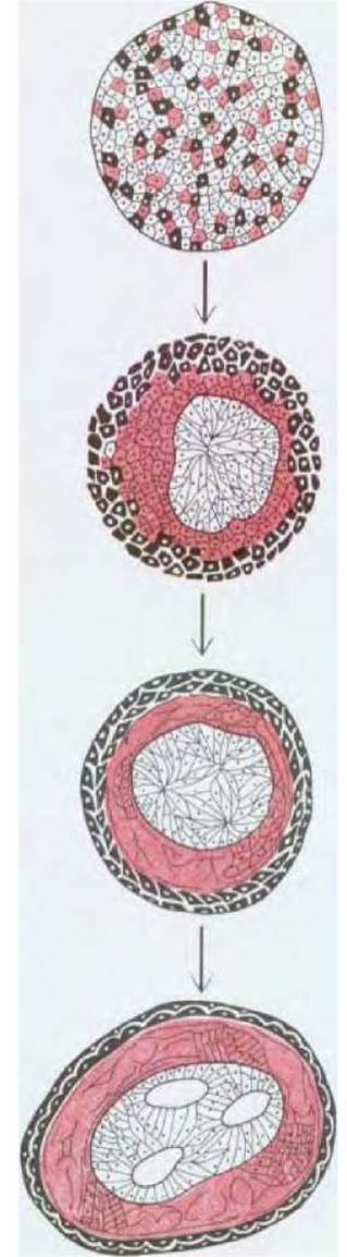
Fig. 16-77. Cells from various Regions of the early amphibian embryo sort themselves out according to their origin. In this classic experiment, shown schematically here, the mesoderm, neural plate, and epidermal cells were dissociated. Upon reaggregation, the cells formed a Structure resembling a normal embryo, with a neural tube on the inside, epidermis on the outside, and mesoderm in between. (P. L. Townes, J. Holtfreter, J. Exp. Zool., 128, 53-120, 1955; modified.)
16.6.1. Selective adhesion stabilizes tissue structures formed by differently determined cells [65, 66]
During animal development, cells move, proliferate, and come into contact with one another, resulting in the intermingling of cell lineages. In chimeric mouse embryos, for instance, cells from two original morulae become mixed, so that the adult Organism contains a haphazard mixture of cells of two genotypes (see Section 16.2.5). However, if cell displacement occurs after determination, it can lead to disruptions in the spatial distribution of cells with different differentiation states. The acquisition of properties dictated by cell position prevents random cell mixing. The compartment phenomenon in *Drosophila* (see Fig. 16.5.15) illustrates one way this is achieved. Presumably, determined cells remain within their respective compartments through selective adhesion to Cells of the same determination state. Cells expressing identical molecular "addresses" likely adhere to one another more strongly than to cells with different molecular addresses. Apparently, the positional information encoded by the activation of genes such as *engrailed* and *Urabithorax* is manifested through the selective deployment of cell-surface adhesion molecules.
In early amphibian embryos, the stabilizing effects of intercellular adhesion on tissue organization—ensuring the proper spatial arrangement of different cell types—are likely strong enough to restore normal architecture even after artificial cell dissociation yields a shapeless, disordered mass. Mesodermal, neural plate, and epidermal cells can sort themselves out from such a mixture to form structured configurations with the epidermis on the outside, the mesoderm beneath it, and a neural tube-like structure on the inside (Fig. 16-77). Studies on chick and mouse embryos suggest that this behavior is determined, at least in part, by a family of Ca2+-dependent cell-adhesion Glycoproteins known as cadherins (see Section 14.3.7). These and other Ca2+-dependent cell-adhesion molecules, such as N-CAM (see Section 14.3.6), are differentially expressed in various Tissues of the early embryo, and Antibodies against these molecules disrupt normal selective adhesion among cells of the same type.
16.6.2. Spatial patterns formed by cell-adhesion molecules regulate the character of morphogenetic movements [66, 67]
Changes in the expression patterns of various cadherins correlate closely with shifts in cell association during Gastrulation, neurulation, and somite formation (Fig. 16-78); such changes in early embryogenesis can be regulated by—and at least partially depend upon—cadherin distribution. In particular, cadherins likely play a crucial role in controlling the formation and dissolution of epithelial layers and cell clusters. Consequently, the movements involved in building the early embryo are governed by chemical differences among cells located in different positions.
This general principle is illustrated by numerous other Examples. During The formation of the adult wing, the *Drosophila* imaginal disc epithelium must flatten, elongate, and fold in a specific manner. Although this process remains largely enigmatic, it is presumably driven by the localized expression of specific cell-surface adhesion molecules, which are now being identified using Monoclonal Antibodies. The distribution of such position-specific cell-surface molecules correlates closely with the folding pattern of the disc during metamorphosis (Fig. 16-79); some of these molecules have been shown to belong to a family of cell-surface receptors called Integrins (Section 14.2.17). Unlike cadherins, integrins mediate cell attachment to Extracellular matrix components. As noted previously, such cell-matrix interactions play a vital role in gastrulation (see Section 16.1.5). Intercellular adhesion molecules, as well as cell-surface and matrix adhesion molecules, can be viewed as Two Types of key machinery for translating positional information into the Spatial Organization of morphogenetic movements.
16.6.3. In vertebrates, the mesoderm is the primary bearer of positional information [68]
It is quite evident that the positional information possessed by cells can manifest in various ways. This is especially well illustrated by The Development of complex organs. A vertebrate limb is formed by six different tissue types that make up connective tissue (bone, tendons, etc.), epidermis, Muscle, Blood vessel linings, nerve cell axons and their glial sheaths, pigment cells, and the hematopoietic tissue of the Bone Marrow. Although all these components possess their own well-defined structure, they make distinct contributions to the limb as a whole. In flies, which are small, the surface-to-volume ratio is very high, and the ectoderm-derived epidermis plays a dominant role both in forming the mechanical exoskeleton and in coordinating spatial pattern formation. In vertebrates, which are substantially larger than insects, the roles of the germ layers are modified. The structural framework of the body is formed by Connective Tissues (derived primarily from the mesoderm); as we shall see, these tissues also play a coordinating role in spatial organization during organogenesis. Indeed, experiments on amphibians early this century demonstrated that the mesoderm plays a critical role in establishing spatial organization at the very earliest stages of body plan formation.
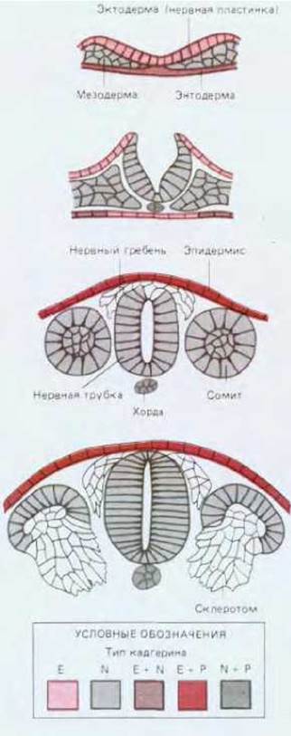
Fig. 16-79. A *Drosophila* imaginal disc treated with monoclonal antibodies recognizing the position-specific antigen PS2. The sharp boundary of the stained region corresponds to the future wing margin, where the dorsal and ventral wing epithelia meet to form a fold. The dashed line marks the compartment boundary along the anteroposterior axis. (V. C. Wilcox, D. L. Bower, R. J. Smith, Cell, 25, 159-164, 1981.)
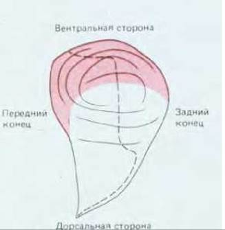
Fig. 16-78. Changes in the expression patterns of three cadherins at several successive stages of early chick or mouse embryo development, shown in transverse sections through the developing neural tube and somites. Spatially restricted cadherins may help regulate the morphogenetic movements involved in forming the neural tube, notochord, somites, neural crest, and sclerotomes. (M. Takeichi, Trends in Genetics, 8, 213-217, 1987.)
As already mentioned (see Sections 16.1.7–16.1.9), the vertebrate Nervous system develops from the ectoderm under the inductive Influence of the underlying mesoderm. If a piece of mesoderm lying directly beneath the prospective neural tube region of one gastrulating amphibian embryo is transplanted under the ectoderm on the ventral side of another embryo, the overlying ectoderm will thicken and fold, forming a segment of neural tube at this ectopic site. The Specific characteristics of this segment will depend on the Water/144.html">Origin of the mesoderm. If the mesoderm was taken from an anterior region, Forebrain structures will form from the ectoderm; mesoderm from a posterior region will induce the formation of a Spinal Cord segment. This suggests that ectodermal cells acquire specific positional values depending on the positional values of the underlying mesodermal cells.
The regulation of spatial organization by underlying connective tissue is observed during the development of many vertebrate organs. For example, the Skin and gut, along with all their glands, appendages, and local specializations, acquire their characteristic architectures through specific interactions in which connective tissue components provide the apposed epithelium with appropriate positional information. Skin covering the limbs serves as an excellent model for studying these phenomena.
16.6.4. The pattern and distribution of epidermal derivatives are controlled by the dermis [66, 69]
Skin consists of two layers: the epidermis (an ectoderm-derived epithelium) and the dermis (a connective tissue composed of fibroblasts largely of mesodermal origin). The epidermis gives rise to keratinized Skin Appendages (Hair, feathers, scales, and claws) as well as numerous glands. Different body regions feature distinct types of keratinized derivatives: the back, wings, and upper legs of a chicken, for instance, bear rows of feathers (Fig. 16-80), whereas the lower legs bear rows of scales. Furthermore, depending on their position within each row, feathers and scales can vary in shape and color. If the epidermis from the leg of a chick embryo—where it normally forms scales—is combined with back dermis—where it normally forms feathers—it will subsequently develop feathers instead of scales (Fig. 16-81); the reciprocal combination yields the opposite result. In general, the dermis controls not only the type of epidermal derivative but also its precise Location. Here we encounter another manifestation of non-equivalence (see Section 16.4.6): the dermis from different body regions appears morphologically identical yet differs in its ability to induce specific differentiation in the overlying epithelium.
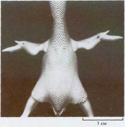
Fig. 16-80. Arrangement of feather germs on the back of a 9-day-incubated chick embryo. Note that the primordia in each row are evenly spaced. (Courtesy of A. Mauger and P. Sengel.)
The molecular mechanisms by which connective tissue controls epithelial differentiation remain unclear, but some success has been achieved in identifying the molecules that govern the morphogenetic movements of cells giving rise to hairs, feathers, or glands. Here too, intercellular adhesion types play a critical role, as does cell-matrix adhesion. Within the connective tissue component, mechanical forces exerted on Collagen secreted by fibroblasts lead to the aggregation of these cells at sites of appendage formation. Simultaneously, changes in the expression patterns of cell-adhesion molecules such as N-CAM and E-cadherin are observed in both the connective tissue and the overlying epithelium, likely regulating cell shape and mutual packing (see Sections 14.3.6 and 14.3.7). At the epithelial-mesenchymal interface, various basement membrane components (see Section 14.2.15)—including Laminin, Proteoglycans, and collagen—are synthesized and degraded, with both Synthesis and Breakdown being tightly controlled to facilitate epithelial outgrowth and folding.
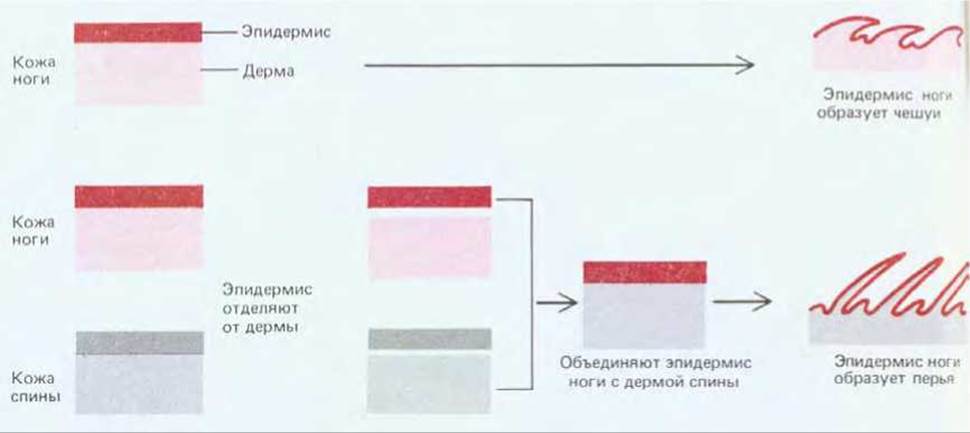
Fig. 16-81. Scheme of experiments demonstrating that the type of skin derivative is determined by the dermis.
16.6.5. Many types of migrating cells colonize the vertebrate limb connective tissue [11, 70]
Connective tissue permeates the entire vertebrate body. In the limbs, connective tissue forms bones and Cartilage, tendons and ligaments, skin, muscle sheaths, the outer layers of blood vessel walls, nerve sheaths, and the interstitial tissue that binds all these components together. These forms of connective tissue are composed of fibroblasts and closely related cells embedded in an extracellular matrix enriched with collagen that they secrete. All of these diverse cells develop from mesenchyme, an uncommitted embryonic tissue filling the limb bud; its origin can be traced back to the lateral plate mesoderm adjacent to the somites of early embryos (see Fig. 16-15). With the exception of the overlying epidermis, all Other components of the limb derive from a population of migrating cells that originate outside the lateral plate. Before reaching their destination and contributing to The structure of the adult animal, these cells must undertake a long journey through the embryonic connective tissue.
Such migration was demonstrated in experiments involving the transplantation of embryonic quail cells into chicken embryos. Although the quail closely resembles the chicken, its cells are easily distinguished in histological preparations because they contain a large, densely staining block of heterochromatin associated with the nucleolus. This nucleolar marker makes it easy to identify the transplanted cells regardless of where they end up within the embryo. If the tissue of a specific group of somites in a chicken embryo is replaced with equivalent tissue from a quail prior to wing bud formation, all the muscle cells of the wings (and only them) will originate from the quail (Fig. 16-82). Evidently, future muscle cells migrate into the limb bud region and remain there, outwardly indistinguishable from other cells but already determined, until the time of their differentiation comes.
Another important example of migrating cells is provided by cells originating from the neural crest, a region located near the neural tube (Fig. 16-83) (see Section 16.11.9). The migratory capacity of these cells was demonstrated in a similar manner by replacing the neural crest in a chick with that of a quail, with the quail neural crest cells being identified by the nucleolar marker. Like glial cells, which form the myelin sheaths of nerve cell axons, pigment cells of the limb derive from neural crest cells. Sensory and autonomic axons of the limbs are outgrowths of Neurons that arose from neural crest cells. (Voluntary motor axons, by contrast, are outgrowths of spinal cord motor neurons).
16.6.6. The Spatial Organization of Limb Connective Tissue Is Independent of the Cells Populating It [71]
If the tissue of the somites, neural crest, and/or neural tube is destroyed in an early embryo before the onset of limb bud formation, limbs lacking specific classes of migrating cells or nerve fibers can readily be obtained. In general, such limbs are quite normal in many respects. For example, one can obtain limbs devoid of muscle cells, yet they possess a normal Skeleton, skin, and sensory nerves; even tendons develop normally, although they subsequently degenerate gradually in the absence of the Muscles that pull on them. Similarly, a limb devoid of nerves and all neural crest derivatives develops a normal skeleton, skin, and muscles, but the muscles, lacking neural stimulation, subsequently degenerate gradually. The internal structures of the limb will not develop normally in the absence of the entire epidermal covering, but partial removal of fairly substantial areas of the epidermis does not disrupt normal development.
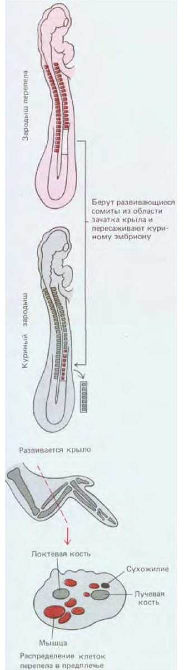
Fig. 16-82. If the somite cells of a chicken embryo are replaced with equivalent quail somite cells after two days of incubation, and histological preparations of the wing region are made a week later, it turns out that the wing muscle cells formed from the transplanted quail somites.
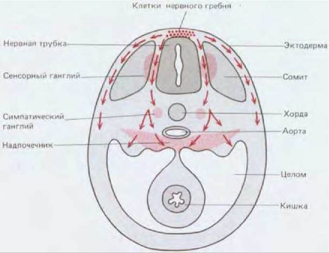
Fig. 16-83. Main migration pathways of neural crest cells in a chicken embryo (schematic transverse section of the mid-body). Cells migrating directly beneath the ectoderm (superficial pathway) give rise to pigment cells of the skin; cells moving along the deep pathway through the somites give rise to sensory and sympathetic ganglia, and in part to the Adrenal Glands (see also Fig. 14-55).
Thus, the spatial organization of connective tissue is virtually independent of other limb components. Moreover, these other limb components possess their own spatial organization, the establishment of which is largely determined by the connective tissue. We have already discussed how this occurs using the epidermis as an example (see Fig. 16-81); using analogous tissue transplantation experiments, it can be demonstrated that the same holds true for migrating cell populations such as muscle cells, nerve cell axons, and pigment cells. Cells of all these types generally form structures that conform to the host limb, regardless of which region of the embryo they originate from. If connective tissue as such acts as a kind of central source and repository of positional information for the vertebrate limb, how does it exert its organizing influence on migrating cells?
16.6.7. Connective Tissue Determines the Migration Pathways and Final Destination of Migrating Cells [11, 72]
In principle, There are five main ways by which connective tissue controls the populations of cells populating it. It can determine:
1) the pathways along which cells move;
2) the sites where migrating cells arrive;
3) the extent of proliferation;
4) the mode of differentiation;
5) the probability of survival.
The Importance of each of these control mechanisms varies depending on the type of migrating cell, but none of them has been studied in sufficient detail. In fact, virtually nothing is known about the Molecular Basis of the connective tissue positional value system upon which its spatial organization is founded. Currently, monoclonal antibodies are just beginning to be employed to reveal position-dependent differences in the distribution of cell surface molecules.
However, certain General Principles of cell migration are beginning to come to light. The behavior of all migrating cells is governed by the MECHANISMS OF CELL adhesion and recognition discussed in Chapter 14; these mechanisms serve as fundamental prerequisites for normal organogenesis. In particular, migrating cells must establish close contact with the extracellular matrix or The surface of other cells. Fibronectin is likely the most crucial (though not the only) substrate component for many migrating cells; antibodies and Peptides that block cell surface fibronectin receptors are capable of disrupting neural crest cell migration in numerous regions of the embryo, as, for example, blocking cell migration during gastrulation (see Section 16.1.5). The universality of certain mechanisms involved in guiding migrating cells to their destinations becomes clearer from studies of mutant mice carrying Steel or dominant-spotting Mutations. In such mutants, pigment, hematopoietic, and primordial Germ Cells fail to reach their destinations in the skin, bone marrow, and Gonads, respectively. The dominant-spotting mutation results in a defect intrinsic to the migrating cells themselves, whereas Steel mutants possess defects in the connective tissue where the migrating cells take up residence.
We are only beginning to comprehend the mechanisms by which limb connective tissue regulates cell migration along specific pathways or, in other words, directs them to specific addresses, but it can be stated with certainty that this guidance is determined by positional information inherent in the connective tissue cells. For instance, the surface of cells with different positional values may possess different properties or secrete different extracellular matrix components. As a cell moves through connective tissue, it continuously extends protrusions, using them to sample the immediate environment and evaluate subtle signals to which these cells are uniquely sensitive through the specific Selection of surface receptor Proteins. Within The Cell, these receptor proteins are linked to the Cytoskeleton, which drives its movement. Protrusions formed at different regions of the cell surface are, as it were, in a constant state of "tug-of-war," resulting in cell translocation toward the direction of strongest substrate adhesion (see Section 11.6.4) until the cell reaches a region where adhesive forces are balanced or so strong that the cell cannot detach from the surface. Chemotaxis plays an important role in this movement, as does the interaction among migrating cells (see Section 14.3); these processes can lead either to the arrest and aggregation of cells in a single area or to their widespread dispersion through mutual repulsion.
16.6.8. The Study of Nervous System Development Poses A number of Special Problems [73]
Our Discussion of cell migration has led us to a topic that has not yet been considered in general: the development of The Nervous System. This process appears to be one of the most enigmatic. The fundamental questions of developmental biology examined in this chapter can be summarized as follows: how different cell types arise in the organism, and how these cells reach their appropriate locations. The nervous system presents an additional problem: how correct connections are formed between cells. Most other cell types can be regarded as point-like objects, each occupying a specific position and possessing specific intrinsic properties. A neuron, however, is not inherently a point-like object: it is enormously extended by a long axon and dendrites that connect it to other cells. If these connections are erroneous, the functioning of the nervous system will be impaired. The processes by which various neuronal types arise and their cell bodies are packed into a regular structure can be explained based on the same principles as for other cells. But the ordered growth of axons and dendrites and the formation of a correct synaptic system represent phenomena of a different order. The leading tip of a growing axon or dendrite crawls in much the same way as a migrating cell: it can be called the migrating organ of a stationary cell. The factors controlling its movement are in some ways the same as those controlling cell movement; these are specific types of adhesion, and so forth. However, as we study how axons relate to other cells, interact with other nerve endings, and acquire The ability to form synapses, new problems arise that require a different approach. Therefore, we defer our Overview of nervous system organization (the driving force of development) to Chapter 19.
The positional values acquired by cells during the spatial ORGANIZATION OF THE embryo are expressed through the adhesive properties of their surface as well as their internal biochemistry. Cells of the same type tend to interact with one another and segregate from different, distinct cells; in this way, spatial organization is stabilized, and cells retain the capacity for spontaneous sorting when artificially mixed. Changes in the pattern of adhesive properties underlie morphogenetic processes such as gastrulation, neurulation, and somite formation. Because the positional value characteristics of a given cell class are manifested through alterations in cell Surface Properties, they can guide the migration of other embryonic cell populations during the assembly of complex tissues or organs. In vertebrates, connective tissue cells are likely the primary bearers of positional information. Dermal connective tissue cells of the skin are capable of controlling the regional specialization of the epidermis, which forms feathers and scales. Similarly, limb connective tissue cells control and coordinate the formation of structures built by migrating cell populations, which include muscle cells (somite derivatives), nerve cell axons (from the Central Nervous System and peripheral ganglia), and pigment cells (neural crest derivatives). Although many general-purpose Cell Adhesion molecules have now been identified, and some have been shown to play central roles in these processes, the molecular mechanisms directing cell migration along specific routes to precise destinations within the limbs remain unknown.
General
Browder L. Developmental Biology, 2nd ed. Philadelphia, Saunders, 1984. Gilbert S.F. Developmental Biology, 2nd ed. Sunderland, MA, Sinauer, 1988. Molecular Biology of Development. Cold Spring Harbor Symp. 50, 1985. Slack J. M. W. From Egg to Embryo: Determinative Events in Early Development.
Cambridge, U.K., Cambridge University Press, 1983. Spemann H. Embryonic Development and Induction. New Haven, Yale University Press, 1938. (Reprinted, New York, Garland, 1988.)
Walbot V. Holder N. Developmental Biology. New York, Random House, 1987.
Weiss P. A. Principles of Development. New York, Holt, 1939.
Cited
1. Browder L. W. ed. Developmental Biology: A Comprehensive Synthesis. Vol. 2: The Cellular Basis of Morphogenesis. New York, Plenum, 1986.
Gerhart J., et al. Amphibian early development. Bioscience, 36, 541-549, 1986.
Slack J. M. W., ed. Early Amphibian Development. J. Embryol. Exp. Morphol., Suppl., 89, 1985.
Trinkaus J. P. Cells into Organs: The Forces that Shape the Embryo, 2nd ed., Englewood Cliffs, NJ, Prentice Hall, 1984.
2. Gerhart J. C. Mechanisms regulating pattern formation in the amphibian egg and early embryo. In Biological Regulation and Development (R. F. Goldberger, ed.), Vol. 2, pp. 133-316, New York, Plenum, 1980.
Gerhart J., Ubbels G., Black S., Hara K., Kirschner M. A reinvestigation of The Role of the grey crescent in axis formation of Xenopus laevis. Nature, 292, 511-516, 1981.
Vincent J. P., Oster G. F., Gerhart J. C. Kinematics of gray crescent formation in Xenopus eggs: the displacement of subcortical Cytoplasm relative to the egg surface. Dev. Biol. 113, 484-500, 1986.
3. Gilbert S. F. Developmental Biology, 2nd ed., pp. 73-111, Sunderland, MA, Sinauer, 1988.
Kirschner M., Newport J. Gerhart J. The timing of early developmental events in Xenopus. Trends Genet., 1, 41-47, 1985.
Wilson E.B. The Cell in Development and Heredity, 3rd ed., pp. 980-1034. New York, Macmillan, 1925. (Reprinted, New York, Garland, 1987.)
4. Furshpan E. J., Potter D. D. Low-resistance junctions between cells in embryos and tissue culture. Curr. Top. Dev. Biol., 3, 95-128, 1968.
Kalt M. R., The relationship between Cleavage and blastocoel formation in Xenopus laevis. II. Electron microscopic observations. J. Embryol. Exp. Morphol., 26, 51-66, 1971.
Warner A. The role of Gap Junctions in amphibian development. J. Embryol. Exp. Morphol., Suppl., 81, 365-380, 1985.
5. Fink R. D., McClay D. R. Three Cell Recognition changes accompany the ingression of sea urchin primary mesenchyme cells. Dev. Biol., 107, 66
74, 1985.
Gustafson Т., Wolpert L. Cellular movement and contact in sea urchin morphogenesis. Biol. Rev., 42, 442-498, 1967.
Hardin J. D., Cheng L. Y. The mechanisms and mechanics of archenteron elongation during sea urchin gastrulation. Dev. Biol., 115, 490-501, 1986. McClay D.R., Wesel G.M. The surface of the sea urchin embryo at gastrulation: a molecular mosaic. Trends Genet., 1, 12-20, 1985.
Wilt F. H. Determination and morphogenesis in the sea urchin embryo. Development, 100, 559-575, 1987.
6. Ettensohn C.A. Mechanisms of epithelial invagination. Q. Rev. Biol., 60, 289-307, 1985.
McClay D. R., Ettensohn C. A. Cell adhesion in morphogenesis. Annu. Rev. Cell. Biol., 3, 319-345, 1987.
Odell G. M., Oster G., Alberch P., Bernside B. The mechanical basis of morphogenesis. I. Epithelial folding and invagination. Dev. Biol., 85, 446462, 1981.
7. Gerhart J., Keller R. Region-specific cell activities in amphibian gastrulation. Annu. Rev. Cell. Biol., 2, 201-229, 1986.
8. Spemann H., Mangold H. Induction of embryonic primordia by implantation of organizers from different species. Roux's Archiv., 100, 599-638, 1924. (English Translation in Foundations of Experimental Embryology, 2nd ed. [B. H. Willier, J. M. Oppenheimer, eds.] New York, Hafner, 1974.)
9. Balinsky B.L Introduction to Embryology, 5th ed., Philadelphia, Saunders, 1981.
Longman J. Medical Embryology, 5th ed. Baltimore, Williams & Wilkins, 1985.
Romer A. S., Parsons T. S. The Vertebrate Body, 6th ed. Philadelphia, Saunders, 1986.
10. Kitchin L. C. The effects of notochordectomy in Amblystotna mexicanum. J. Exp. Zool, 112, 393-411, 1949.
Smith J. С., Watt F. W. Biochemical Specificity of Xenopus notochord. Differentiation, 29, 109-115, 1985.
11. Le Douarin N. The Neural Crest. Cambridge U.K., Cambridge University Press, 1982.
Newgreen D. F., Erickson C. A. The migration of neural crest cells. Int. Rev. Cytol, 103, 89-145, 1986.
12. Bumside B. Microtubules and microfilaments in amphibian neurulation. Am. Zool,
13. 989-1006, 1973. Gordon R. A review of the theories of vertebrate neurulation and their relationship to the mechanics of neural tube birth defects. J. Embryol. Exp. Morphol., Suppl., 89, 229-255, 1985.
Karfunkel P. The mechanisms of neural tube formation. Int. Rev. Cytol., 38, 245-271, 1974.
13. Blackhaw S.E., Warner A. E. Low resistance junctions between mesoderm cells during development of trunk muscles. J. Physiol., 255, 209-230, 1976.
Keynes R. J., Stern C. D. Mechanisms of vertebrate segmentation. Development, 103, 413-429, 1988.
14. Slack J.M. W. From Egg to Embryo: Determinative Events in Early Development. Cambridge, U.K., Cambridge University Press, 1983.
15. DiBerardino M.A. Orr N.H., McKinnel R.G. Feeding tadpoles cloned from Ram erythrocyte nuclei. Proc. Nat. Acad. Sci. USA., 83, 8231-8234, 1986.
Gurdon J. B. The Control of Gene Expression in Animal Development. Cambridge, Harvard University Press, 1974.
Gurdon J. B. Transplanted nuclei and Cell Differentiation. Sci. Am., 219(6), 24-35, 1968.
McKinnel R. G. Cloning-Nuclear Transplantation in Amphibia. Minneapolis, University of Minnesota Press, 1978.
16. Davidson E.H. Gene Activity in Early Development, 3rd ed., pp. 411-524, Orlando, FL, Academic Press, 1986.
Jeffery W.R. Spatial distribution of mRNA in the cytoskeletal framework of Ascidian eggs. Dev. Biol., 103, 482-492, 1984.
Satoh N. Towards a molecular understanding of differentiation mechanisms in Ascidian embryos. Bioessays, 7, 51-56, 1987.
Wilson E.B. The Cell in Development and Heredity, 3rd ed., pp. 1035-1121, New York, Macmillan, 1928. (Reprinted, New York: Garland. 1987.)
17. Gurdon J. B. Embryonic induction-molecular Prospects. Development, 99, 285-306, 1987.
Kimelman D., Kirschner M. Synergistic induction of mesoderm by FGF and TGF-beta and the identification of an mRNA coding for FGF in the early Xenopus embryo. Cell, 51, 869-877, 1987.
Rosa F., et al. Mesoderm induction in amphibians: the role of TGF-beta2-like factors. Science, 239, 783-785, 1988.
Slack J. M. W, Darlington B. G., Heath J. K., Godsave S. F. Mesoderm induction in early Xenopus embryos by heparin-binding growth factors. Nature, 326, 197-200, 1987.
Weeks D. L., Melton D. A. A maternal mRNA localized to the vegetal hemisphere in Xenopus eggs codes for a growth factor related to TGF-beta. Cell, 51, 861-867, 1987.
18. Austin C.R., Short R. V., eds. Embryonic and Fetal Development, 2nd ed. Reproduction in Mammals, Ser., Book 2, Cambridge, U. K., Cambridge University Press, 1982.
Hogan В., Constantini F., Lacy E. Manipulating the Mouse Embryo: A Laboratory Manual. Cold Spring Harbor, NY, Cold Spring Harbor Laboratory, 1986.
Rugh R. The Mouse: Its Reproduction and Development. Minneapolis, Burgess, 1968.
19. Gardner R.L. Clonal analysis of early mammalian development. Philos. Trans. R. Soc. Lond. (Biol.), 312, 163-178, 1985.
McLaren A. Mammalian Chimeras. Cambridge, U. K., Cambridge University Press, 1976.
20. Johnson M. H., Chisholm J. C., Fleming T. P., Houliston E. A role for cytoplasmic determinants in the Development of the mouse early embryo? J. Embryol. Exp. Morphol., Suppl. 97, 97-121, 1986.
Kelly S. J. Studies of the developmental potential of 4-/and 8-cell stage mouse blastomeres. J. Exp. Zool., 200, 365-376, 1977.
Tarkowski A. K. Experiments on the development of isolated blastomeres of mouse eggs. Nature, 184, 1286-1287, 1959.
21. Illmensee K., Stevens L.C. Teratomas and chimeras. Sci. Am., 240(4), 120-132, 1979.
Papaioannou V. E., Gardner R. L., McBurney M. W., Babinet C., Evans M. J. Participation of cultured teratocarcinoma cells in mouse embryogenesis. J. Embryol. Exp. Morphol., 44, 93-1.04, 1978.
Robertson Е. J. Pluripotential stem cell lines as a route into the mouse germ line. Trends Genet., 2, 9-13, 1986.
22. Weiss P. A. Principles of Development, pp. 289-437, New York, Holt, 1939.
23. SpemannH. Über die Determination der ersten Organanlagen des Amphibienembryo I-VI Arch. Entw. Mech. Org., 43, 448-555, 1918.
24. Hardeman E. C., Chiu C.-P., Minty A., Blau H. M. The pattern of Actin expression in human fibroblast x mouse muscle heterokaryons suggests that human muscle regulatory factors are produced. Cell, 47, 123-130, 1986.
25. Barton S. C., Surani M. A. H., Norris M. L. Role of paternal and maternal genomes in mouse development. Nature, 311, 374-376, 1984.
Monk M. Memories of mother and father. Nature, 328, 203-204, 1987.
Reik W. Collick A., Norris M. L., Barton S. C., Surani M. A. Genomic Imprinting determines methylation of parental alleles in Transgenic Mice. Nature, 328, 248-251, 1987.
Sapienza C., Peterson A. C., Rossant J., Balling R. Degree of methylation of transgenes in dependent on gamete of origin. Nature, 328, 251-254, 1987.
Swain J. L., Stewart T. A., Leder P. Parental legacy determines methylation and expression of an autosomal transgene: a molecular mechanism for parental imprinting. Cell, 50, 719-727, 1987.
26. Kimmel С. Б., Varga R. M. Cell Lineage and developmental potential of cells in the zebra fish embryo. TIG, 4, 68-74, 1988.
Gardner R. L., Lawrence P. eds., Single Cell Marking and Cell Lineage. Philos.
Trans. R. Soc. Lond. (Biol.), 312, 1985. Price J. Retroviruses and the study of cell lineage. Development, 101, 409-419, 1987.
Wolfram S. Cellular automata as models of complexity. Nature, 311, 419-424, 1984.
27. Edgar L. G., McGhee J. D. DNA Synthesis AND the control of embryonic GENE EXPRESSION IN C. elegans. Cell, 53, 589-599, 1988.
Wilkins-A.S. Genetic Analysis of Animal Development. New York, Wiley, 1986. Wood W. В., et al. The Nematode Caenorhabditis elegans. Cold
Spring Harbor, NY, Cold Spring Harbor Laboratory, 1988.
28. Kenyon C. Cell lineage and the control of Caenorhabditis elegans development. Philos. Trans. R. Soc. Lond. (Biol.), 312, 21-38, 1985.
Sulston J. E., Horvitz H. R. Post-embryonic cell lineage of the nematode, Caenorhabditis elegans. Dev. Biol, 56, 110-156, 1977.
Sulston J. E., Schierenberg E., White J. C., Thompson J. N. The embryonic cell lineage of the nematode Caenorhabditis elegans. Dev. Biol., 100, 64-119, 1983.
29. Ambros V., Horvitz H. R. The lin-14 locus of Caenorhabditis elegans controls the time of expression of specific post-embryonic developmental events. Genes Dev., 1, 398-414, 1987.
Chalfle M., Horvitz H. R., Sulston J. E. Mutations that lead to reiteration of the cell lineages of C. elegans. Cell, 24, 59-69, 1981. Ellis H. M., Horvitz H. R. Genetic control of programmed cell death in the nematode C. elegans. Cell, 44, 817-829, 1986.
Sternberg P. W., Horvitz H. R. The genetic control of cell lineage during nematode development. Annu. Rev. Genet, 18, 489-524, 1984.
30. Cooke J. Properties of the primary organization field in the embryo of Xenopus laevis. IV. Pattern formation and the regulation following early inhibition of mitosis. J. Embryol. Exp. Morphol., 30, 49-62, 1973.
Satoh N. Towards a molecular understanding of differentiation mechanisms in Ascidian embryos. Bioessays, 7, 51-56, 1987.
Stephens L., Hardin J., Keller R., Wilt F. The effects of aphidicolin on morphogenesis and differentiation in, the sea urchin embryo. Dev. Biol., 118, 64-69, 1986.
31. Kimble J.E. Strategies for control of pattern formation in Caenorhabditis elegans. Philos. Trans. R. Soc. Lond. (Biol.), 295, 539-551, 1981. Priess J.R., Schnable H., Schnabel R. The glr-1 locus and cellular interactions in early C. elegans embryos. Cell, 51, 601-611, 1987.
Sternberg P. W., Horvitz H. R. Pattern formation during vulval development in C. elegans. Cell, 44, 761-772, 1986.
Sulston J. E., White J. G. Regulation and cell autonomy during postembryonic development Caenorhabditis elegans. Dev. Biol., 78, 577-597, 1980.
32. Artavanis-Tsakonas S. The molecular biology of the Notch locus and the fine-tuning of differentiation in Drosophila. Trends Genet, 4, 95-100, 1988.
Ferguson E. L., Sternberg P. W., Horvitz H. R. A genetic pathway for the specification of the vulval cell lineages of Caenorhabditis elegans. Nature, 326, 259-267, 1987.
Greenwald I. The lin-12 locus of Caenorhabditis elegans. Bioessays, 6, 70-73, 1987.
33. Hughes S. M., Lillien L. E., RaffM. C., Rohrer H., Sendtner M. Ciliary neurotrophic factor induces type-2 astrocyte differentiation in culture. Nature, 335, 70-73, 1988.
RaffM. C., Abney E.R., Fok-Seang J. Reconstitution of a developmental clock in vitro: a critical role for astrocytes in the timing of oligodendrocyte differentiation. Cell, 42, 61-69, 1985.
Raff M. C., Miller R., Noble M. A glial progenitor cell that develops in vitro into an astrocyte or an aligodendrocyte depending on the culture medium. Nature, 303, 390-396, 1983.
Richardson W.D., Pringle N. Mosley M., Westermark В., Dubois-Dalcq M. A role for platelet-derived growth factor in normal gliogenesis in the central nervous system. Cell, 53, 309-319, 1988.
Temple S., Raff M. C. Clonal analysis of oligodendrocyte development in culture:evidence for a developmental clock that counts cell divisions. Cell, 44, 773-779, 1986.
34. Malacinski G. M., Bryant S. V., eds. Pattern formation: A Primer in Developmental Biology. New York, Macmillan, 1984.
Meinhardt H. Models of Biological Pattern Formation. New York, Academic Press, 1982.
Theories of Biological Pattern Formation. Philos. Trans. R. Soc. Lond. (Biol.), 295, 425-617, 1981.
35. Bode P. M., Bode H. R. Patterning in Hydra. In Pattern Formation: A Primer in Developmental Biology (G. Malacinski, S.V. Bryant, eds.), pp. 213-241, New York, Macmillan, 1984.
Jqffe L. F. The role of ionic currents in establishing developmental pattern. Philos. Trans. R. Soc. Lond. (Biol.), 295, 553-566, 1981.
Lenhoff H.M., Lenhoff S.G. Trembleys polyps. Sci. Am., 258 (4), 86-91, 1988. Lohs-Schardin M. Dicophalic - a Drosophila mutant affecting polarity in follicle organization and embryonic patterning. Wilhelm Roux's Arch. 191, 28-36, 1982.
Robinson K. R., Cone R. Polarization of fucoid eggs by a calcium ionophore gradient. Science, 207, 77-78, 1980.
36. Wolpert L. Positional information and pattern formation. Curr. Top. Dev. Biol., 6, 183-224, 1971.
37. Lewis J., Slack J. M. W., Wolpert L. Threshold in development. J. Theor. Biol., 65, 579-590, 1977.
38. Crick F.H.C. Diffusion in embryogenesis. Nature, 225, 420-422, 1970.
39. Sounders J. W. Jr., Gasseling M. Т., Cairns J. M. The differentiation of prospective thigh mesoderm grafted beneath the apical ectodermal ridge of the wing bud in the chick embryo. Dev. Biol., 1, 281-301, 1959.
40. Bryant S. V., Gardiner D. M., Muneoka K. Limb development and regeneration. Am. Zool., 27, 675-696, 1987.
Butler E. G. Regeneration of the urodele forelimb after reversal of its proximodistal axis: J. Morphol., 96, 265-282, 1955.
Goss R.J. Principles of Regeneration. New York, Academic Press, 1969. Wallace H. Vertebrate Limb Regeneration. New York, Wiley, 1981.
41. Fallon J. F., et al, eds. Limb Development and Regeneration, Parts A and B. Prog. Clin. Biol. Res., 110, 1983.
Wolpert L. Pattern formation in biological development. Sci. Am. 239(4), 154-164, 1978.
42. Maden M. Retinoids and the control of pattern limb development and regeneratioa Trends Genet., 1, 103-107, 1985.
Thaller C., Eichele G. Identification and spatial distribution of retinoids in the developing chick limb bud. Nature, 327, 625-628, 1987.
Tickle C. The number of polarizing region cells required to specify, additional digits in developing chick wing. Nature, 289, 295-298, 1981.
Tickle C., SummerbellD., WolpertL. Positional signalling and specification of digits in chick limb morphogenesis. Nature, 254, 199-202, 1975.
43. Bohn H. Tissue interactions in the regenerating cockroach leg. In Insect Development (P. A. Lawrence, ed.), Royal Entomological Society of London Symposium No. 8, pp. 170-185, Oxford, U. K., .Blackwell, 1976.
Bryant P. J., Bryant S. V., French V. Biological regeneration and pattern formation. Sci. Am., 237(1),66-81, 1977.
Bryant P. J., Simpson P. Intrinsic and extrinsic control of growth in developing organs. Q. Rev. Biol., 59, 387-415, 1984.
Lewis J. Simpler rules for epimorphic regeneration: the polar-coordinate model without polar coordinates. J. Theor. Biol., 88, 371-392, 1981.
44. Akam M. The molecular basis for metameric pattern in the Drosophila embryo, Development, 101, 1-22, 1987.
Garcia-Bellido A., Lawrence P. A., Morata G. Compartments in animal development. Sci. Am., 241(1), 102-111, 1979. Ingham P. W. The Molecular Genetics of embryonic pattern formation in Drosophila. Nature, 335, 25-34, 1988.
Scott M. P., O'Farrell P. H. Spatial programming of gene expression in early Drosophila embryogenesis. Annu. Rev. Cell Biol., 2, 49-80, 1986.
45. Martinez-Arias A., Lawrence P. A. Parasegments and compartments in the Drosophila embryo. Nature, 313, 639-642, 1985.
46. Foe V.E., Alberts B.M. Studies of nuclear and cytoplasmic behavior during the five mitotic cycles that precede gastrulation in Drosophila embryogenesis. J. Cell Sci., 61, 31-70, 1983.
Sander K. Morphogenetic movements in insect embryogenesis. In Insect Development (P. A. Lawrence, ed.), Royal Entomological Society of London Symposium 8, pp. 35-52. Oxford, U.K., Blackwell, 1976.
Technau G. M. A single cell approach to problems of cell lineage and commitment during embryogenesis of Drosophila melanogaster. Development, 100, 1-12, 1987.
47. Anderson K. V. Dorsal-ventral embryonic pattern genes of Drosophila. Trends Genet., 3, 91-97, 1987.
48. Driever W., Nusslein-Volhard C. The bicoid protein determines position in the Drosophila embryo in a concentration-dependent manner. Cell, 54, 95-104, 1988.
Driever W. Nüsslein-Volhard C. A gradient of bicoid protein in Drosophila embryos. Cell, 54, 83-93, 1988.
Lawrence P. A. Background to bicoid. Cell, 54, 1-2, 1988.
Nüsslein-Volhard C., Frohnhöfer H.G., Lehmann R. Determination of anteroposterior polarity in Drosophila. Science, 238, 1675-1681, 1987.
Sander K. Embryonic pattern formation in insects: Basic Concepts and their experimental foundations. In Pattern Formation: A Primer in Developmental Biology (G. Malacinski, S. V. Bryant, eds), pp. 245-268, New York, Macmillan, 1984.
49. Nüsslein-Volhard C., Wieshaus E. Mutations affecting segment number and polarity in Drosophila., Nature, 287, 795-801, 1980.
50. Gaul U., Jäckle H. How to fill a gap in the Drosophila embryo. Trends Genet. 3, 127-131, 1987.
Gaul U., Jäckle H. Pole region-dependent Repression of the Drosophila gap gene Kruppel by maternal gene products. Cell, 51, 549-555, 1987.
Hafen E., Küroiwa A., Gehring W.J. Spatial distribution of the transcripts from the segmentation gene fushi tarazu during Drosophila embryonic development. Cell, 37, 833-841, 1984.
51. Jäckle H., Tautz D., Schuh R., Seifert E., Lehmann R. Cross-regulatory interactions among the gap genes of Drosophila. Nature, 324, 668-670, 1986.
52. Ish-Horowicz D., Howard K. R., Pinchin S. M., Ingham P. W. Molecular and genetic Analysis of the hairy locus in Drosophila. Cold Spring Harbor Symp., 50, 135-144, 1985.
Weir M. P., Kornberg T. Patterns of engrailed and fushi tarazu transcripts reveal novel intermediate stages in Drosophila segmentation. Nature, 318, 433-439, 1985.
53. DiNardo S., Sher E., Heemskerk-Jongens J., Kastis J. A., O'Farrell P. H. Two-tiered regulation of spatially patterned engrailed gene expression during Drosophila embryogenesis. Nature, 332, 604-609, 1988.
Ingham P. W., Baker N. E., Martinez-Arias A. The products offtz and eve genes act as a positive and negative regulators of engrailed and wingless expression in the Drosophila blastoderm. Nature, 331, 73-75, 1988.
Martinez-Arias A., Baker N. E., Ingham P. W. Role of segment polarity genes in the definition and maintenance of cell states in the Drosophila embryo. Development, 103, 157-170, 1988.
Rijsewijk F., et al. The Drosophila homolog of the mouse mammary oncogene int-1 is identical to the segment polarity gene wingless. Cell, 50, 649-657, 1987.
54. Lewis E. B. A gene complex controlling segmentation in Drosophila. Nature, 276, 565-570, 1978. Morata G., Lawrence P. A. Homeotic genes, compartments and cell determination in Drosophila. Nature, 265, 211-216, 1977.
Struhl G. A homeotic mutation transforming leg to antenna in Drosophila. Nature, 292, 635-637, 1981.
Wakimoto В. Т., Kaufman Т. С. Analysis of larval segmentation in lethal genotypes associated with Antennapedia gene complex in Drosophila melanogaster. Dev. Biol., 81, 51-64, 1981.
55. Akam M. E. Segments, lineage boundaries and the domains of expression of homeotic genes. Philos. Trans. R. Soc. Lond. (Biol.), 312, 179-187, 1985.
Harding K., Wedeen C., McGinnis W., Levine M. Spatially regulated expression of homeotic genes in Drosophila. Science, 229, 1236-1242, 1985.
56. Akam M. The molecular basis for metameric pattern in the Drosophila embryo. Development, 101, 1-22, 1987.
Lawrence P. A., Morata G. The elements of the bithorax complex. Cell, 35, 595-601, 1983.
Struhl G., White R. A. Regulation of the Ultrabithorax gene of Drosophila by other bithorax complex genes. Cell, 43, 507-519, 1985
57. Ashburner М., Wright Т. F., eds. The Genetics and Biology of Drosophila. vol 2C, London, U.K., Academic Press, 1978.
Gehring W., Nothiger R., The imaginal discs of Drosophila. In Developmental Systems: Insects (S. Counce, C.H. Waddington, eds.), vol. 2, pp. 211-290, New York, Academic Pess, 1973.
Hadorn E. Transdetermination in cells. Sci. Am., 219(5), 110-120, 1968.
58. Nöthiger R. Clonal analysis in imaginal discs. In Insect Development (P. A. Law rence, ed.) Royal Entomological Society of London Symposium No. pp. 109-117, Oxford, U.K., Blackwell, 1976.
Stern C. Genetic Mosaics and Other Essays. Cambridge, Harvard University Press, 1968.
Struhl C. Genes controlling segment specification in the Drosophila Thorax. Proc Natl. Acad. Sci. USA, 79, 7380-7384, 1982.
59. Crick F. H. C., Lawrence P. A. Compartments and polyclones in insect development Science, 189, 340-347, 1975.
Garcia-Bellido A., Lawrence P. A., Morata G. Compartments in animal development. Sci. Am., 241(1), 102-111, 1979.
Kornberg Т., Siden I., O'Farrell P., Simon M. The engrailed locus of Drosophila: in situ localization of transcripts reveals compartment-specific expression. Cell, 40
45-53, 1985. Simpson P., Morata G. Differential mitotic rates and patterns of growth in compartments in the Drosophila wing. Dev. Biol., 85, 299308, 1981.
60. Desplan G., Theis J. O'Farrell P.H. The sequence specificity of homeodomain-DNA interaction. Cell, 54, 1081-1090, 1988.
Hiromi Y., Gehring W.J. Regulation and function of the Drosophila segmentation gene fushi tarazu. Cell, 50, 963-974, 1987.
RobertsonM. A genetic switch in Drosophila morphogenesis. Nature, 327, 556-557, 1987.
61. Bender W., et al. Molecular genetics of the bithorax complex in Drosophila melanogaster. Science, 221, 23-29, 1983.
Peifer M., Karch F., Bender W. The bithorax complex: control of segmental identity, Genes Dev., 1, 891-898, 1987.
Struhl G., Akam M. Altered distributions of Ultrabithorax transcripts in extra sex combs mutant embryos of Drosophila. EMBO J., 4, 3259-3264, 1985.
62. Desplan C., Theis J. O'Farrell P. H. The sequence specificity of homeodomain-DNA interaction. Cell, 54, 1081-1090, 1988.
Gehring W.J. Homeo boxes in the study of development. Science, 236, 1245-1252, 1987.
Scott M. P., Carroll S. B. The segmentation and homeotic gene network in early Drosophila development. Cell, 51, 689-698, 1987.
63. Beeman R. W. A homeotic gene cluster in the red flour beetle. Nature, 327, 247-249, 1987.
McGinnis W., Garber R. L., Wirz J., Kuroiwa A., Gehring W.J. A homologous protein-coding sequence in Drosophila homeotic genes and its conservation in other Metazoans. Cell, 37, 403-408, 1984.
Müller M. M., Carrasco A. E., De Robertis E. M. A homeo-boxcontaining gene expressed during oogenesis in Xenopus. Cell, 39, 157-162, 1984.
64. Dressier G. R., Gruss P. Do multigene families regulate vertebrate development? Trends Genet., 4, 214-219, 1988.
Hogan В., Holland P., Schofield P. How is the mouse segmented? Trends Genet, 1,1 67-74, 1985. HollandP. W. H., Hogan B. L. M. Expression of homeotic genes during mouse development; a review. Genes Dev., 2, 773-782, 1988.
Kieny M., Mauger A., Sengel P. Early regionalization of the somitic mesoderm as studied by the development of the axial Skeleton of the chick embryo. Dev. BioL 28, 142-161, 1972.
Keynes R.J., Stern C.D. Mechanisms of vertebrate segmentation. Development, 103, 413-429, 1988.
65. Gardner R.L. Cell mingling during mammalian embryogenesis. J. Cell Sci., Suppl 4, 337-356, 1986.
Steinberg M.S. Does differential adhesion govern self-assembly processes in histogenesis? Equilibrium configurations and The Emergence of a hierarchy among populations of embryonic cells. J. Exp. Zool., 173, 395-434, 1970.
Townes P.L., Holtfreter J. Directed movements and selective adhesion of embryonic amphibian cells. J. Exp. Zool., 128, 53-120, 1955.
66. Edelman G. M. Cell adhesion molecules in the regulation of animal form and tissue pattern. Annu. Rev. Cell Biol, 2, 81-116, 1986.
McClay D. R., Ettensohn C. A. Cell adhesion in morphogenesis. Annu. Rev. Cell Biol., 3, 319-345, 1987. Takeichi M. The cadherins: cell-cell adhesion molecules controlling animal morphogenesis. Development, 102, 639-655, 1988.
67. Bagaert Т., Brown N.. Wilcox M. The Drosophila PS2 antigen is an invertebrate integrin that, like the fibronectin receptor, becomes localized to muscle attachments. Cell, 51, 929-940, 1987.
Duband J. L., et al. Adhesion molecules during somitogenesis in the avian embryo. J. Cell Biol., 104, 1361-1374, 1987.
Leptin M., Wilcox M. The Drosophila position-specific Antigens: clues to their morphogenetic role. Bioessays, 5, 204-207, 1986.
Nose A., Nagafuchi A., Takeichi M. Expressed recombinant cadherins mediate cell sorting in model systems. Cell, 54, 993-1001, 1988.
68. Spemann H. Embryonic Development and Induction, pp. 260-296, New Haven, Yale University Press, 1938 (Reprinted, New York, Garland, 1988.)
Wessells N. K., Tissue Interactions and Development. Menlo Park, CA, Benjamin-Cummings, 1977.
69. Bernfield M., Banerjee S. D., Koda J. E., Rapraeger A. C. Remodelling of the basement membrane as a mechanism of morphogenetic tissue interaction. In The Role of the Extracellular Matrix in Development (R. L. Trelstad, ed.), pp. 545-572, Symp. Soc. Dev. Biol., 42, New York, Liss, 1984.
Ekblom P. W., Vestweder D., Kemler R. Cell-matrix interactions and cell adhesion during development. Annu. Rev. Cell Biol., 2, 27-47, 1986. Harris A. K., Stopak D., Warner P. Generation of spatially periodic patterns by a mechanical instability: a mechanical alternative to the Turing model. J. Embryol. Exp. Morphol., 80, 1-20, 1984.
Sengel P. Morphogenesis of Skin. Cambridge, U. K., Cambridge University Press, 1976.
70. Chevallier A., Kieny M., Mauger A. Limb-somite relationship: origin of the limb musculature. J. Embryol. Exp. Morphol., 41, 245-258, 1977. Christ В., Jacob H. J., Jacob M. Experimental analysis of THE ORIGIN OF the wing musculature in avian embryos. Anat. Embryol., 150, 171-186, 1977.
Stopak D., Harris A. K. Connective tissue morphogenesis by fibroblast traction. I. Tissue culture observations. Dev. Biol., 90, 383-398, 1982.
71. Bryant S. V., Gardiner D. M., Muneoka K. Limb development and regeneration. Am. Zool., 27, 675-696, 1987.
Chevallier A. Role of the somitic mesoderm in the development of the thorax in bird embryos. II. Origin of thoracic and appendicular musculature. J. Embryol. Exp. Morphol., 49, 73-88, 1979.
Kieny M., Chevallier A. Autonomy of tendon development in the embryonic chick wing. J. Embryol. Exp. Morphol., 49, 153-165, 1979.
Martin P., Lewis J. Normal development of the skeleton in chick limb buds devoid of dorsal ectoderm. Dev. Biol., 118, 233-246, 1986.
72. Bronner-Fraser M. An antibody to a receptor for fibronectin and laminin perturbs cranial neural crest development in vivo. Dev. Biol., 117, 528-536, 1986.
Chabot В., Stephenson D. A., Chapman V.M. Besmer P., Bernstein A. The protooncogene c-kit encoding a transmembrane Tyrosine kinase receptor maps to the mouse W locus. Nature, 335, 88-89, 1988.
Dufour S., Duband J. L., Konblihtt A. R., Thiery J. P. The role of fibronectins in embryonic cell migration. Trends Genet., 4, 198-203, 1988.
Mackie E. J., et al. The distribution of tenascin coinsides with pathways of neural cell migration. Development, 102, 237-250, 1988.
Weston J. Phenotypic diversification in neural crest-derived cells: the time and stability of commitment during early development. Curr. Top. Dev. Biol., 20, 195-210, 1986.
Wylie C. C., Stott D., Donovan P. J. Primordial germ cell migration. Developmental Biology: A Comprehensive Synthesis. Vol. 2: The Cellular Basis of Morphogenesis (L.W. Browder, ed.), pp. 433-448, New York, Plenum, 1986.
73. Purves D., Lichtman J. W., Principles of Neural Development. Sunderland, MA, Sinauer, 1985.
Last update: 12/08/2026
Editorial and Educational Adaptation: This material has been compiled based on the primary/original source text. The project team performed an editorial review, corrected technical inaccuracies, structured sections, and adapted the content for an educational format.
What was processed:
- elimination of formatting defects (OCR errors, structural breaks, corrupted characters);
- editorial organization of content;
- standardization of terminology in accordance with academic sources;
- verification of factual statements against the original source text.
All mentions of the author, publication year, and origin of the primary text have been preserved in accordance with the source.