BIOCHEMISTRY AND MOLECULAR BIOLOGY - W. ELLIOTT - 2002
CHAPTER 20. DNA SYNTHESIS AND REPAIR
DNA Synthesis is a complex process whose mechanisms are better understood in bacterial Cells, such as E. coli, than in Eukaryotic cells. Nevertheless, the underlying processes are largely similar in both cases.
Each time a Cell divides, its DNA content must be doubled or, as is commonly said, the chromosome(s) must be replicated. This ensures that each daughter cell receives a complete set of DNA. It is hardly necessary to emphasize Structure/19.html">The Importance of precise Replication, since a single incorrect base in a Gene can lead to the synthesis of a dysfunctional protein.
General Principles of METABOLISM/36.html">DNA replication
The core of a chromosome consists of a single continuous double-stranded DNA molecule, which replicates in a semi-conservative manner. The two original strands, referred to as parental strands, separate from each other, with each serving as a template for the synthesis of a new strand. Consequently, each new double helix contains one old strand and one new strand. This was conclusively proven in a classic experiment (Fig. 20.1).
Class="center">Fig. 20.1. Demonstration of the semi-conservative Nature of DNA replication by Meselson and Stahl. Cellular DNA was labeled by growth in a medium containing the heavy nitrogen isotope N15 as a nitrogen source; this was incorporated into the DNA, rendering both parental strands "heavy". The cells were then transferred to a medium containing the ordinary natural isotope N14. Density gradient analysis showed that During the first division following the transfer, each DNA molecule contained one "heavy" and one "light" strand. The appearance of DNA molecules with both "light" and "heavy" strands indicated a semi-conservative mode of replication. Continuation of the experiment through subsequent divisions confirmed this result. Newly synthesized strands are highlighted in color.

The formation of a new strand is governed by THE PRINCIPLE OF base complementarity (G with C, and A with T), such that The base sequence of the parental strand uniquely determines the base sequence of the new daughter strand.
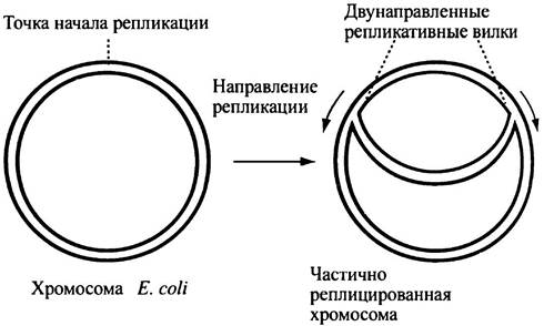
Because Watson-Crick Base Pairs in DNA are held together by Hydrogen Bonds, the synthesis of new molecules can only occur when the strands unwind, making the bases accessible for new interactions with complementary bases. The E. coli chromosome is circular and contains about four million base pairs. At a specific site, termed the origin of replication, the strands separate to form two replication forks that move in opposite directions. DNA is synthesized at a rate of approximately 500 base pairs per second, with parental DNA unwinding and new strand synthesis occurring simultaneously (Fig. 20.2). The two forks meet on the opposite side of the circle. Consequently, the two newly replicated rings remain intertwined. This topological entanglement was initially raised as an argument against the aforementioned model of DNA replication. One can only marvel at the insight of Crick, who postulated that evolution must have provided a mechanism to untangle the rings. It was later discovered that this function is performed by the enzyme topoisomerase II (see p. 245).
Fig. 20.2. Bidirectional Replication of the E. coli chromosome. Newly synthesized strands are highlighted in color.
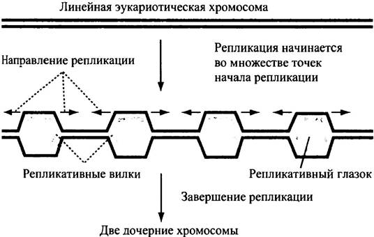
In eukaryotes, DNA synthesis at the Replication fork proceeds at a rate of roughly 50 base pairs per second—far too slow to replicate massive chromosomal DNA within the time allotted for Cell Division. In reality, replication of eukaryotic DNA is initiated simultaneously at numerous sites (likely numbering more than a thousand). Two replication forks emanate from each such site in both directions (Fig. 20.3); otherwise, the process proceeds similarly to replication in E. coli.
Fig. 20.3. Schematic diagram illustrating the formation of multiple bidirectional replication forks in A eukaryotic chromosome.
Regulation of DNA Replication Initiation in E. coli
Prior to E. coli cell division, complete duplication of the chromosome must occur; cell division itself takes place somewhat later, about 20 minutes afterward. The exact mechanism coordinating cell division and DNA replication remains unclear. Both processes require Protein Synthesis AND a substantial increase in cell size. As shown previously (see Fig. 20.2), E. coli possesses a single origin of DNA replication (oriC), from which replication proceeds bidirectionally. An entire E. coli chromosome is referred to as a replicon; in eukaryotes, a replicon is a stretch of DNA whose replication is controlled by a single origin of replication.
THE ORIGIN OF replication features a specific base sequence rich in A-T pairs, which presumably facilitates strand Separation (recall that A-T pairs share two hydrogen bonds, whereas G-C pairs have three, making the former less tightly bound). During initiation, numerous molecules of a protein designated as DnaA bind to this region, triggering strand separation. This, in turn, assists the primary unwinding enzyme, helicase—operating at each replication fork—in initiating and sustaining progressive unwinding of the strands in both directions. Helicase (also known as DnaB, i.e., the protein encoded by the dnaB gene) will be discussed in detail during the Description of the DNA synthesis mechanism.
Initiation of DNA Replication in Eukaryotes
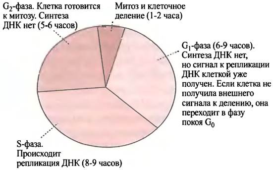
Eukaryotic cells feature a more complex Cell Cycle than E. coli cells, with DNA synthesis restricted to a specific period known as the S phase (synthesis phase) (Fig. 20.4). The duration of cell cycles varies widely among different cell types; in Animal Cell Cultures, the S phase occupies roughly 8 hours of a total 24-hour cell cycle. The S phase is preceded by the G1 phase (gap in DNA synthesis). To enter division, mammalian cells must receive an appropriate external signal, which is delivered by protein molecules called growth factors.
Fig. 20.4. The Introduction/5.html">Eukaryotic Cell cycle. Cycle duration varies across different cell types. Shown here are the cycle phases for a rapidly dividing mammalian cell culture (total cycle time 24 hours).
These factors bind to cell-surface receptors that transmit a division signal into The Cell interior (see Chapter 26). As noted earlier, animal Chromosomes contain numerous origins of replication from which replication proceeds bidirectionally. Although the precise regulation of replication initiation remains unknown, it is crucial that each replicon is activated only once during a single cell division cycle.
Unwinding of the DNA Double Helix and Supercoiling
The separation of DNA strands by helicase presents a topological (physical) challenge. To explain this phenomenon, we must first examine THE CONCEPT OF DNA Supercoiling.
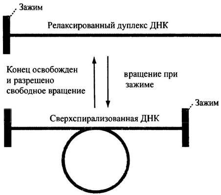
Two DNA strands (the DNA duplex) form a right-handed helix, with 10 base pairs per turn. An isolated short fragment of linear DNA that can rotate freely around its long axis spontaneously adopts a relaxed configuration known as the relaxed form. Imagine that one end of the duplex is fixed and cannot rotate, while additional turns are introduced at the opposite end. In this case, the helix begins to wind tighter, the number of turns in this DNA segment increases, and the number of base pairs per turn decreases. This phenomenon is termed positive supercoiling, or overwinding. Conversely, if the duplex is twisted in the opposite direction, the helix unwinds: the number of turns per unit length decreases, while the number of bases per turn increases. The DNA enters an underwound state, or negative supercoiling. Both forms—overwound and underwound—represent a strained state of the molecule. To relieve this strain, The Double Helix must twist around itself, forming a plectonemic coil, or superhelix (Fig. 20.5). Supercoiling is easy to visualize by taking a piece of doubly twisted rope. Suppose someone holds one end of the rope, or secures it so it cannot rotate freely, and twists the rope along its axis. This creates a helix twisted under the torsional stress. If the end of the supercoiled rope is released, it immediately returns to its original (relaxed) form.
Fig. 20.5. Schematic diagram illustrating how twisting a DNA fragment without freedom of rotation leads to supercoiling, which relieves torsional stress. Cellular DNA is firmly anchored and lacks rotational freedom. However, if free rotation were possible, the superhelix would transition into a relaxed state. If rotation occurs in the unwinding direction, the superhelix is negative; if rotation is performed in the opposite direction, the superhelix is positive (differences between these two forms are not shown)
What happens to DNA during replication?
After all, a DNA strand within a cell cannot rotate freely around its axis. In E. coli, the closed chromosomal ring securely "fixes" the DNA. In eukaryotes, massive DNA molecules can form loops (see Fig. 19.7, d) anchored by protein structures; this creates ring-like arrangements that render these segments incapable of free rotation. Yet, separating DNA strands inherently requires Rotation of the duplex. This induces supercoiling, leading to the accumulation of positive supercoils ahead of the replication fork; as the helix tightens, further unwinding becomes difficult. If this torsional stress is not relieved, it will obstruct strand separation, and DNA replication will halt.
A very simple experiment will convince you of this. Take a short piece of double-stranded rope and pull its ends in opposite directions: the rope will quickly unwind without accumulating positive supercoils. In this process, the rope strands easily and completely separate from each other. Now, coil a long piece of the same rope into a ring, or secure one of its ends so the rope cannot rotate freely, and attempt to pull its strands apart. Positive supercoils will rapidly "jam" the resulting fork and prevent subsequent separation. The exact same thing happens during DNA replication in the cell unless something prevents it.
It follows that for DNA synthesis to continue, the positive supercoils ahead of the replication fork must be relaxed. As needed, this is achieved through transient breaks in the polynucleotide chain.
How are positive supercoils ahead of the replication fork eliminated?
This process is catalyzed by a group of Enzymes known as topoisomerases. They act on DNA by isomerizing or altering its topology. There are two classes of topoisomerases (Type I and Type II). We will first examine their MECHANISM OF ACTION and then explain their role in DNA replication.
Type I enzymes cleave one of the two strands of the supercoiled double helix, allowing the entire duplex to rotate around the single phosphodiester bond of the opposite strand, which acts as an efficient swivel in the DNA molecule. Once rotation is complete, the enzyme reseals the duplex (Fig. 20.6). Crucially, the enzyme does not hydrolyze the phosphodiester bond it attacks. Instead, it transfers the bond from the 3'-OH group of deoxyribose to the OH group of a Tyrosine residue within the enzyme molecule. This process is readily reversible because no energy is lost during the Cleavage. Notably, topoisomerase I does not consume ATP energy. The supercoiled DNA molecule is under torsional stress, and its energy level is higher than that of the relaxed form, regardless of the type of supercoiling—positive or negative.
Fig. 20.6. Reaction catalyzed by topoisomerase I. Tyr-OH represents a tyrosine residue in the enzyme molecule

Topoisomerase I is only capable of relaxing supercoiled DNA and cannot introduce new turns. At the same time, E. coli possesses a specialized topoisomerase I that can relax only negatively supercoiled DNA. However, while playing an important role, this alone does not solve the DNA unwinding problem.
Relaxation of the positive supercoils generated ahead of the E. coli replication fork occurs through the active introduction of negative supercoils, which "neutralize" the positive ones. This process is mediated by topoisomerase II.
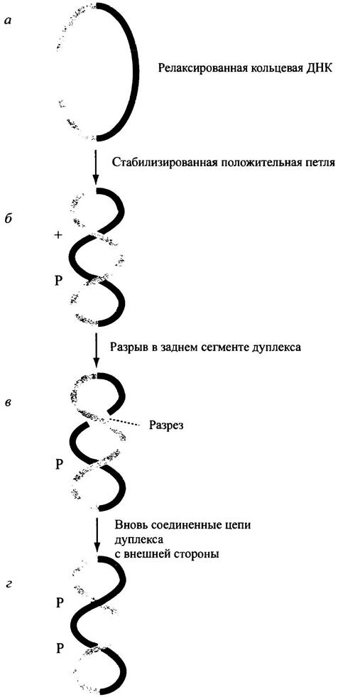
Topoisomerase II breaks both DNA strands of the double duplex, transferring the bonds to itself, which makes the Cleavage of the polynucleotide chains readily reversible. The enzyme physically passes the circular DNA duplex through the break without rotating the strands. This process requires ATP energy; Conformational Changes in the protein likely occur precisely due to ATP Hydrolysis. In E. coli, topoisomerase II, known as gyrase, introduces negative supercoils into DNA (Fig. 20.7). For simplicity, the figure illustrates the formation of negative supercoils in relaxed circular DNA (see Fig. 20.7, a). However, the same principle applies to DNA at the replication fork in E. coli, where positive supercoiling arises from DNA strand unwinding. It has been established that the incorporation of negative supercoils in this region corresponds to the relaxation of positive supercoiling. Gyrase interacts with DNA in such a way that the DNA wraps around the protein. This induces overwinding (positive supercoiling) in the Regions of the molecule bound to the protein (see Fig. 20.7, b). However, as long as covalent bonds remain unbroken, structural integrity is preserved; consequently, the local positive superhelix formed in response to protein binding can be counterbalanced by negative supercoiling elsewhere in the DNA molecule, reducing net supercoiling to zero. Next, the enzyme cleaves both DNA strands (see Fig. 20.7, c) and passes the double strand from the inside to the outside, after which it reseals both breaks, converting the positive loop into a negative one (see Fig. 20.7, d). In other words, a negative supercoil is inserted. The physical translocation of DNA is driven by ATP hydrolysis. The negatively supercoiled state is also under torsional stress; therefore, the introduction of negative supercoils is an energy-dependent process. Research has shown that DNA bond cleavage does not occur via hydrolysis, but rather through the temporary transfer of single-stranded DNA ends to the enzyme without any loss of Free energy; thus, the bond-breaking process is easily reversible.
Fig. 20.7. The reaction of incorporating negative supercoils into circular DNA, catalyzed by gyrase. This process can convert positively supercoiled DNA into negatively supercoiled DNA
What is the Biological Role of topoisomerases?
From an evolutionary perspective, solving topological problems is crucial for cell survival, and this is accomplished by Two Types of topoisomerases (I and II) with distinct Mechanisms of action. In many organisms, The properties of these enzymes vary slightly. At least 11 variants of topoisomerases have been described.
In E. coli, topoisomerase I relaxes only negative supercoils. Meanwhile, gyrase (topoisomerase II) actively introduces negative supercoils, and because this relaxes positive supercoils, it thereby ensures the continuation of DNA synthesis.
In eukaryotes, topoisomerase I can relax both types of supercoils—positive and negative—whereas topoisomerase II relaxes only positive supercoils and cannot introduce negative ones (Table 20.1). Thus, during DNA replication in both PROKARYOTES AND EUKARYOTES, tangling of the strands during unwinding is prevented by the induction of positive supercoils.
Table 20.1. Topoisomerases of E. coli and eukaryotes: types and mechanism of action
Type* |
Action on DNA |
Effect on supercoiling |
Topoisomerase I |
||
E. coli |
Cleaves One DNA strand |
Relaxes negative supercoiling |
Eukaryotes |
Cleaves one DNA strand |
Relaxes positive and negative supercoiling |
Topoisomerase II (gyrase) |
||
E. coli |
Cleaves both DNA strands; ATP-dependent |
Relaxes positive supercoils; introduces negative supercoils; unlinks intertwined circular DNA molecules |
Eukaryotes |
Cleaves both DNA strands; ATP-dependent |
Relaxes positive supercoils, but cannot introduce negative supercoils |
* Both types of topoisomerases are also found in other organisms
However, DNA supercoiling is critically important for processes other than replication alone. Meticulously isolated cellular DNA exists in the form of a negative superhelix. In relaxed DNA, there are 10.5 base pairs per turn of the double helix, whereas in cellular DNA there are 12, meaning it is underwound. The degree of supercoiling is comparable across DNA isolated from various cell types, highlighting the importance and regulation of this phenomenon. It is known that in E. coli, negative supercoiling is essential for both replication and Transcription (see Chapter 21), which implies that strand separation occurs more readily in the supercoiled state. It is also hypothesized that protein-DNA interactions at negatively supercoiled sites help reduce conformational strain and increase the net energy yield required for binding, thereby facilitating the attachment of Proteins to DNA.
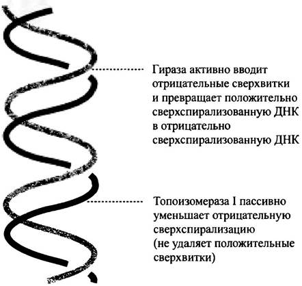
To sum up, it should be emphasized once again that in E. coli cells, the required negative DNA supercoiling is generated by gyrase (topoisomerase II), whereas topoisomerase I relaxes negative supercoils but does not act on positive ones. The balance between these two opposing activities (gyrase and topoisomerase I) maintains the necessary degree of supercoiling (Fig. 20.8); the proper ratio of the two enzyme activities is essential for cell viability. To control this, two specific enzymes are synthesized; the degree and sign of supercoiling are regulated by a factor whose synthesis is controlled by a corresponding gene. The requirement of gyrase for bacterial viability is confirmed by the fact that the antibiotic nalidixic acid inhibits their reproduction by inactivating gyrase. Eukaryotes lack this enzyme, but nalidixic acid can be used to treat certain infections in humans.
Fig. 20.8. The balanced action of gyrase and topoisomerase I in E. coli. As a result of the joint action of these enzymes, the degree of negative supercoiling necessary for DNA Replication and Transcription is achieved. Gyrase is an ATP-dependent enzyme. It is believed that the amount and ratio of the synthesized enzymes are controlled by the degree of supercoiling of gene promoters.
Interestingly, thermophilic Bacteria living at very high temperatures contain reverse gyrase, whose mechanism of action is directly opposite to that of E. coli gyrase. This enzyme activity can be explained by the fact that positively supercoiled DNA is better protected against strand separation, which occurs at high temperatures.
Eukaryotic DNA within the cell, much like prokaryotic DNA, is underwound or negatively supercoiled. However, unlike prokaryotes, no eukaryotic topoisomerase capable of actively introducing negative supercoils into DNA is known. What happens in eukaryotic cells?
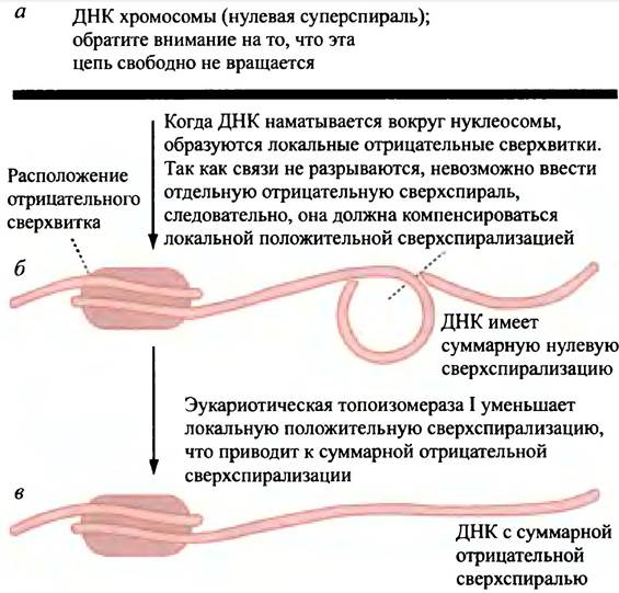
During Chromatin formation, DNA is wrapped around the nucleosome core, and the DNA molecule is underwound in the region of contact with the protein. Such nucleosomal turns have no breaks, and the DNA in the chromosome cannot rotate freely. Therefore, local negative supercoiling on the nucleosome is compensated by positive supercoiling elsewhere, so that the net structural changes sum to zero (Fig. 20.9, b). Eukaryotic topoisomerases relax the positively supercoiled fragment by incorporating a negative supercoil (Fig. 20.9, c). Thus, the prokaryotic type of gyrase is not needed here.
Fig. 20.9. The mechanism by which eukaryotic DNA acquires negative supercoils. Eukaryotic cells lack an enzyme capable of actively incorporating negative supercoils in the manner of prokaryotic gyrase.
Thus, we have helicases that unwind the strands, and topoisomerases that allow them to separate by reducing the positive supercoiling caused by the double helix rotating like a swivel mechanism during strand unwinding. But that is not all: SSB proteins are also required for helix unwinding.
What is SSB?
These are single-strand binding proteins (hence the abbreviation SSB). They exhibit a high affinity for single-stranded DNA — regardless of base sequence — and interact with it along the entire length of the separated strands. The energy released upon binding prevents the unwound strands from re-associating, thereby protecting the single-stranded form from reversible annealing. When the strands serve as a template for the synthesis of new DNA, the SSB proteins are removed.
Current Status
So far, we have dealt with the general aspects of DNA replication — the semi-conservative mechanism based on Watson-Crick base-pair complementarity, the biological aspects of the cell cycle, replication initiation, and The Mechanism of DNA unwinding. The foregoing demonstrates The complexity of the DNA replication process. We have gained an understanding of DNA strand separation, but have not yet learned about the mechanism of DNA synthesis at the replication fork. Let us return to this problem. First of all, we must understand: how do NUCLEOTIDES link together during the synthesis of a new DNA strand? The enzymes catalyzing this process are called DNA polymerases, because they polymerize nucleotides to form a DNA molecule.
Main Enzymatic Reactions Catalyzed by DNA Polymerases
We will examine the main chemical reaction of DNA formation, but first let us dwell on a few aspects.
✵ In E. coli, there are three DNA polymerases, designated Pol I, Pol II, and Pol III.
✵ DNA synthesis occurring at the replication fork is catalyzed by Pol III or its eukaryotic analogues; Pol I plays an important role in both DNA Replication and Repair. Pol II is less studied; it is hypothesized to be involved in a specific type of DNA Repair.
✵ The substrates for the DNA polymerase reaction are four deoxynucleoside triphosphates: dATP, dCTP, dGTP, and dTTP. During replication, daughter strands are formed as copies of the parent strands.
✵ To copy a template, the polymerase requires a single-stranded DNA template. The term copying is used in the sense of complementarity: G on the template is "copied" (transcribed) into C in the daughter strand; correspondingly, A is copied into T, C into G, and T into A.
✵ It is very important to remember: DNA polymerase can only elongate an already existing strand called a primer. The primer is complementary to the template and consists of 2–10 nucleotides; synthesis cannot begin without it. DNA polymerase does not initiate the formation of a new strand, as it cannot join two free nucleotides together.
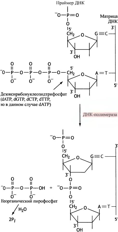
The polymerase attaches nucleotides to the free 3'-OH end of the primer, releasing inorganic pyrophosphate in the process (Fig. 20.10). Phosphate hydrolysis increases the negative value of ΔG° for polynucleotide chain synthesis, thereby driving the polymerization reaction forward. The incorporation of a nucleotide into the new DNA chain is accompanied by the formation of hydrogen bonds with the template. This process releases energy, making it thermodynamically favorable.
Fig. 20.10. The reaction catalyzed by DNA polymerase. The scheme shows The addition of an adenine deoxynucleoside (from ATP) to the 3'-end of the primer; the choice of the incorporated base is determined by the base in the template strand. Synthesis proceeds in the 5' —> 3' direction, i.e., the chain elongates in this direction.
The order in which the four deoxynucleoside triphosphates interact with DNA polymerase is determined by the bases of the parental strand being copied.
DNA synthesis always proceeds in the 5' —> 3' direction of the growing chain. This means that the growing chain is elongated in the 5' —> 3' direction; a nucleotide is added to the free 3'-OH end of the preceding nucleotide. Recall that when we speak of synthesis in the 5' —> 3' direction, we are always referring to the direction of elongation or polarity of the new strand. The template strand has the opposite polarity. We discussed the polarity of DNA strands in the previous chapter (see p. 235).
Complications and yet more complications in DNA synthesis
How is a new strand initiated?
DNA polymerase is unable to initiate the formation of a new chain on its own; therefore, synthesis must be primed at any starting point.
The solution to this problem is quite remarkable and relies on the fact that DNA chains are initiated with the help of RNA. RNA possesses a structure analogous to that of single-stranded DNA, except that ribose serves as its sugar component, and thymine (T) is replaced by uracil (U). RNA is synthesized by RNA polymerase via a chemical mechanism nearly identical to that of DNA, with the exception that ribonucleoside triphosphates ATP, CTP, GTP, and UTP are employed for the synthesis. In this case, however, the fundamental difference is that RNA polymerase is capable of initiating new chains. It can join two nucleotides together (a template is still required for this process), whereas DNA polymerase cannot.
Once a short stretch of An RNA primer (for example, consisting of 5 nucleotides) has been synthesized by a specialized RNA polymerase known as primase, DNA polymerase steps in and continues to elongate the chain.
Let us now examine the antiparallel arrangement of strands in the DNA molecule.
How is The problem of strand polarity resolved during DNA replication?
Let us recall the previous chapter, which discussed the polarity of DNA strands. DNA is replicated at each replication fork, which gradually progresses along the chromosome. In E. coli, two molecules of DNA polymerase III (Pol III) operate at each replication fork—one for each strand. These two molecules associate into a single asymmetric dimer and form the holoenzyme. The polymerase molecules must move in the same direction (Fig. 20.11). However, the 5' —> 3' direction of one parental strand is opposite to the movement of the fork, whereas in the other strand, it coincides with it. Because DNA polymerase can only synthesize in the 5' —> 3' direction, the template strand must run in the 3' —> 5' direction, which is opposite to the direction of synthesis. This suits the synthesis of one new strand (on the left in Fig. 20.11) perfectly, but what about the other? It runs in the direction of fork movement (5' —> 3'); consequently, the Synthesis of the daughter strand must proceed in the opposite direction. Figure 20.11 highlights what initially seems like an insoluble problem: the polymerase on the right-hand strand must move upward on the page, which indeed it does, yet the DNA must be synthesized in a downward direction. The left-hand strand is termed the leading strand, and the other is the lagging strand.
Fig. 20.11. Distribution of strand polarity during DNA replication
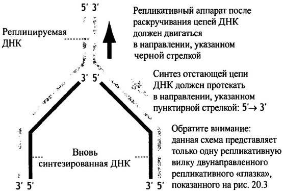
But How is the synthesis of the lagging strand initiated? For the leading strand, the situation is clear-cut: the primase lays down a simple primer at the origin of replication, and DNA polymerase extends the chain. However, this approach does not work for the lagging strand. The problem is solved in the following manner: as the DNA strands are unwound, multiple short DNA chains are synthesized on the right-hand strand through repeated priming by primase (RNA polymerase) (Fig. 20.12). At first glance, this seems entirely incredible!
Fig. 20.12. The replication fork. The leading strand is synthesized continuously, whereas the lagging strand is synthesized in discrete short fragments (Okazaki fragments)
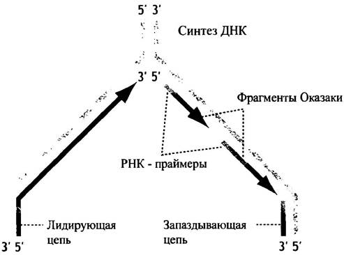
The short DNA pieces associated with RNA primers on the lagging strand are called Okazaki fragments, named after the researcher who discovered them. Nevertheless, the diagram still fails to clarify how DNA polymerase can synthesize DNA in a direction opposite to its own movement. Furthermore, the lagging strand consists of a series of disjointed short fragments attached to primers. To begin with, let us examine how the replication machinery operates at the replication fork.
The enzymatic complex of the E. coli replication fork
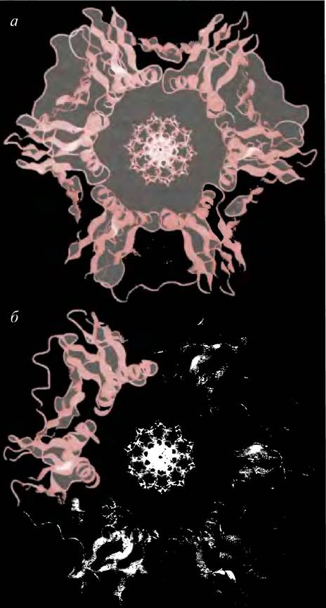
The functional complex of proteins at the replication fork is depicted in Fig. 20.13. The Key Enzymes are helicase, which unwinds the double helix; primase, which synthesizes primers on segments of the lagging strand; and, of course, the Pol III complex. The unwinding activity of helicase is ATP-dependent; the enzyme moves along the DNA strand (presumably via conformational changes) and separates the two strands of the double helix. Single-stranded DNA is stabilized upon the binding of SSB proteins. In E. coli, two interconnected Pol III molecules are present at the replication fork, one synthesizing the leading strand and the other the lagging strand. This is essentially the same enzyme, but the holoenzyme dimer is asymmetric due to an additional subunit on the lagging-strand side. Pol III is a highly processive enzyme: once bound to a DNA strand, it remains attached over thousands of bases, which is advantageous for the leading strand. Its fixation to the template strand is facilitated by ring-shaped sliding "clamps" (β-subunits) composed of two halves. These form a sliding ring behind Pol III that is linked to the enzyme, preventing the latter from dissociating from the DNA template strand. The sliding clamp features a central channel that allows DNA to thread through freely. The structure of the clamp is illustrated by the ribbon model (Fig. 20.14).
Fig. 20.13. Arrangement of enzymes at the E. coli replication fork (simplified diagram)
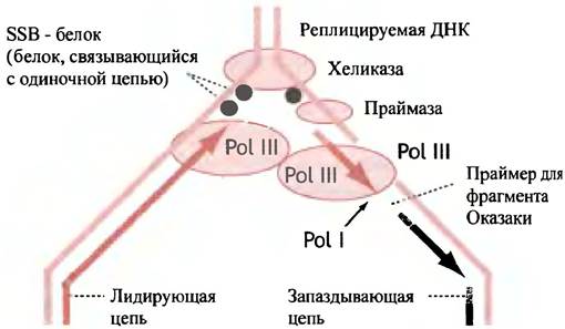
Fig. 20.14. Ribbon models of the sliding clamps of Yeast and E. coli. (a) The yeast clamp, which ensures DNA polymerase processivity, is a trimer; (b) the E. coli clamp, which associates with DNA polymerase III, is a dimer. Individual subunits within each ring are shown in different shades. The strands of the β-sheet are depicted as flat ribbons, and the α-Helix as a spiral. A model of B-form DNA is placed in the center of each structure, as the ring can encircle the duplex
On the lagging strand, the enzyme (along with the sliding clamp) halts upon encountering the primer synthesized by the primase for the preceding fragment.
It remains unclear whether the Pol III complex dissociates in order to re-bind after encountering a new primer used for the synthesis of the next Okazaki fragment, or if the process unfolds via a different mechanism altogether.
Let us now turn to the topological problem that arises during the 5' —> 3' synthesis of Okazaki fragments, given that the replication machinery lacks a reverse gear. The core principle is straightforward: if the lagging strand loops around, a short segment of it will be oriented in the same direction as the leading strand. Consequently, the replication apparatus can move in the direction of the fork while simultaneously synthesizing both new strands.
Although the principle is simple, the mechanics of moving the loop along the parental strand during the synthesis of Okazaki fragments are far more complex. To address this, Kornberg proposed a model known as the trombone model (or loop model) (Fig. 20.15).
Fig. 20.15. The looping model for Okazaki fragment synthesis. Darker arrows indicate the direction of DNA synthesis. According to the model, the loop must be dismantled when a new Okazaki fragment meets the previous one, after which a new loop must form. In this model, both strands can be synthesized in the 5' —> 3' direction as the replication apparatus moves along with the fork. It is impossible to describe the precise details of loop formation
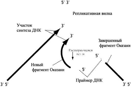
As the loop expands and the replicative machinery advances, newly synthesized DNA strands will encounter the 5' ends of RNA from the preceding Okazaki fragment. At this point, the polymerase must dissociate, the loop must collapse, and a new one must form. Let us now examine how Okazaki fragments are joined into a continuous DNA strand.
What happens to Okazaki fragments?
In E. coli, Pol III dissociates from the DNA upon reaching the RNA primer of the preceding Okazaki fragment, leaving a nick at the junction between the DNA and RNA. This is where DNA polymerase I (Pol I) comes into play. It is a remarkable enzyme that combines three distinct enzymatic activities within a single molecule.
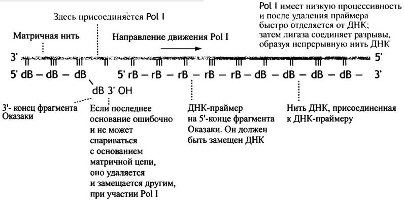
Looking at the diagram in Fig. 20.12, we can see that individual DNA pieces (Okazaki fragments) must be converted into a continuous DNA strand. Each fragment begins with an RNA segment that must be removed, followed by gap filling and the ligation of the separate DNA fragments. Pol I binds to the gaps between completed Okazaki fragments, adding nucleotides to the 3'-OH end of the preceding fragment and moving in the 5' —> 3' direction, just like Pol III (during DNA synthesis, nucleotide addition always proceeds in the 5' —> 3' direction). As it advances, the leading edge of Pol I encounters the RNA of the previous Okazaki fragment and excises its ribonucleotides, while the trailing region of the enzyme concurrently incorporates deoxyribonucleotides, filling the gap. The "forward" activity is termed 5' —> 3' exonuclease: "exo" because the enzyme removes nucleotides from the end of the molecule, "nuclease" because it hydrolyzes Nucleic Acids, and "5' —> 3'" indicates the direction of movement from the 5' end of the RNA toward the 3' end. Note that Pol III, unlike Pol I, lacks 5' —> 3' exonuclease activity and cannot cleave the RNA primer upon encountering an Okazaki fragment. Therefore, it dissociates from the DNA at this point, leaving Pol I to perform the remaining steps.
However, that is not all. In addition to its polymerase function, the "rear" side of the Pol I molecule exhibits 3' —> 5' exonuclease activity. This exonuclease hydrolyzes nucleotides from the 3' end of the DNA strand just synthesized by Pol I, cutting them away in the 5' direction—that is, opposite to the action of the 5' —> 3' exonuclease.
Paradoxical as it may seem, the enzyme simultaneously degrades and synthesizes DNA. This dual action can be readily explained. The 3' —> 5' exonuclease hydrolyzes the terminal nucleotide of a newly formed DNA strand only when its base fails to pair correctly with the corresponding base on the template strand. Conversely, the DNA polymerase adds nucleotides only when the preceding Base of the daughter strand is properly paired with its template counterpart. In this manner, the enzyme proofreads the inclusion of the previous base, excising it if mismatched and replacing it with the correct one. This process is known as mismatch correction (or proofreading), which ensures high fidelity during synthesis.
DNA polymerase I (unlike Pol III) has low processivity. It is held loosely on the template strand and dissociates rather quickly after removing the RNA, lacking the sliding clamp that anchors Pol III to the DNA. This makes biological sense; otherwise, it would continuously replace long stretches of newly synthesized DNA. Following its dissociation, nicks remain in the strand, which are then sealed by an enzyme called DNA ligase.
This enzyme catalyzes the formation of a phosphodiester bond between the 3'-OH group of one DNA fragment and the 5'-phosphate of the next. The process requires an input of energy. In some prokaryotes and eukaryotes, ATP serves as the energy source. The Mechanism of enzyme (E) action involves accepting AMP from ATP, releasing pyrophosphate, and transferring the AMP to the 5'-phosphate of the DNA. Subsequently, the DNA-AMP intermediate reacts with the DNA-3'-OH group, releasing AMP and sealing the nick:
E + ATP —> E-AMP + PPi;
E-AMP + P-5-DNA —> E + AMP-P-5'-DNA;
DNA-3’-OH + AMP-P-5-DNA —> DNA-3’-O-P-5'-DNA + AMP.
The AMP is linked to both the enzyme and the DNA via a 5'-phosphate group.
In E. coli, NAD+ is utilized instead of ATP. It can serve as a source of AMP just as ATP does, and this is indeed the pathway used by E. coli. Next, Pol I binds to the DNA at a specific site (Fig. 20.16). The polymerase adds nucleotides to the 3' end of the DNA fragment, proofreading each incorporated base as it moves in the 5' —> 3' direction. The RNA and a portion of the DNA are excised, and the resulting gap is filled with DNA. Pol I then dissociates, and ligase joins the two DNA fragments together. Thus, a series of Okazaki fragments are assembled into a continuous new DNA strand.
Fig. 20.16. Schematic diagram of DNA polymerase I action in Processing Okazaki fragments. dN — Deoxyribonucleotide; rN — Ribonucleotide
Proofreading of base mismatches by polymerase III
The base sequence in a newly synthesized DNA strand must be replicated with high fidelity, as the incorporation of even a single incorrect base can be lethal. Nevertheless, given the vast number of bases involved, errors are inevitable.
When Pol III encounters a template base such as G, its high Specificity ensures that, in the vast majority of cases, only dCTP will be bound to the enzyme's Active Site and incorporated into the growing DNA strand. The same holds true for other bases. The mechanism by which Pol III selects the correct nucleotide to match the template base is based on the Watson-Crick model, wherein all complementary base pairs share a uniform geometric shape distinct from any other pair configuration; this geometry is precisely recognized by the enzyme (Fig. 20.17). Hydrogen bonding between correct base pairs and base-stacking interactions within the same strand also contribute to accurate Selection by rendering the process energetically favorable.
Fig. 20.17. Geometric characteristics of Watson-Crick base pairs and "erroneous" pairs. Data derived from X-ray crystallography of double-stranded oligodeoxyribonucleotides (B-form DNA). Note the strict geometric conformity of Watson-Crick pairs versus the unstable states of "wobble" A-C and G-T pairs, or the G-A pair (in anti and syn Conformations, respectively). The term "wobble pairs" will be explained on page 287. Distances (in nm) between deoxyribose C-1' atoms and the glycosidic Bond Angles between the base and deoxyribose are indicated.
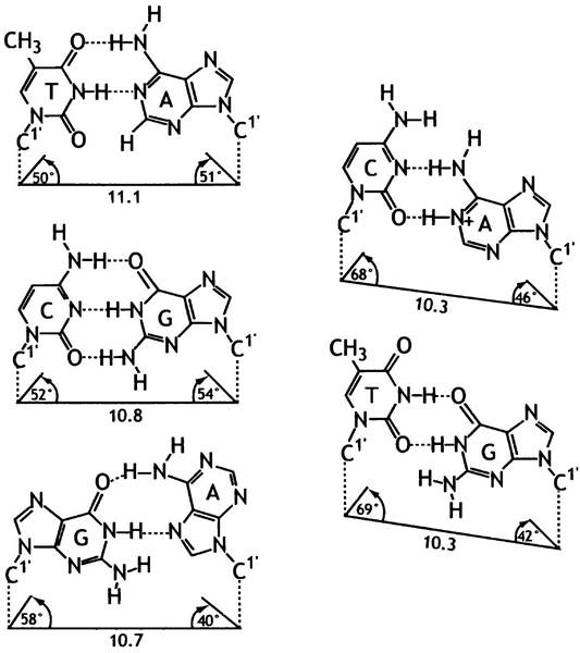
However, this mechanism has limits in precision. Although Watson-Crick pairing is the primary determinant, within any population of nucleotides, a small fraction of molecules—such as dATP—will transiently adopt an imino tautomer form (=NH instead of -NH2), allowing them to pair incorrectly with C. Consequently, Pol III may commit errors during DNA copying at a frequency of approximately one in 105 to 106 bases. If left unchecked, this would result in an unacceptably high mutation rate. Yet, in E. coli, the actual final error rate during DNA replication is significantly lower, roughly 10-10 per replication round.
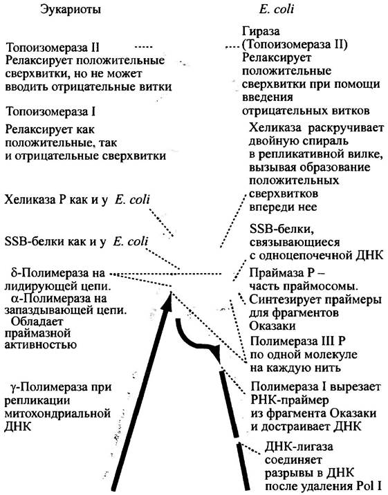
It turns out that Pol III also possesses 3' —> 5' exonuclease activity, which enables the removal of the most recently incorporated base if it is mismatched. This proofreading mechanism minimizes reading errors to a bare minimum. Most of the events occurring during DNA replication in E. coli and eukaryotes are illustrated in Fig. 20.18.
Fig. 20.18. DNA replication in E. coli and eukaryotes
Methyl-directed Mismatch Repair
Despite employing the aforementioned proofreading mechanisms that ensure high fidelity in DNA replication, errors are still bound to occur given the massive number of incorporated nucleotides. E. coli possesses an additional mechanism used to correct base-pairing errors in the newly synthesized DNA strand. If a mismatch escapes the polymerase, it causes a distortion in the duplex.

The error resides in the new strand, and the mismatch repair system must be able to recognize it specifically. Crucially, in such cases of mispairing, the base in the parental strand is correct by definition, whereas the complementary base in the new strand is incorrect. The repair system must clearly distinguish between the two strands
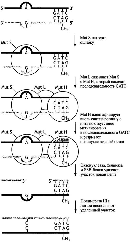
(parental and daughter) in order to excise the mutated base from the newly synthesized strand and correct the error. How is strand discrimination achieved? In E. coli DNA, the adenine within the sequence GATC is consistently methylated by the enzyme Dam methylase. Base methylation does not disrupt base pairing or DNA Structure because it occurs some time after the synthesis of the new strand. Thus, for a certain period, the parental strand remains methylated, while the newly synthesized strand does not. The mismatch repair system involves fewer components than the DNA replication machinery. A single Mut S protein recognizes the helical distortion; Mut H, bound to Mut S with the assistance of Mut L, locates the unmethylated GATC sequence on one of the duplex strands (thereby identifying the newly synthesized strand) and cleaves it at that site (Fig. 20.19). The cleavage site can be thousands of bases away from the actual mismatch. Subsequently, through the concerted action of a helicase, SSB proteins, and an exonuclease, the segment of the strand from the nick to the mismatch is excised, and DNA polymerase III fills the resulting gap in the DNA. DNA ligase completes the repair by sealing the nick. Correcting a single base may require the replacement of thousands of bases (see Fig. 20.19). This mismatch repair system increases replication fidelity a thousandfold. A similar mismatch repair system exists in eukaryotes. Humans possess proteins analogous to the E. coli Mut S and Mut L proteins, and Mutations in their genes are associated with an increased risk of Cancer.
Fig. 20.19. The methyl-directed mismatch repair pathway
DNA Damage and Repair in E. coli
While the mechanism described above ensures DNA replication with an exceptionally high degree of fidelity, essential for the continuation of life, a major challenge remains. The base sequence of DNA must remain strictly invariant, as the information it carries is vital to organisms throughout their entire lifespan. Yet, from a chemical standpoint, DNA is merely a conventional molecule susceptible to chemical alterations that could trigger numerous mutations if not for continuous repair processes. Some damage occurs spontaneously. The glycosidic bond linking purine and (to a lesser extent) pyrimidine bases to deoxyribose residues is unstable, frequently leading to depurination and depyrimidination, processes in which large numbers of Purines and Pyrimidines are cleaved from the DNA. Furthermore, cytosine and adenine undergo deamination, converting into uracil and hypoxanthine, respectively (see formulas on p. 217 and Fig. 18.7). DNA is also subject to attack by a multitude of chemical compounds.
Ionizing radiation, reactive oxygen species (see p. 212), carcinogens, and UV radiation can all damage DNA and consequently induce mutations.
Overall, damage affecting both strands of a double-helical DNA molecule precisely at the same position in both strands is relatively rare,
although it does happen. When a region of one strand is damaged, the intact second strand can always serve as a template to guide the repair of the affected portion. Cells possess numerous repair systems capable of handling virtually any type of DNA damage, as maintaining the integrity of this molecule is of paramount biological importance. DNA must be repaired at all costs.
Various types of repair systems exist.
✵ Direct Repair. Ultraviolet irradiation of DNA can cause covalent linkage between two adjacent thymine bases (on the same strand), forming a T-dimer.
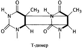
In E. coli, these aberrant bonds are cleaved via a light-activated mechanism, thereby restoring the thymine residues to their original state. This system is widespread and of great importance in plants. Alkyl groups on bases (generated by the Action of Certain mutagens) can be directly removed by a so-called "suicide enzyme," which acts as an acceptor for these groups and is subsequently destroyed in the process. Strictly speaking, this is a specific protein reagent rather than a true enzyme.
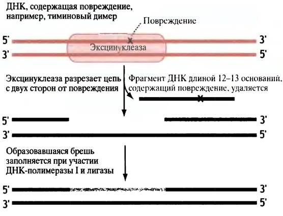
✵ Nucleotide Excision Repair. Damage that distorts the double helix, such as T-dimers, can also be repaired by excising short single-stranded nucleotide sequences containing the lesion and subsequently replacing them with the correct sequence. The opposite DNA strand serves as a template for this process. E. coli features a specialized endonuclease known as excinuclease, or the uvrABC complex, named after the three genes encoding the enzyme. It cleaves the DNA on both sides of the damage and removes a single-stranded fragment of 12–13 nucleotides (Fig. 20.20). DNA polymerase I binds to the nick and adds nucleotides to the 3'-end of the broken strand, after which a ligase seals the nick. The operation of this system relies on the accurate recognition of the damaged DNA strand.
Fig. 20.20. The nucleotide excision repair pathway in E. coli
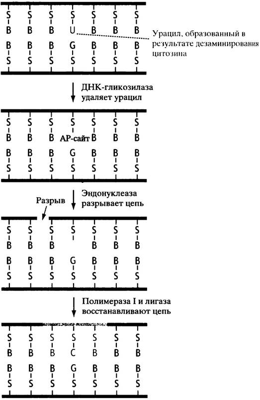
✵ Base excision repair and AP site repair. Deamination converts cytosine to uracil and adenine to hypoxanthine. DNA glycosylases recognize the abnormal base and cleave it off, leaving an AP site (apurinic-apyrimidinic site)—a deoxyribose sugar lacking a base (Fig. 20.21). AP sites can also form spontaneously because the purine-deoxyribose bond is relatively unstable. Repair of AP sites involves incision of the polynucleotide chain adjacent to the damage site, followed by replacement of the missing segment through the action of DNA polymerase I and ligase. The uracil derived from cytosine must also be removed (which likely explains why T is incorporated into DNA instead of U). Recall that T differs from U by the presence of an additional methyl group (see p. 217). If normal DNA contained U, it would be impossible to distinguish a normal U from a "false" U generated by the deamination of C.
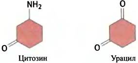
Fig. 20.21 Formation and repair of an AP site: S — sugar; B — base. In the example shown, the AP site is formed following the removal of uracil by a glycosylase. However, such sites can also arise via spontaneous hydrolysis of purine bases (and, to a lesser extent, pyrimidine bases)
Using the base T instead of U in DNA solves this problem. As will be discussed in the next chapter, U is utilized in RNA, but since RNA has a relatively short lifespan and a much smaller size, errors in it are not as critical. This is presumably why RNA is not repaired. A clear correlation is observed: while E. coli cells may contain numerous DNA nicks requiring Pol I binding and repair synthesis, they maintain only two replication forks that require Pol III. Consequently, an E. coli cell contains only 10–20 molecules of Pol III and a significantly larger number of Pol I molecules.
Organization OF THE Eukaryotic Replication Fork
The general principles of DNA replication in E. coli also apply to eukaryotes, albeit with a few modifications. The main difference is that instead of the Pol III dimer found in E. coli,
different polymerases are involved in the synthesis of the leading and lagging strands.
Five DNA polymerases have been described in eukaryotes, designated by Greek letters. Polymerases α and δ are involved in nuclear DNA replication (analogous to Pol III in E. coli). Pol α forms a complex with primase but lacks 3' —> 5' exonuclease activity, whereas polymerase δ possesses 3' —> 5' exonuclease activity. It appears that polymerases α and δ together form a replication complex in eukaryotes for leading-strand synthesis. Yeast δ-polymerase is associated with a sliding circular clamp known as PCNA (Proliferating Cell Nuclear Antigen), which ensures its processivity, much like the E. coli enzyme. Fig. 20.14 shows the ribbon structure of both the yeast and E. coli clamps. Despite their structural similarity, their Amino acid sequences differ significantly; furthermore, the E. coli clamp is a dimer, whereas the yeast protein is a trimer. This indicates that these proteins evolved independently.
Two other eukaryotic DNA polymerases, β and ε, perform a repair function, while γ-polymerase is involved in Mitochondrial DNA replication.
The Replication Problem at Eukaryotic Chromosome Ends
Eukaryotic chromosomes are linear, which creates a problem that does not arise during the replication of circular chromosomes.
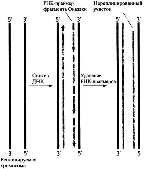
Let us consider the replication of the chromosome shown in Fig. 20.22. It replicates bidirectionally from a single initiation site located in the center (though eukaryotic chromosomes actually have many such sites). The 5'-end region of the leading strand is replicated completely. However, this does not happen during lagging-strand synthesis because the RNA primer involved in Okazaki fragment formation is located at the 3' end of the template strand (see Fig. 20.22). Once the primer is removed, these ends remain unreplicated, and there is no mechanism to fill them in using the DNA-synthesizing complex described above. RNA primers are required to fill in the lost segments, but there are no templates available to generate them. Consequently, with each cell division, the chromosomes would progressively shorten; further predictions are difficult since the mechanism of DNA synthesis implies complete replication of double-stranded DNA. The solution to the problem is that specialized repeating DNA sequences are located at the ends of eukaryotic chromosomes—telomeric DNA (the chromosome ends containing these sequences are called telomeres). Although the ends still cannot be replicated by the DNA-synthesizing complex described earlier, complete replication of the "true" chromosome is achieved using a primer at the 3' end of the template strand located opposite the telomeric DNA. Moreover, in rapidly dividing cells, telomeres are added to ensure that the chromosome never risks shortening.
Fig. 20.22. Schematic representation of the shortening of newly formed linear chromosomes during replication. Bidirectional replication of a very short DNA fragment is shown. Note that primer removal from Okazaki fragments occurs continuously (shown here as a separate event). A typical chromosome has multiple origins of replication. The synthesized DNA and the RNA primers of the Okazaki fragments are highlighted in light tones.
How Is Telomeric DNA Synthesized?
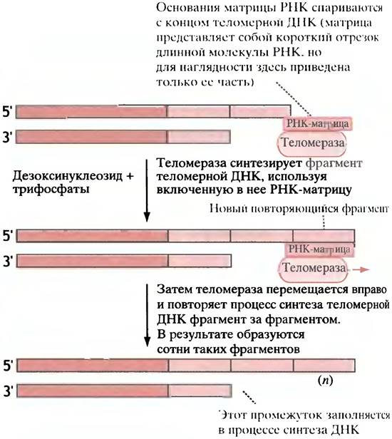
A telomere consists of short repeating segments (the base sequence of these segments varies among species). Humans have hundreds of tandem repeats of the sequence TTAGGG. The enzyme telomerase adds these sequences (one after another) to the 3' end of pre-existing telomeric DNA, thereby elongating it. Telomerase has two distinctive features: 1) it uses RNA as a template for DNA synthesis, making it a Reverse Transcriptase (see Chapter 23); 2) this template is built into the enzyme's structure. In humans, the RNA contains a sequence comprising 1.5 repeats complementary to TTAGGG. The RNA template hybridizes with the end of the telomeric DNA (Fig. 20.23), and another segment is then gradually added, nucleotide by nucleotide. Following this, the enzyme shifts and binds to the end of the newly formed segment. In this way, the telomere is continuously extended. A complementary strand is then synthesized on the elongated telomere of the single strand, giving the telomere a double-stranded structure. However, the exact mechanism of this structure's formation remains unclear, though DNA polymerase is thought to be involved in the process.
Fig. 20.23. Mechanism of telomeric DNA synthesis by telomerase. Chromosomal DNA (the "true" chromosome) is indicated by dark lines, and pre-existing telomeric DNA at one end of the chromosome by light lines. Telomerase incorporates a short RNA molecule containing a sequence complementary to the species-specific repeating segment. The enzyme attaches via the interaction of the RNA with the terminal bases of the pre-existing telomere and adds one repeating segment (base by base sequentially). Synthesis always proceeds in the 5' —> 3' direction. The enzyme shifts so that the RNA template remains base-paired with the terminal bases of the new repeating segment, after which the next segment is added, and so on. The newly synthesized telomeric DNA serves as a template for constructing the opposite strand, ensuring that the telomere is present on both strands.
The necessity of telomeres has been demonstrated using YACs, or yeast artificial chromosomes. These comprise three DNA segments essential for Chromosome replication: centromeres, replication origins, and telomeres. Research has shown that once integrated into yeast cells, YACs can persist for several generations, but gradually disappear as the telomeric ends are lost.
This does not occur in prokaryotes, because their circular chromosome always retains a template accessible for primer formation. The existence of telomeres in eukaryotes is critical. Telomerase has been detected in embryonic cells, which undergo continuous and rapid division, where the ongoing elongation of chromosomes is necessary to compensate for their progressive shortening.
However, in somatic cells that divide infrequently, the addition of telomeres is less critical, and indeed, telomerase has not been found in them. If this is the case, a logical Conclusion follows: somatic cells receive their allocated 'portion' of telomeric DNA, which is sufficient for the lifetime of the cell and its progeny. In most cells, telomeres shorten with age, and this may be a major factor determining lifespan. When somatic cells undergo malignant transformation, they enter a state of unlimited division, thereby becoming immortal—with no limit to their proliferative capacity in culture. Interestingly, the presence of telomerase has been confirmed in cancer cells.
DNA Damage Repair in Eukaryotes
The dealkylating 'suicide' enzyme described in E. coli has also been discovered in human cells. Eukaryotes likewise utilize excision repair. For instance, individuals with the genetic disorder xeroderma pigmentosum typically lack The ability to perform excision repair of pyrimidine dimers. Exposure of the Skin to UV radiation induces the covalent binding of two adjacent thymine bases (on the same strand), forming a thymine dimer. Sun exposure in patients with xeroderma pigmentosum leads to skin cancer. Thus, excision repair is of paramount importance to humans. Genes encoding proteins homologous to the Mut S and Mut L of E. coli (see p. 254) have been described, and mutations in these genes have been linked to cancer development.
Eukaryotic repair systems play a vital role in maintaining genome integrity. Today, many researchers are investigating The connection between human Aging and a decline in DNA repair capacity, including an increased mutation frequency.
Transposons, or Jumping Genes
It turns out that certain DNA fragments can relocate from one site to another within the same chromosome or insert into a different chromosome. The existence of jumping genes was first demonstrated by B. McClintock during her genetic studies on maize. She discovered that regulatory gene elements move across The Genome, influencing Gene Expression and resulting in phenotypic variations. This work went largely unnoticed for 30 years until studies on E. coli confirmed the ability of genes to move and insert elsewhere in the genome, for which McClintock was awarded the Nobel Prize. These mobile DNA sequences were named transposons, or jumping genes.
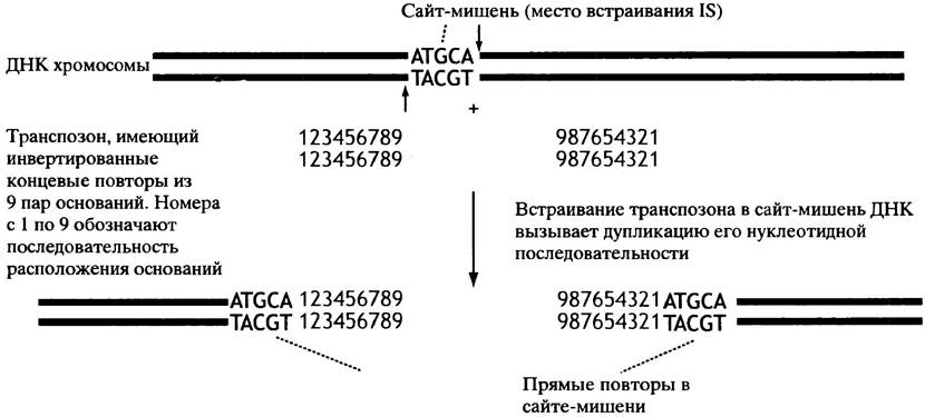
The simplest type of E. coli transposon is known as an insertion sequence (IS). It represents a DNA segment containing the gene for transposase—the enzyme responsible for mobilization. At first glance, the presence of an IS seems puzzling: a gene that codes for nothing other than its own ability to move from place to place! An IS can integrate into virtually any region of a chromosome, as it exhibits no sequence-specific insertion sites. When a transposon inserts into a gene, it inactivates the latter by disrupting its nucleotide sequence; however, the frequency of transposition events is quite low. In addition to the transposase gene, an insertion sequence features inverted terminal repeats at its ends. These consist of short fragments oriented as shown in Fig. 20.24.
Fig. 20.24. Integration of a transposon (IS type) into a chromosome. Break points in the recipient DNA sequence are indicated by arrows
Various mechanisms of transposition are possible. One involves the prior replication of the transposon rather than its direct excision and transfer. A copy of the transposon leaves its original site and integrates into a new Location. The Emergence of numerous repetitive sequences within the genome provides a foundation for Homologous Recombination. It is likely that chromosomal rearrangements mediated by transposons are critically important for the evolutionary process.
Is the described mechanism the only way DNA is synthesized?
It is well known that evolution has employed various pathways to achieve the same ends. For instance, in synthesizing DNA strands, some Viruses use proteins as promoters instead of RNA. Retroviruses, however, rely on a fundamentally different mechanism: their genomic RNA is copied into DNA (see Chapter 23). Interestingly, Mitochondria possess their own DNA, which must replicate when their population increases. At the same time, mitochondria lack DNA repair systems. This is presumably because their chromosome is significantly shorter, thereby reducing the probability of replication errors; furthermore, since a cell contains A large number of mitochondria, a minor number of defects is not critically important. Nevertheless, the absence of repair means that mutations in mitochondrial DNA can accumulate over time and potentially drive aging processes. A wide range of Genetic Disorders are known to be associated with mitochondrial DNA mutations.
Questions for Chapter 20
1. WHAT IS A replicon?
2. What topological problems arise when parental DNA strands are separated during replication?
3. How are the problems specified in the previous question resolved in E. coli and eukaryotes?
4. Explain, using diagrams, the action of topoisomerases I and II.
5. Eukaryotes lack a topoisomerase capable of introducing negative supercoils into DNA. At the same time, eukaryotic DNA does contain such supercoils. Explain how they are formed.
6. What is the substrate for DNA synthesis?
7. Why does DNA synthesis utilize TTP rather than UTP, which is used in RNA?
8. A. Can a DNA strand be synthesized using only four nucleoside triphosphates? Explain your answer. B. In what direction does DNA synthesis occur? Explain your answer.
9. What thermodynamic forces drive DNA synthesis?
10. E. coli DNA polymerase III is highly processive. Explain what this means, how high processivity is achieved, and why it evolved.
11. E. coli DNA polymerase I is a complex enzyme. What activities do its subunits possess, and what is their role in DNA synthesis?
12. By what mechanism does E. coli DNA polymerase III achieve a high degree of fidelity during DNA synthesis?
13. The activity of E. coli polymerase III alone is insufficient to achieve the required level of reading accuracy. When an incorrect nucleotide is incorporated, it must be replaced. Consequently, the repair system must recognize the mismatched base. How does this happen? Does such a mechanism exist in human cells?
14. Explain what a thymine dimer is, how it is formed, and how it is repaired.
15. Explain how eukaryotic chromosomes shorten with each successive replication cycle.
16. Explain how the shortening of DNA is prevented.
Last update: 06/08/2026
Editorial and Educational Adaptation: This material has been compiled based on the primary/original source text. The project team performed an editorial review, corrected technical inaccuracies, structured sections, and adapted the content for an educational format.
What was processed:
- elimination of formatting defects (OCR errors, structural breaks, corrupted characters);
- editorial organization of content;
- standardization of terminology in accordance with academic sources;
- verification of factual statements against the original source text.
All mentions of the author, publication year, and origin of the primary text have been preserved in accordance with the source.