Human Biochemistry, Volume 2 - Murray R. 1993
Structure, Function, and Replication of Information Macromolecules
DNA Organization and Replication
DNA Synthesis and Replication
The primary Functional Significance of METABOLISM/36.html">DNA Replication lies in supplying progeny with Genetic information. To ensure the genetic stability of an Organism and a species, DNA must be replicated completely and with extremely high fidelity. The DNA replication process is remarkably complex and involves numerous Enzymes. The first enzymological study of DNA replication was conducted by Arthur Kornberg, who discovered an enzyme in Escherichia coli now known as DNA polymerase I. This enzyme exhibits several Types of Enzymatic activity and features a complex Structure. As substrates, DNA polymerase I utilizes deoxyribonucleoside triphosphates—derivatives of adenine, guanine, cytosine, and thymine. The polymerase activity first demonstrated for DNA polymerase I is characteristic of other polymerases in prokaryotic and Eukaryotic Cells, though It is important to recall that The primary function of E. coli DNA polymerase I has been established as DNA Repair.
Initiation of DNA Synthesis
The initiation of DNA synthesis (Fig. 38.13) requires short RNA sequences (10–200 NUCLEOTIDES in length) that function as primers. Synthesis begins with a reaction between the 3'-hydroxyl group of the RNA primer and the a-phosphate group of a deoxyribonucleoside triphosphate, during which a deoxyribonucleoside residue is attached to the RNA primer with the simultaneous release of pyrophosphate. The 3'-hydroxyl group of the incorporated deoxyribonucleoside monophosphate then carries out a nucleophilic attack on the a-phosphate group of the next incoming deoxyribonucleoside triphosphate, also accompanied by the Cleavage of pyrophosphate. Naturally, the Selection of each successive nucleotide at every step of synthesis is dictated by the DNA template strand According to the rules first proposed by Watson and Crick (Fig. 38.14). Thus, if an adenine deoxyribonucleoside monophosphate residue is located at the corresponding position of the template strand, thymidine triphosphate will enter the reaction, and its a-phosphate group will be attacked by the 3'-hydroxyl group of the terminal residue of the growing chain. The reaction occurs only if the incorporated nucleotide forms a complementary pair with the next nucleotide of the DNA template strand and, through hydrogen bonding, assumes a position where the 3'-hydroxyl group of the growing chain attacks the new nucleotide and incorporates it into the polymer. The DNA sequences attached to the RNA primers were named Okazaki fragments after the Japanese scientist who discovered them (Fig. 38.15). In mammals, following The formation of a significant number of Okazaki fragments, the replication complex proceeds to remove the RNA primers and fill the resulting gaps with appropriate deoxyribonucleotides. Subsequently, DNA ligase seals the fragments together to form a continuous DNA strand.
Class="center">
Fig. 38.13. Initiation of DNA synthesis by An RNA primer and subsequent attachment of the second deoxyribonucleoside triphosphate.

Fig. 38.14. Template function of the DNA strand during RNA primer-initiated Synthesis of the complementary strand.
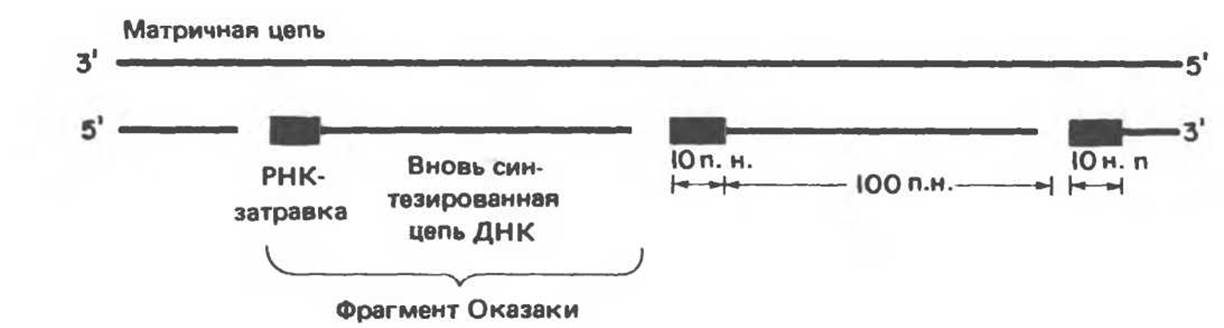
Fig. 38.15. Discontinuous polymerization of deoxyribonucleotides and formation of Okazaki fragments.
Polarity of Replication
As already noted, DNA molecules consist of two antiparallel strands. DNA Introduction/23.html">Replication in Prokaryotes and eukaryotes occurs simultaneously on both strands. However, an enzyme capable of directing DNA synthesis in the 3'→5' direction does not exist; consequently, the newly synthesized strands would appear unable to grow in the same direction simultaneously. Despite this paradox, a single enzyme carries out virtually synchronous synthesis of both strands. In this process, the strand synthesized in the 5'→3' direction (the "leading" strand) is continuous. Synthesis of the second (the "lagging") strand is carried out in fragments of 150–200 nucleotides. Successive initiation events for these fragments, which at any given moment also proceed in the 5'→3' direction, occur as the Replication fork generally progresses in a single direction. A diagram of "semidiscontinuous" DNA synthesis is presented in Fig. 38.16.

Fig. 38.16. The process of semidiscontinuous, Simultaneous replication of both strands of double-stranded DNA.
During the replication of mammalian nuclear DNA, the majority of RNA primers are removed by the end of the process; however, during the replication of the Mitochondrial Genome, small RNA fragments remain integrated within the closed circular DNA molecule.
DNA Polymerization and Repair Enzymes
Mammalian Cell nuclei contain a class of polymerases known as polymerase alpha (Pol a), which are responsible for chromosomal replication. A single Pol a molecule is capable of incorporating about 100 nucleotides per second into the growing chain, functioning roughly ten times slower than bacterial DNA polymerase. This reduced speed may be explained by steric hindrance from nucleosomes. How DNA polymerase overcomes nucleosomes remains unknown. Nevertheless, it is established that upon completion of replication, the respective nucleosomes are distributed randomly between both daughter strands.
Mammalian cell nuclei also contain a DNA polymerase with a lower molecular weight than Pol a—polymerase beta (Pol ß), which is not involved in routine replication but is essential for DNA repair (see below). Another enzyme, the Mitochondrial DNA polymerase gamma (Pol
y), carries out the replication of the circular mitochondrial genome.
Complete replication of the mammalian genome takes about 9 hours—the time required to double the genetic material of a dividing diploid cell. Such a rate indicates that replication initiates simultaneously at many sites, termed origins of replication and designated as ori. There are approximately 100 such sites. Replication proceeds bidirectionally, and both strands are replicated simultaneously. This results in the formation of so-called "replication bubbles" on the chromosome (Fig. 38.17).
The sites that act as origins of replication in eukaryotes have not been precisely defined. More comprehensive data in this regard are available for Yeast and several animal Viruses. It is safe to say that initiation processes are controlled both spatially and temporally, since adjacent ori clusters are initiated synchronously. It is hypothesized that functional Chromatin domains replicate as discrete units, implying that origins of replication are positioned in a highly specific manner relative to Transcription units.

Fig. 38.17. Formation of "replication bubbles" during DNA synthesis. The bidirectional nature of replication and the putative arrangement of strand-unwinding Proteins at the replication forks are shown.

Fig. 38.18. Hypothetical MECHANISM OF ACTION of a single-stranded DNA-binding protein at the replication fork. As the second strand is synthesized, the protein is released and binds to newly formed single-stranded DNA regions. (Courtesy of B. Alberts.)
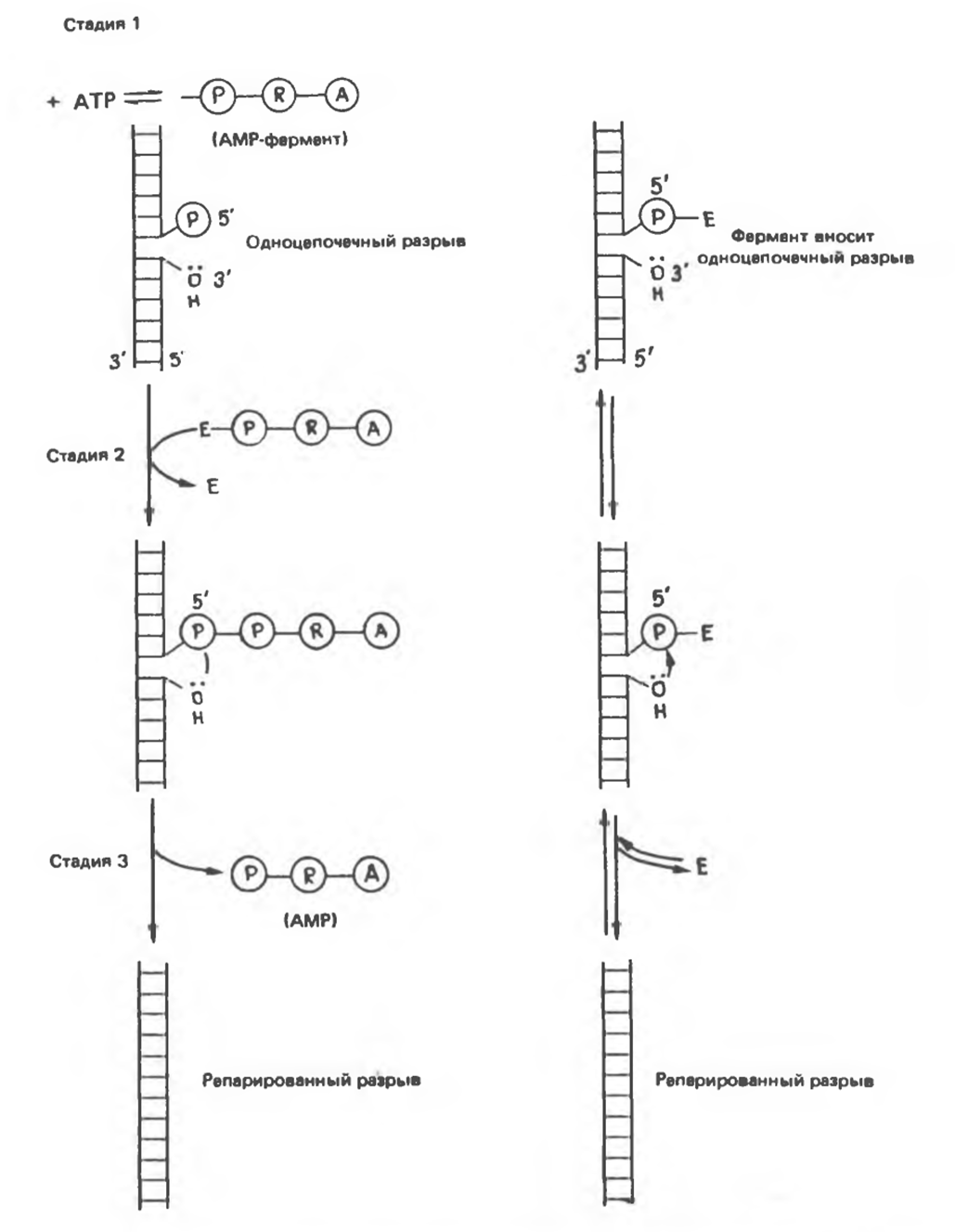
Fig. 38.19. Comparison of Two Types of reactions repairing single-strand DNA breaks. The reactions on the left are catalyzed by DNA ligase, and those on the right by DNA topoisomerase. (Slightly modified and reproduced, with permission from Lehninger A. L.: Biochemistry, 2nd ed. Worth, 1975.)
During replication, double-stranded DNA must unwind into individual strands so that each can serve as a template. DNA strand Separation is facilitated by specific proteins that stabilize the single-stranded structure as the replication fork advances. These stabilizing proteins bind stoichiometrically to the single strand without interfering with the ability of nucleotides to act as templates (Fig. 38.18). Along with strand separation, the helix must also unwind (1 turn for every 10 nucleotides), accompanied by the twisting of the newly synthesized daughter strands. Given the timeframe of prokaryotic replication, one can calculate that the DNA molecule would have to unwind at a rate of 400,000 rpm, which is entirely impossible. Consequently, multiple "swivels" must exist along the length of the DNA molecule. Swivel Functions are performed by a special enzyme (DNA topoisomerase), which introduces breaks into one of the strands of the unwinding double helix. These breaks are rapidly resealed by the same enzyme without additional Energy Expenditure, because the necessary Energy is stored in the form of a high-energy covalent bond formed between the sugar-phosphate backbone of the DNA strand and the topoisomerase. The scheme of this process presented in Fig. 38.19 can be compared to The sequence of events in DNA strand break sealing catalyzed by DNA ligase. DNA topoisomerases are also responsible for unwinding supercoiled DNA. Supercoiled DNA is a highly ordered structure formed by circular or ultra-long DNA molecules when they wrap around a histone core (Fig. 38.20).
One class of animal viruses (Retroviruses) possesses enzymes capable of synthesizing a DNA molecule using an RNA template. RNA-dependent DNA polymerase, or Reverse Transcriptase (as this enzyme is called), first synthesizes an RNA-DNA hybrid, using the viral ribonucleic acid genome as a template. Then, the enzyme RNase H removes the RNA strand, and the remaining DNA strand, in turn, serves as a template for the synthesis of the second DNA strand. This gives rise to cDNA—a double-stranded DNA copy containing the information initially presented in the form of the retrovirus RNA genome.
Regulation of DNA Synthesis
In animal and human cells, DNA replication occurs only during a specific period of the Cell life cycle. This period is called the synthesis phase (the so-called S phase). The S phase is separated from mitosis by the pre-synthetic (G1) and post-synthetic (G2) periods (Fig. 38.21). The primary regulation of a cell's own DNA synthesis lies in the fact that replication occurs at a strictly defined time and predominantly in cells preparing to divide. Cyclic purine nucleotides and, possibly, the substrates of DNA synthesis themselves are involved in regulating The Cell's entry into the S phase. The Mechanism of this regulation remains unknown. Many oncoviruses can weaken or disrupt the internal signaling pathways that control cell entry into the S phase. In this case, too, the mechanism remains unclear, although it may involve the phosphorylation of specific host cell protein molecules.
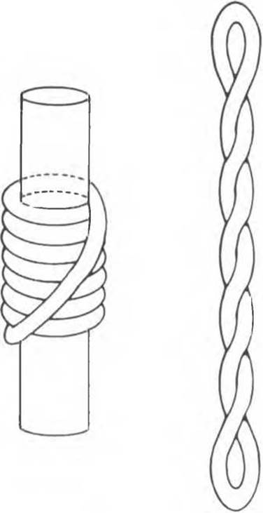
Fig. 38.20. DNA Supercoiling. A left-handed toroid (solenoid) superhelix (left) converts to a right-handed one upon removal of the cylindrical core. A similar transition occurs during nucleosome disruption caused by histone extraction with concentrated salt solutions.
In the S phase, mammalian cells contain a higher amount of polymerase $\alpha$ than in non-synthetic periods of the Cell Cycle. Furthermore, The activity of enzymes involved in generating DNA synthesis substrates (deoxynucleoside triphosphates) increases during the S phase. The activity of these enzymes drops upon exiting the S phase and remains at a low level until a signal to resume DNA synthesis arrives. Complete and strictly single-round replication of nuclear DNA occurs during the S phase. It appears that replicated chromatin is somehow marked, thereby preventing further replication until the cell passes through mitosis. One can hypothesize that methyl groups act as such covalent markers (i.e., DNA marking is achieved through its methylation).
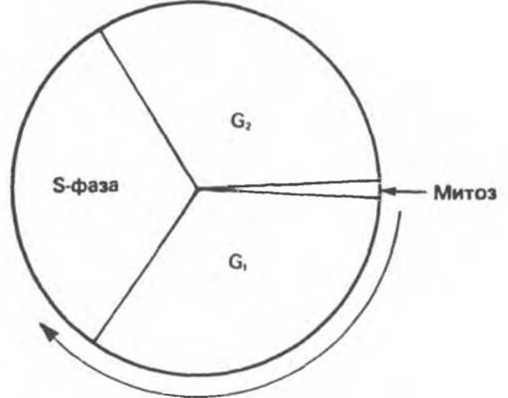
Fig. 38.21. Mammalian Cell Division cycle. The DNA synthesis phase (S phase) is separated from mitosis by the G1 and G2 periods. (The arrow indicates the direction of the cell cycle development.)
As a rule, each given chromosome pair replicates simultaneously and during a strictly defined interval of the S phase. The Nature of the signals regulating DNA synthesis at this level is unknown, but each individual chromosome apparently possesses such a regulatory mechanism.
DNA Degradation and Repair
The transmission of genetic information in an unaltered state is a crucial prerequisite for the survival of both the individual organism and the species as a whole. Consequently, an evolutionary system must have developed that enables the cell to correct DNA Lesions caused by replication errors or environmental damaging agents. It has been estimated that, As a result of Damage caused by these factors, an average of six nucleotide substitutions occur per year in the human germline genome. Somatic cells presumably experience a comparable number of Mutations per year.
As described in Chapter 37, the primary condition for accurate replication is the correct pairing of nucleotides. The fidelity of complementary interactions depends on whether purine and Pyrimidine nucleotides are in the tautomeric form favorable for pairing (Fig. 34.7). At equilibrium, the concentration of favorable tautomeric
forms of nucleotides exceeds that of unfavorable tautomers by $10^4$–$10^5$-fold. This is clearly insufficient to ensure error-free recognition. Therefore, bacterial and mammalian cells possess a specialized system for monitoring nucleotide pairing fidelity. This step is checked twice: first, during the incorporation of deoxynucleoside triphosphates into the growing chain, and second, after incorporation, using an energy-dependent mechanism to remove incorrectly inserted nucleotides from the newly synthesized DNA strand. Thanks to this control, incorporation errors occur no more than once every $10^8$–$10^6$ Base Pairs. In *E. coli* cells, this mechanism is provided by DNA polymerase possessing 3'→5' exonuclease activity. At the same time, mammalian DNA polymerases lack pronounced proofreading nuclease activity.
Environmental Physical and Chemical factors cause four types of damage in DNA (see Table 38.2). Damaged sites can be repaired, replaced via recombination, or remain unchanged. In the latter case, mutations arise that potentially lead to cell death. The possibility of repair and replacement is based on the redundancy of information encoded in The structure of double-stranded DNA: a defective region in One DNA strand can be repaired using the undamaged complementary strand.
The key moment in all recombination and repair events is the recognition of a defect, accompanied by either Direct Repair or marking for subsequent correction. The thermal lability of the purine $N$-glycosidic bond leads to DNA depurination at a frequency of about 5,000–10,000 per cell (per day) at 37°C. Depurination sites are recognized by specialized enzymes that specifically fill the gap without breaking the phosphodiester backbone of the molecule.
Table 38.2. Types of DNA Damage
|
I. Affecting single nucleotides A. Depurination B. Deamination of cytosine to uracil C. Deamination of adenine to hypoxanthine D. Alkylation of bases E. Nucleotide insertion or deletion F. Incorporation of a base analog |
|
II. Affecting a nucleotide pair A. UV-induced formation of thymine dimers B. Cross-linking by bifunctional alkylating agents |
|
III. Strand breaks A. Ionizing radiation B. Radioactive disintegration of the DNA backbone |
|
IV. Cross-links A. Between bases of the same strand or different strands B. Between DNA and protein molecules (e.g., Histones) |
Both cytosine and adenine bases spontaneously deaminate to form uracil and hypoxanthine, respectively. Since normal DNA contains neither uracil nor hypoxanthine, it is hardly surprising that specific $N$-glycosylases recognize these abnormal bases and remove them. The resulting break serves as a signal for the action of repair purine- or pyrimidine-specific endonucleases, which cleave the phosphodiester bond near the corresponding damaged site. Subsequently, through the sequential action of an exonuclease, repair DNA polymerase, and DNA ligase, the gap is filled, and the original correct structure is restored (Fig. 38.22). This chain of events is termed Excision Repair. The Repair of DNA containing alkylated bases and base analogs proceeds in a similar manner.
The restoration of deletions and removal of insertions occur through recombination processes, which can proceed either with or without replication.
Ultraviolet radiation induces the formation of pyrimidine-pyramidine dimers. This process predominantly affects stacked thymine bases of the same strand (Fig. 38.23). The cell employs two mechanisms to remove thymine dimers: excision repair and photoreactivation. This repair method involves the photoactivation by visible light of a specific enzyme that reverses the process leading to the Formation of the dimeric structure.
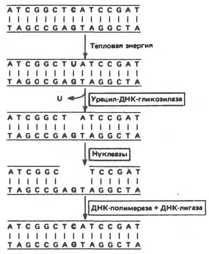
Fig. 38.22. The enzyme uracil-DNA glycosylase removes uracil arising from the Spontaneous deamination of cytosine. (Courtesy of V. Alberts.)
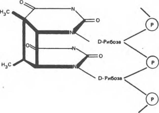
Fig. 38.23. Thymine-thymine dimer formed by the linking of adjacent thymine bases.
Single-strand DNA breaks caused by ionizing radiation can be repaired by direct ligation or recombination. The mechanisms involved in repairing cross-links between bases on opposite strands or between DNA and proteins remain poorly understood.
Thus, the repair of damage caused by ionizing radiation and base alkylation is carried out via excision and resynthesis of short DNA segments. The removal of ultraviolet-induced damage and cross-links is achieved in a similar manner, but in this case, longer stretches of DNA are involved. In mammalian cells, the occurrence of repair replication is evidenced by unscheduled DNA synthesis, i.e., the incorporation of radioactive precursors into DNA outside of the S-phase.
When excision repair processes are intensified in response to damaging agents, the activity of the enzyme poly(ADP-ribose) polymerase increases in mammalian cells. In the presence of the coenzyme NAD+, this enzyme catalyzes the ADP-ribosylation of chromatin proteins. Mono-ADP-ribosylation predominantly occurs, although the attachment of homopolymeric chains (ADP-ribose)n is occasionally observed. The exact function of poly(ADP-ribose) polymerase or its product — (ADP-ribose)n — during excision repair is not yet fully understood. A temporal correlation is observed between the intensification of repair processes and the increase in enzyme activity. Specific inhibition of this enzyme's activity prevents the sealing of breaks in the DNA chain. The elevation in poly(ADP-ribose) polymerase activity is apparently triggered by DNA fragmentation within The Nucleus. Such fragmentation can be induced primarily by physical agents (e.g., X-radiation); furthermore, it may occur at an inappropriately high rate during the repair of ultraviolet damage or damage caused by alkylating agents. The increase in enzyme activity caused by DNA breaks can be so substantial that it may lead to the depletion of intracellular NAD+ reserves.
Xeroderma pigmentosum is an autosomal recessive genetic disorder. The clinical syndrome includes sensitivity to sunlight (ultraviolet radiation), leading to multiple Skin Cancer lesions and a fatal outcome. This disease appears to be associated with defects in repair processes. In cultured cells from xeroderma pigmentosum patients, The rate of photoreactivation of thymine dimers is reduced. However, the underlying genetic defects leading to xeroderma pigmentosum comprise at least 7 complementation groups, indicating the multifactorial Etiology of this condition.
In xeroderma pigmentosum cells from most (if not all) complementation groups, abnormalities are observed in the rate and magnitude of the poly(ADP-ribose) polymerase response to ultraviolet irradiation. In cells belonging to at least one complementation group, the decrease in enzyme activity appears to be associated with an inability to cleave the DNA chain near the damaged site,
since The addition of deoxyribonuclease to such defective cells normalizes the level of poly(ADP-ribose) polymerase activity.
Patients with ataxia-telangiectasia (an autosomal recessive disorder leading to cerebellar ataxia and lymphoreticular neoplasia) exhibit increased sensitivity to X-irradiation. Patients with Fanconi anemia (an autosomal recessive anemia accompanied by an increased incidence of malignancies and chromosomal instability) likely have a defective cross-link repair system. All three described syndromes are characterized by an elevated tumor incidence. It is quite possible that other human diseases caused by defects in DNA Damage Repair systems will be identified in the near future.
Bauer W. R. et al. Supercoiled DNA, Sci. Am. (July), 1980, 243, 118.
Cantor C.R. DNA choreography, Cell, 1981, 25, 293. Igo-Kemenes T., Hörz W., Zachau H.G. Chromatin, Annu. Rev. Biochem., 1982, 51, 89.
Jelinek W. R., Schmid C. W. Repetitive sequences in eukaryotic DNA and their expression, Annu. Rev. Biochem., 1982, 51, 813.
Jongstra J. et al. Induction of altered chromatin structures by simian virus 40 enhancer and promoter elements, Nature, 1984, 307, 708.
Kornberg A. DNA Replication, Freeman, 1980.
Lindahl T. DNA repair enzymes, Annu. Rev. Biochem., 1982, 51, 61.
Loeb L. A., Kunkel T. A. Fidelity of DNA synthesis, Annu. Rev. Biochem., 1982, 51, 429.
McGhee J. D., Felsenfeld G. Nucleosome structure, Annu. Rev. Biochem., 1980, 49, 1115.
Nossal N. G. Prokaryotic DNA replication systems, Annu. Rev. Biochem., 1983, 52, 581.
Last update: 06/08/2026
Editorial and Educational Adaptation: This material has been compiled based on the primary/original source text. The project team performed an editorial review, corrected technical inaccuracies, structured sections, and adapted the content for an educational format.
What was processed:
- elimination of formatting defects (OCR errors, structural breaks, corrupted characters);
- editorial organization of content;
- standardization of terminology in accordance with academic sources;
- verification of factual statements against the original source text.
All mentions of the author, publication year, and origin of the primary text have been preserved in accordance with the source.