Biological Membranes - A. N. Ogurtsov 2012
Electrogenesis of Biomembranes
Action Potential
Properties of the Action Potential
Electrical nerve impulses (action potentials) in a living Organism transmit information from receptors to Brain Neurons and from brain neurons to Muscles. A living organism is an electrified system, and without electricity, there is no life.
The Action Potential was discovered before the Resting Potential. Animal electricity has been known for a long time. Discharges of the electric eel (occurring at voltages up to 600 V, with a current of about 60 A and a duration of about a millisecond) were used in medicine as far back as Ancient Rome to treat Gout, headache, and Epilepsy. The electrical Nerve Impulse was discovered by Luigi Galvani, a professor of anatomy in Bologna. The results of his electrophysiological experiments are presented in his book "Treatise on the Effects of Electricity on Muscular Motion" (1791).
Galvani discovered that Muscle contractions in the limbs of a dissected frog could be induced by an electrical impulse, and that the living system itself is a source of electrical impulses. Galvani's discovery played an outstanding role in The Development of physics, electrical engineering, electrochemistry, physiology, biophysics, and medicine.
However, the immense popularity of Galvani's ideas led to their vulgarization (galvanization of corpses, galvanism of touches and glances, etc.), which aroused skepticism among scientists toward Galvani's experiments. Galvani's younger contemporary, physics professor Alessandro Volta, was a fierce opponent of the idea of animal electricity (except for the special cases of electric fish: the electric eel and the electric ray). In his experiments, he excluded the biological object and demonstrated that an electric current could be obtained through the contact of a series of metals separated by an electrolyte (the voltaic pile). Thus, a chemical source of current was discovered (which was, however, later named a galvanic Cell in honor of his scientific opponent).
In the 19th century, a primitive notion took hold that electrical currents propagate along nerves like wires. However, In the second half of the 19th century, Helmholtz showed that the propagation speed of a nerve impulse is only 1-100 m/s, which is significantly lower than the speed of an electrical impulse propagating through wires (3 ∙ 108m/s). Therefore, by the end of the 19th century, the hypothesis of the electrical Nature of the nerve impulse was rejected by most physiologists. It was suggested instead that a chemical reaction propagates along nerve fibers.
Later, it was shown that the slow Propagation of the electrical nerve impulse is due to the slow charging of capacitors, which are represented by cell membranes, through high resistances. The membrane charging time constant т = RC is large because the membrane capacitance C and the nerve fiber resistance R are large.
That the nerve impulse is indeed an electrical current impulse was only proven by the mid-20th century, primarily through the work of the English physiologist Alan Hodgkin and his colleagues. In 1963, Alan Lloyd Hodgkin, Andrew Fielding Huxley, and John Carew Eccles were awarded the Nobel Prize in Physiology or Medicine "for their discoveries concerning the ionic mechanisms involved in Excitation and Inhibition in the peripheral and central portions of the nerve cell membrane".
An action potential is an electrical impulse caused by A change in the ionic permeability of the membrane and associated with the propagation of an excitation wave along nerves and muscles.
Experiments investigating the action potential were conducted (mainly by Hodgkin and his colleagues) on giant squid axons using the microelectrode technique with high-impedance voltage meters, as well as the labeled atom (tracer) method.
In the experiments investigating the action potential, two microelectrodes inserted into the axon were used (Figure 113).
Class="center">
Figure 113 - Schematic of the experiment investigating the action potential
An impulse with an amplitude V from a square-wave generator G is applied to the first microelectrode, changing the Membrane Potential. The membrane potential is measured using the second microelectrode with a high-impedance voltage recorder R.
A negative stimulating impulse causes a shift in the membrane potential only for a short time, after which the membrane potential quickly decays, and the resting potential is restored (Figure 114). An increase in the amplitude of the negative stimulating impulse is accompanied by membrane hyperpolarization.
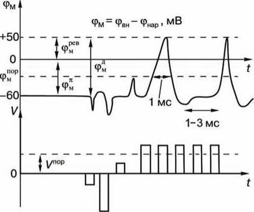
Figure 114 - Action potential: V - square wave pulse amplitude; Vпоp - threshold amplitude value; t - time, φМ - membrane potential; φвн, фнар - potentials on the inner and outer surfaces of the membrane; φМД - action potential amplitude; φМ - resting potential level; φМпор - threshold membrane potential value; φМрев - reversal membrane potential
An action potential is also not generated when the stimulating impulse is positive (depolarizing) but its amplitude is less than the threshold value Vпор.
However, if the amplitude of the positive, depolarizing impulse exceeds the value Vпор, φм becomes greater than φпорМ, and a process develops in the membrane that results in a sharp increase in the membrane potential, and the membrane potential φм even reverses its sign—becoming positive (φвн > φнар)—resulting in membrane depolarization.
After reaching a certain positive value φревМ—the reversal potential—the membrane potential returns to the resting potential value φПМ, undergoing something like a damped oscillation. In nerve fibers and skeletal muscles, the duration of the action potential is about 1 ms (and in The cardiac muscle, about 300 ms).
After the excitation is removed, certain residual phenomena are observed in the neuronal membrane for another 1–3 ms, during which the membrane is refractory (unexcitable).
A new depolarizing potential V > Vпop can trigger a new action potential only after the membrane has completely returned to its resting state. Furthermore, the amplitude of the action potential φДМ = |φПМ | + |φревМ| does not depend on the amplitude of the depolarizing potential (provided that V > Vпop).
If the membrane is polarized at rest (the Cytoplasm potential is negative relative to the extracellular medium), then upon excitation, membrane depolarization occurs (the potential inside The Cell becomes positive), and after the excitation is removed, membrane repolarization takes place.
Characteristic Properties of the action potential:
1) the presence of a threshold value for the depolarizing potential;
2) the "all-or-none" law, meaning that if the depolarizing potential exceeds the threshold, an action potential develops, the amplitude of which is independent of the stimulating impulse amplitude, and no action potential occurs if the depolarizing potential amplitude is below the threshold;
3) There is a refractory period of membrane inexcitability during the Development of the action potential and aftereffects following the cessation of excitation;
4) at the moment of excitation, the membrane resistance decreases sharply (in the squid axon, the membrane resistance decreases 40-fold, from 0.1 Ohm∙m2 at rest to 0.0025 Ohm∙m2 during excitation).
Last update: 13/08/2026
Editorial and Educational Adaptation: This material has been compiled based on the primary/original source text. The project team performed an editorial review, corrected technical inaccuracies, structured sections, and adapted the content for an educational format.
What was processed:
- elimination of formatting defects (OCR errors, structural breaks, corrupted characters);
- editorial organization of content;
- standardization of terminology in accordance with academic sources;
- verification of factual statements against the original source text.
All mentions of the author, publication year, and origin of the primary text have been preserved in accordance with the source.