Orthopedics - Oleksa A.P. 2006
Joint Diseases
Gout
Gout (gout, Gicht) results from an abnormally elevated concentration of uric acid in human Body Fluids. The clinical syndrome of gout comprises four core elements:
1. Acute recurrent inflammation of The Musculoskeletal System (gouty attacks).
2. Tophaceous deposits (gouty nodules).
3. Chronic, progressively worsening joint changes (arthropathy).
4. Pathological alterations in the Urinary System (calculi, chronic nephritis).
Biochemical Blood analysis reveals characteristic hyperuricemia. The pathognomonic sign of gout is the presence of uric acid crystals in Tissues, which can sometimes be visualized through thinned Skin near the joints (Fig. 323) or via histological examination of tophi (Fig. 324).
Uric acid is the end product of purine METABOLISM in The Human Body. Purines are derived from the Catabolism of tissue and dietary Nucleoproteins. Synthesis begins with The formation of 5-phosphoribosyl pyrophosphate. Purines are subsequently metabolized into uric acid. It is believed that the majority of uric acid is synthesized in the Liver, about 25% enters the Small Intestine directly, and the remainder enters the bloodstream. In the intestines, bacterial action drives the further breakdown of uric acid (25%), known as uricolysis. Some of these breakdown products are excreted in the feces, while others are reabsorbed into the bloodstream and eliminated via the Kidneys and Lungs. The portion of uric acid that enters the bloodstream is excreted in the urine as uric acid and partially as urea.
Uric acid is a weak acid. At the normal pH of body fluids, it dissociates. The urate ion binds with a cation to form a monosodium salt. In an acidic environment, this salt dissociates back into free uric acid. Urea is more readily soluble than the acid form. Both uric acid and its salt can create a supersaturated solution from which crystals precipitate, a process facilitated by shifts in environmental pH.
In body fluids, uric acid exists exclusively in the form of urates. Because it is bound to Proteins, it passes through the Glomerular Filtration barrier of the kidneys. Since the pH in the renal tubules is lower than that in the blood, free uric acid is formed, with its quantity dependent on urinary acidity. This free uric acid is reabsorbed and enters the bloodstream.
Normally, The amount of uric acid filtered in the renal glomeruli is estimated to exceed 6.7 mg per minute (Gutman A., 1965), and the amount reabsorbed can reach up to 15 mg per minute.
Using isotopic tracers (Benedict J.D. et al., 1953; Sorensen L.B., 1962), it has been established that in healthy individuals, body fluids contain no more than 6 mg of uric acid per 100 mL, with a total body pool averaging 1,200 mg. The body produces 500–1100 mg of uric acid daily, and urinary excretion averages 420 mg on a low-purine diet (Gutman A.), up to 730 mg on a normal diet (Cottet J., Canarelli), and even higher when consuming purine-rich foods (such as meat, veal, or liver)—although Wyngaarden J.B. (1964) notes that a high-purine diet does not play a primary role in The Development of hyperuricemia.
The normal level of uric acid in body fluids depends on regular Purine Biosynthesis, normal nucleoprotein breakdown, and proper renal excretion of uric acid. Metabolic Disorders or renal diseases lead to hyperuricemia.
Class="center">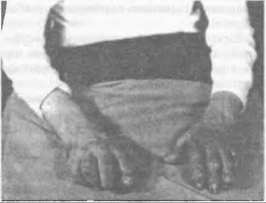
Fig. 323. Trophic skin changes in gout, showing translucent uric acid crystals near the JOINTS OF THE hands.
However, uricaemia of unclear aetiology has also been observed (Ciba T., 1962; Ravault P. et al., 1964; Ryckewaert A. et al., 1966) in coronary artery disease, arterial Hypertension, rheumatoid Arthritis, hypoparathyroidism, and hyperthyroidism. Uric acid levels tend to be higher in obese individuals and are more common in men than in women.
Thus, while hyperuricemia is the definitive cause of gout, it is not synonymous with the disease and can occur in other conditions. Among individuals with chronic hyperuricemia, only 20% develop clinical gout, and about 2% develop renal or hematological disorders (Wyngaarden J., 1961; Seegmiller J. et al., 1963).
Depending on the underlying cause, gout is classified as primary or secondary. Secondary gout refers to cases that develop as a consequence of another confirmed medical condition, accounting for about 10% of all gout patients. Most commonly, secondary gout is triggered by blood disorders (hemolytic anemia, leukemia, drug- or cytostatic-induced Cell breakdown) or chronic Kidney diseases (renal failure, Pregnancy toxemia, diuretic and sulfathiazole medications, etc.).
It is worth noting that hyperuricemia very rarely results from Diuretics, the administration of which is actually used to relieve a gouty attack.
In secondary gout, crystal deposition in superficial tissues is less frequent, and the condition clinically manifests as recurrent acute joint inflammation (gouty attacks). However, the clinical picture may be dominated by the primary underlying disease and its complications rather than the manifestations of gout.
Primary gout typically occurs in men As a result of impaired purine biosynthesis and hyperuricemia. Yet, in approximately 90% of patients with primary gout, the underlying ROOT cause cannot be clinically established.
Gout may be genetically inherited in a dominant or, less frequently, recessive manner (Blumberg B.S., 1965). Among all gout patients, 10–45% have close relatives who also suffer from the disease, while asymptomatic hyperuricemia is detected in 25–72% (Talbott J., 1997; Wyngaarden J., 1968). Some authors attribute the onset of primary gout to selective Renal Dysfunction. Two main hypotheses exist: one proposed by Nugent et al. and Liddle et al., suggesting that the primary cause of hyperuricemia is inadequate renal excretion of uric acid; and a second, proposed by Gutman and Yu, stating that hyperuricemia may arise from a deficiency in renal glutaminase, which normally breaks down glutamine—resulting in its accumulation and enhanced uric acid synthesis. Inadequate ammonium production has also been proposed to explain the low urinary pH (Cottet J. and Canarelli G., 1965) observed in the majority of gout patients (Gutman A., Yu T.F., 1963; Vries de A. et al., 1964).
Gout most frequently affects men aged 30 to 50 years. The primary symptoms of gout are caused by the penetration of urate and uric acid crystals into tissues. The initial manifestation is acute inflammation within the musculoskeletal system. A prolonged disease course eventually leads to tophus formation and arthropathy. Occasionally, an attack may be preceded by Renal Colic, though this alone does not indicate gout, even in the presence of hyperuricemia. A definitive Diagnosis of gout is made only when a classical attack occurs. This means that renal changes do not always signal gout, though they often appear sooner and more frequently than tophi and arthropathy. Research by McCarty and others demonstrated that microscopic analysis of synovial fluid in all gout patients revealed urate crystals, whereas none were found in patients with rheumatoid arthritis, even though many of the latter had hyperuricemia. During a gout attack and immediately afterward, all crystals were located inside leukocytes, whereas during remission, they were typically extracellular.
Based on his research, Seegmiller proposed the following hypothesis regarding the Pathogenesis of a gouty attack. Acute inflammation results from a supersaturated solution of urates in the periarticular fluid. A more acidic environment promotes crystal precipitation within tissues, triggering the phagocytosis of these crystals. During phagocytosis, leukocytes produce more lactic acid than normal, which increases tissue acidity, thereby attracting fresh crystals and further stimulating phagocytosis. This creates a vicious cycle in which phagocytosis acts as a driver of inflammation up to a certain point. However, phagocytosis cannot always eliminate all urate crystals, causing them to accumulate in the tissues (Fig. 324).
Factors that provoke gouty attacks include adverse meteorological conditions, such as barometric pressure fluctuations and high humidity.
Clinical manifestations of gout. The disease typically begins abruptly in the middle of the night with sharp pain in the big toe, waking the patient. Body Temperature rises to 38–39°C, and fever may develop. The toe swells significantly, the skin turns bluish-red, and active movement becomes impossible due to intense pain, while passive movement sharply exacerbates joint pain. By morning, the pain subsides and body temperature drops. The following night, the attack recurs, and this pattern can persist for a week or longer. Onset involving multiple joints is rare. With each successive attack, the intensity of clinical symptoms gradually diminishes, and the process goes into remission for several weeks or months before recurring in other joints and tissues. Recurrences of acute inflammation are characteristic of the clinical course of gout.
If the first attack begins in the joint of the first toe (Fig. 325), subsequent regular attacks involve other joints of the FOOT, the ankle, sometimes the knee, and later the finger joints, elbow, and wrist. As a rule, the hip and shoulder joints are rarely or never affected by gout.
It should be noted that over time (over the years), The Nature of the attacks changes. Relapses become more frequent in patients with elevated plasma uric acid levels (above 9-10 mg%), last longer each time, and the inflammatory symptoms in multiple joints become increasingly pronounced. While the initial attack may leave no traces on the affected area, each subsequent flare-up increases the amount of crystals deposited in the tissues, forming characteristic nodules (tophi) around the joints. Over time, these may even become visible through thinned skin, especially on the fingers (Fig. 323). Clinically, the disease manifests as chronic tophaceous gout.
A pathognomonic sign of gout is the formation of identical crystal nodules in the Cartilage of the auricular concha (Fig. 326).
Before another gout attack, immediate clinical precursors sometimes occur: renal colic, paresthesias or a feeling of tension in the periarticular tissues, and generalized weakness. The exact trigger for a subsequent attack is unknown, but it is believed that alcohol consumption, heavy fatty meals before Sleep, infectious inflammatory processes, physical and psychological trauma, and sharp changes in atmospheric pressure may contribute to it.
Additional Diagnostic Methods. Gout presents with characteristic radiological findings. Radiographs reveal round or oval marginal defects (punched-out erosions, geodes) with clear contours, as if punched out with a die, featuring sclerotic margins in the articular ends of the hand or foot bones. These are sites of urate deposits, which are radiolucent. When large joints are affected by gout, radiographs typically do not show such changes.
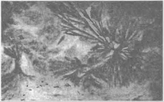
Fig. 324. Uric acid crystals under histological examination (x500, by E. Maiyk) of a gouty tophus.
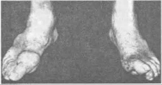
Fig. 325. Gouty inflammation of the first metatarsophalangeal joints of both feet: left — affected joint, right — scars at the site of crystal rupture in a similar joint in gout.
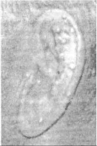
Fig. 326. Gouty tophi in the auricle.
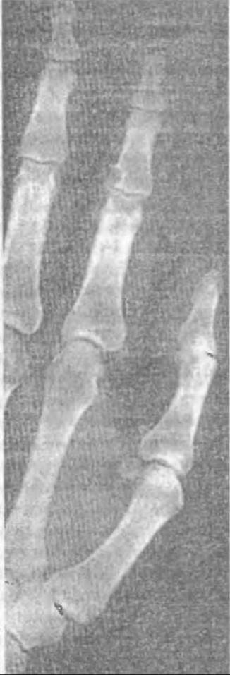
Fig. 327. Radiograph of a gouty geode in the middle phalanx of the second finger.
Laboratory tests. During a gout attack, the ERYTHROCYTE SEDIMENTATION RATE (ESR) and plasma mucoid levels increase significantly, and C-reactive protein appears.
Occasionally, dysproteinemia can be detected—hypoalbuminemia and atypical deviations in the globulin fraction, along with short-term neutrophilic leukocytosis (10-15 thousand/mm³). An accelerated ESR is a sufficient indicator of inflammation, remaining elevated as long as clinical signs of inflammation persist. Treatment resolves the inflammation and decreases the ESR, though in chronic gout, it may decline slowly (over several weeks). During an attack, blood uric acid levels may not show changes, while intracellular leukocytic urate crystals can sometimes be detected in the synovial fluid.
Gouty arthropathy
Chronic pathological joint changes in gout develop as a result of urate deposition in the articular cartilage, bones, and periarticular tissues. When a significant amount of exudate accumulates in the joint, urates are released and settle in the superficial layers of the cartilage, after which crystals penetrate deeper into the cartilage, causing its delamination and destruction. This can eventually lead to the destruction of the subchondral plate and penetration into the bone epiphyses.
From the synovial fluid and directly from the blood, urate crystals are deposited in the synovial membrane. In the chronic course of the disease, this leads to the proliferation of synovial villi with the formation of a pannus that impairs cartilage Nutrition and penetrates deep into its Structure.
Initially, small round defects form near the articular surfaces (Fig. 327); these enlarge, and their size depends on the amount of deposited urate crystals and the degree of bone destruction. This can cause subluxations and dislocations in the joints.
In some inadequately treated patients, crystals erode the bone to such an extent that significant defects form at the articular ends.
In chronic disease progression and frequently recurring flare-ups in untreated patients, severe destructive joint changes occur. Such patients become disabled due to both renal failure (urolithiasis) and arthropathy.
The course of gout tends to be more severe in patients whose first attack occurred before the age of 30, and generally milder when the initial attack happens after 60.
The proportion of women with gout does not exceed 10%, and their first attacks most commonly appear during menopause, differing in no way from the clinical course in men.
The average life expectancy of individuals with gout does not differ from the general population; however, they often suffer from comorbid conditions (arterial hypertension, Diabetes Mellitus, etc.) that can become the cause of death.
Treatment. The management of a gout patient encompasses three complementary measures:
1) treatment during gout attacks and for their Prevention;
2) treatment to reduce uric acid levels in the body;
3) treatment of affected joints and kidneys.
The treatment plan depends on the frequency and intensity of gout attacks, the degree of kidney damage, uric acid levels, and the extent of urate crystal deposition.
During an acute attack, colchicine is considered the drug of choice because it is specific to gout and can serve as a diagnostic test to clarify joint disorders of undetermined Etiology.
The MECHANISM OF ACTION of colchicine has not yet been fully elucidated, but based on laboratory experiments, Seegmiller J.E. et al. (1963) concluded that its anti-inflammatory effect in gout consists in the inhibition of phagocytosis.
Colchicine is available in 0.5, 0.6, and 1.0 mg tablets (dragees) and for intravenous administration as 3 mg in 2 ml of an aqueous solution. It is dosed at 1 mg and prescribed as follows:
1) on the first day — four times: the first three doses every two hours, and the fourth dose before bedtime; if colchicine is administered by injection, a single slow intravenous dose of 3 mg is given;
2) on the second day — three times 1 mg at equal time intervals, or four times if no improvement has occurred after the first day;
3) on the third day — in the same doses two or three times at equal intervals;
4) on the fourth and fifth days — twice or once, with the last dose always to be taken before bedtime; the total dose of colchicine over the five days should not exceed 15 mg;
5) from the sixth day, 1 mg of colchicine is taken once before bedtime, even if all signs of inflammation have disappeared.
Maintenance therapy is carried out for several weeks or longer, depending on the severity of the disease. If colchicine is used in a timely manner, inflammation resolves within three to five days. Sometimes biochemical parameters improve simultaneously as well.
Diarrhea may occur on the 3rd–4th day of treatment following the intake of colchicine, in which case the dose should be reduced. Cases of alopecia, leukopenia, thrombocytopenia, gastric ulceration, hepatitis, and even death have been described, which are associated with excessively high prescribed doses of colchicine.
In cases of gastric or duodenal ulcer, the patient is prescribed colcemid (demecolcine), a derivative of colchicine. However, The Use of colcemid in similar doses requires caution due to its potent cytostatic effect, and therefore it is administered only for short periods.
It should be noted that phenylbutazone relieves inflammation faster than colchicine, but it may cause even more complications than colchicine. Consequently, it is prescribed for severe attacks or when prolonged treatment with colchicine is insufficiently effective.
Phenylbutazone is so potent that a single intramuscular injection of 0.6 relieves the acute manifestations of gout. Sometimes repeated injections are required over the next two days, and then every other day — for a total of no more than six injections. Colchicine must not be used while taking phenylbutazone to avoid inducing granulocytopenia. If absolutely necessary, phenylbutazone is administered in the same dose every 7–10 days for several weeks.
During an acute attack, colchicine can be replaced by indomethacin tablets of 25 mg, starting with a dose of 200 mg per day, gradually reducing it by 50 mg daily, after which prophylactic doses of colchicine are prescribed.
Corticosteroids are used very rarely, although during acute inflammation or relapses, alongside colchicine treatment, local para-articular administration of hydrocortisone (25 mg) or Kenalog-40 once every 4–5 days is sometimes performed, yielding good results.
Salicylates are unsuitable for treating gout at doses up to 3.0 g per day because they do not inhibit uric acid excretion.
After the acute inflammation subsides, It is important to prevent its recurrence using prophylactic doses of colchicine and reducing uric acid levels in the body by prescribing uricosuric agents and drugs that decrease its synthesis.
As stated, from the sixth or seventh day of colchicine treatment, the dose is reduced to 1.0–0.5 mg per day and the course is continued until the ESR decreases and clinical manifestations disappear. However, if prodromal symptoms of an attack appear while taking prophylactic doses, without waiting for its clinical manifestation, the dose is increased to the therapeutic level for 2–3 days and then reduced to the maintenance dose.
Colchicine eliminates inflammation, but does not affect the synthesis and amount of uric acid in the body; therefore, uricosuric drugs and those that inhibit its synthesis are prescribed.
Uricosurics are prescribed to patients who have developed crystal deposits in tissues, geode formations, and arthropathy, and when the uric acid level reaches 8–10 mg%.
When the uric acid level is 6–8 mg% in the absence of crystal nodules, the use of uricosuric agents is relatively indicated. Furthermore, uricosuric drugs are not prescribed for primary hyperuricemia without clinical manifestations of gout. Several months after starting uricosuric therapy, the blood uric acid level decreases significantly, and crystal nodules may even disappear. Therefore, uricosuric agents are sometimes taken for years.
Indications for prescribing drugs that inhibit uric acid synthesis include renal failure, urolithiasis, high serum uric acid levels, and the ineffectiveness of diuretics. Allopurinol is prescribed at 100 mg twice daily, and if necessary, 200 mg three times daily. The maximum daily dose is 600 mg.
Allopurinol inhibits xanthine oxidase function, thereby reducing The production of uric acid. When taking colchicine, the dose of allopurinol is gradually increased at weekly intervals.
It should be noted that under The Influence of this treatment, small tophi in the tissues may disappear, but large ones persist, and pathological changes that have developed in the joints remain unchanged. Therefore, tophi are sometimes removed and arthropathic joint deformities are corrected surgically.
Patients should consume a low-calorie diet and avoid foods containing significant amounts of purines (liver, meat broths, fish, herring, etc.). Alcoholic beverages should also be avoided.
Patients may undergo the same physiotherapeutic and balneological Procedures as those used for degenerative-dystrophic joint conditions.
Last update: 10/08/2026
Editorial and Educational Adaptation: This material has been compiled based on the primary/original source text. The project team performed an editorial review, corrected technical inaccuracies, structured sections, and adapted the content for an educational format.
What was processed:
- elimination of formatting defects (OCR errors, structural breaks, corrupted characters);
- editorial organization of content;
- standardization of terminology in accordance with academic sources;
- verification of factual statements against the original source text.
All mentions of the author, publication year, and origin of the primary text have been preserved in accordance with the source.