Molecular Biology of the Cell - Volume 3 - Alberts B., Bray D., Lewis J., Raff M., Roberts K., Watson J. 1994
From Cells to Multicellular Organisms
Cellular Mechanisms of Development
Drosophila and the Molecular Genetics of Pattern Formation
Classical genetics is entirely built on the premise that The Structure of any Organism is controlled by its genes. Although The Role of DNA in heredity has been known for over a century, the Genetic control mechanisms responsible for shaping the normal body structure of adult animals have remained elusive until recently. Over the past few years, however, this gap in our knowledge has begun to close. New breakthroughs have come primarily from studying The Development of the fruit fly, Drosophila. Experiments on Drosophila led to the discovery of a Class of development-controlling genes whose specific function is to establish the Spatial Organization of the body. Combining classical and molecular genetic approaches has helped elucidate the functions of these genes. Below, we will demonstrate that the general strategy for body plan patterning in vertebrates is remarkably similar to that of Drosophila, and furthermore, vertebrates possess close homologues of the genes that control this process in flies.
The initial insights into the existence of such a genetic system emerged from the discovery of Mutations that cause bizarre disruptions in the body plan of adult Drosophila. For instance, the Antennapedia mutation causes legs to grow on the HEAD instead of antennae (Fig. 16-53); likewise, the bithorax mutation replaces the halteres with an extra pair of wings. Such mutations, which transform certain body parts into structures that normally belong elsewhere, were termed homeotic, and the normal genes subject to such mutations are called homeotic selector genes. The discovery of homeotic mutants spurred ingenious experiments showing that the normal fly body is built rather like a patchwork quilt, with each patch expressing a distinct combination of homeotic selector genes. The products of these genes act as molecular addresses, providing Cells with initial positional values. A homeotic mutation thus disrupts the normal delivery of information to all cells within a given region: the cells are misinformed about their Location, leading them to develop into structures that normally reside in other PARTS OF THE body.
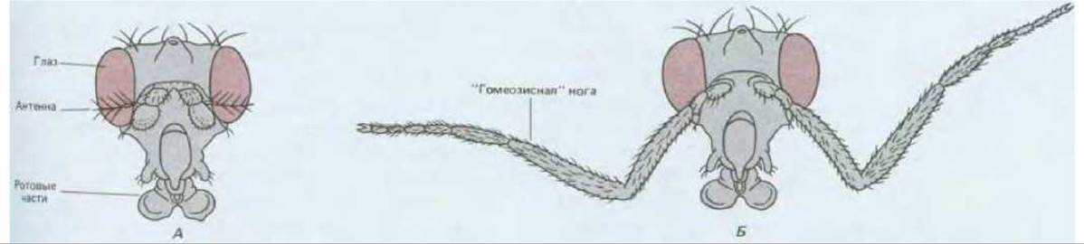
Fig. 16-53. Head of a normal adult Drosophila (A) and a Drosophila carrying the homeotic Antennapedia mutation (B). An extreme form of this mutation; usually, only a part of the antenna is transformed into a leg structure. (The mutant Antennapedia illustration is based on a photograph kindly provided by Peter Lawrence.)
Homeotic selector genes represent just one component of the complex genetic machinery responsible for assembling the insect's normal mosaic body plan. In this section, we will describe the system as a whole and briefly review some of its underlying molecular mechanisms, drawing on METABOLISM/2.html">THE CONCEPT OF pattern formation discussed in the previous section. We will see that this system consists of three classes of genes that control pattern formation:
1) egg-polarity genes act first, their products establishing the spatial coordinates of the embryo by Setting up a morphogen gradient within the egg;
2) segmentation genes interpret the positional information provided by the primary morphogen gradients, carving up the embryo into a series of segments—the fundamental modules that make up the body of all insects; and
3) segmentation genes, through their products, influence the expression of homeotic selector genes, which differentiate the segments from one another. Through the combined activity of homeotic selector and segmentation genes, the cells of each segment memorize specific positional values via imprinting, which governs their subsequent behavior. Finally, within each segmented region of the body, local Cell-cell communications fine-tune the minutest details of the adult organism, likely guided by the intercalation rule.
16.5.1. The Insect Body Is Formed by Modifying a Basic Plan Featuring Repeating Segments [45]
The development of Drosophila from egg to adult is illustrated in Fig. 16-54. Embryogenesis begins upon Fertilization and takes about 24 hours, at the end of which the embryo hatches into a larva. Larval development proceeds through three stages known as instars; each instar ends with a molt, during which the old cuticle is shed and a new, larger one is formed. At the end of the third instar, the larva pupates. Inside the pupal case, a dramatic restructuring of all Tissues takes place, and 9 days after fertilization, the adult fly, or imago, emerges.
The body of the fly consists of a head, three thoracic segments designated T1–T3, and nine abdominal segments designated A1–A9. Each segment is characterized by a general ground plan while possessing distinct features of its own. Segment T1, for example, bears a pair of legs; T2 bears a pair of legs and a pair of wings; and T3 bears a pair of legs and a pair of halteres—small, club-shaped balancers essential for flight (which evolved from the second pair of wings found in more primitive insects). This quasi-repeating segmentation is more pronounced in the larva, where the segments appear more uniform; at earlier embryonic stages, the head rudiments and regions destined to form the adult structures also appear segmented (Fig. 16-55). Segment boundaries in this context are somewhat conventional, governed by specific descriptive conventions. When discussing Gene Expression patterns, we will see that it is standard to speak of 14 parasegments (designated P1–P14), which represent half the number of traditionally defined segments (Fig. 16-56). Finally, both extremities of the animal bear highly specialized structures of non-segmental origin.

Fig. 16-54. Diagram of Drosophila development from egg to adult fly.

Fig. 16-55. Emergence of Drosophila body segments during embryonic development, shown in side-view drawings (A–C) and corresponding scanning electron micrographs (D–F). (A, D) After 2 hours of development, the embryo is at the syncytial blastoderm stage (see Fig. 16-57). No segmentation is visible yet, although a presumptive fate map outlining future segments can already be drawn (colored areas). (B, E) After 5–8 hours of development, the embryo undergoes germ band elongation: Gastrulation has occurred, segments begin to appear, and the segmented body axis elongates, bending at the tail end to conform to the egg casing. (C, F) After 10 hours of development, the body axis shortens and straightens out again, and all segments are clearly defined; the head structures visible on the larval surface at this stage temporarily retract inward, only to reappear after pupariation. (D, E: courtesy of Rudi Turner and Anthony Mahowald; F: courtesy of Jane Petschek.)
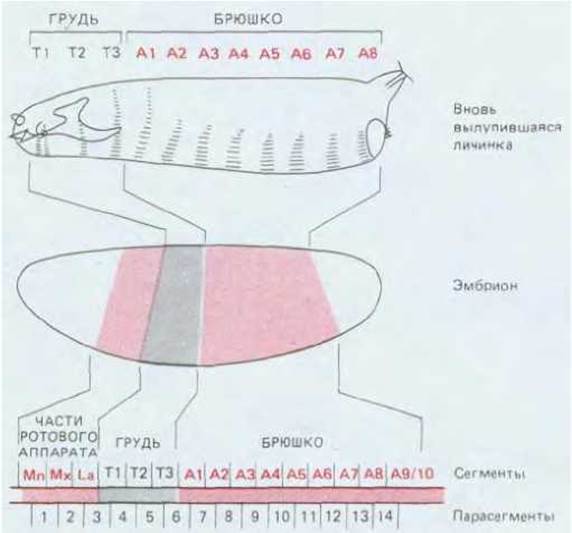
Fig. 16-56. Segments of the Drosophila larva and their correspondence to blastoderm regions. Note that the terminal Regions of the blastoderm correspond to unsegmented structures that largely form the Internal Organs of the larva, as well as the segmented mouthpart rudiments of the adult fly. Segmentation in Drosophila can be described using either "segments" or "parasegments"; their relationship is indicated at the bottom of the figure. The exact number of abdominal segments remains debated: eight are well-established, but a ninth likely exists. Mn, Mandible; Mx, Maxilla; La, labium.
The overall body plan—featuring two specialized termini and a series of modulated repeats of a basic unit—arises from processes that unfold within the egg and early embryo During the first hours after fertilization.
16.5.2. Drosophila Development Begins with The formation of a Syncytium [46]
A Drosophila egg is about 400 µm long and 160 µm in diameter, and it exhibits strong polarity. As in many other insects, early development is rather unusual: successive rounds of nuclear division not accompanied by cytoplasmic division give rise to a syncytium. The early nuclear divisions are synchronous and rapid, occurring every 8 minutes. The first 9 divisions produce a cluster of nuclei, most of which migrate from the center of the egg to the periphery, where they form a monolayer known as the syncytial blastoderm. Following the next 4 nuclear divisions, Plasma Membranes grow inward from the egg surface to enclose each Nucleus. This converts the syncytial blastoderm into the cellular blastoderm, which contains about 5,000 individual cells (Fig. 16-57). A small cluster of nuclei located at the very posterior tip of the egg cellularizes a few cycles earlier. These pole cells are the primordial Germ Cells that will give rise to future generations of eggs and sperm. Just as in amphibian Cleavage, the high rate of DNA Replication suppresses Transcription; consequently, up to the cellular blastoderm stage, development relies primarily (though not entirely) on maternal mRNAs and Proteins stockpiled in the oocyte prior to fertilization. Once cellularization is complete, cell divisions adopt a more conventional, asynchronous pattern, slow down significantly, and transcription rates increase dramatically.
The Drosophila embryo at the cellular blastoderm stage corresponds to the hollow blastula of amphibians or sea urchins, with the key difference that amphibian and sea urchin blastulae are fluid-filled, whereas the center of the insect embryo at this stage is packed with yolk. Gastrulation begins immediately after cellularization. Although insect gastrulation exhibits highly specialized geometry, the overall outcome is similar: through coordinated cell movements along the embryonic axis, an endodermal gut tube forms, surrounded by mesoderm, which occupies the space between the gut tube and the outer ectodermal germ layer. During gastrulation, the long axis of the embryo first stretches ("germ band elongation") and then shortens again ("germ band retraction"); this elongation causes the body axis to curve to fit within the confines of the egg envelope (Fig. 16-55).

Fig. 16-57. Development of the Drosophila egg from fertilization to the cellular blastoderm stage. [H. A. Schneiderman, in Insect Development (P. A. Lawrence, ed.), pp. 3–34, Oxford, U.K., Blackwell, 1976.]

Fig. 16-58. Presumptive fate map of the Drosophila embryo at the cellular blastoderm stage (lateral view and cross-section), revealing the relationship between the dorsoventral differentiation of major tissue types and the spatial arrangement of segments along the anteroposterior axis. The thickened line outlines the region that gives rise to segmented structures. During gastrulation, cells along the ventral midline invaginate to form the mesoderm, whereas cells destined to form the digestive tract invaginate near both ends of the embryo. (V. Hartenstein, G. M. Technau, J. A. Campos-Ortega, Wilhelm Roux Arch. Dev. Biol., 194:213–216, 1985.)
By observing cell behavior during complex gastrulation movements, a presumptive fate map can be drawn for the monolayer of cells located On the surface of the blastoderm (Fig. 16-58). The fate map appears simplest in transverse sections through the middle of the embryo; here, the prospective mesoderm lies ventrally, while the prospective ectoderm is situated on either side of it. In this case, as in vertebrates, an elongated cluster of neural cells—derived from a portion of the ectoderm—runs along the entire body axis. In insect development, we find stages corresponding not only to gastrulation and neurulation, but also to somite formation: upon the completion of gastrulation, grooves and ridges appear on The surface of the embryo, marking the subdivision of the body into parasegments along the anteroposterior axis (Fig. 16-55). More precise assays show that the fundamental features of this segmental organization are already determined at the cellular blastoderm stage, prior to the onset of gastrulation.
16.5.3. The Embryonic Body Plan Is Controlled by Two Orthogonal Coordinate Systems [47]
To simplify orientation on the blastoderm, it is customary to use two coordinates, analogous to latitude and longitude. Genetic tests have also demonstrated that spatial organization is established through two mechanisms: one determines organization along the dorsoventral axis, and the other along the anteroposterior axis. The dorsoventral axis is controlled by a group of approximately 20 genes; mutations in any of these genes result in embryos that are either “dorsalized” (i.e., lacking ventral structures) or “ventralized” (i.e., lacking dorsal structures). Another group of about 50 genes—which includes the segmentation genes and homeotic selector genes mentioned earlier—controls spatial organization along the anteroposterior axis. Mutations in these genes lead to the development of embryos in which certain elements along the anteroposterior axis are either missing or duplicated. Although many genes from both groups have now been cloned and their protein products identified, we will restrict our Discussion of embryonic spatial organization to the anteroposterior axis alone.
16.5.4. The Anteroposterior Polarity of the Embryo Is Controlled by Signals from Both Ends of the Drosophila Egg [48]
The initial polarity of the egg is governed by the distribution of maternal substances stored during oogenesis while the oocytes are in the Ovary (Fig. 16-39). If a tiny puncture is made at the anterior pole of a Drosophila egg to allow a small amount of Cytoplasm from the extreme anterior tip to leak out, the embryos lose the capacity to form head structures. Furthermore, if posterior cytoplasm from another egg is injected in place of the lost anterior cytoplasm, a second set of abdominal structures with reversed polarity arises in the anterior half of the recipient egg (Fig. 16-59).
Mutations leading to such defects in the anterior or posterior regions of the embryo have been identified. The egg-polarity genes discovered through these mutants represent the first elements of the hierarchical genetic system involved in establishing spatial organization along the anteroposterior axis. Egg-polarity genes belong to the class of genes transcribed in the maternal genome during oogenesis; their stored products begin to function shortly after fertilization. For this reason, the embryonic phenotype is determined by the maternal alleles rather than by the combination of paternal and maternal alleles present in the embryo itself. Genes expressed in this manner are referred to as maternal-effect genes.
Mothers homozygous for the bicoid egg-polarity mutation produce embryos lacking a head and thoracic structures, causing the abdominal structures to occupy an unusually large fraction of the entire body. Conversely, the oskar egg-polarity mutation results in embryos completely devoid of abdominal segments. (The non-segmented structures at both body ends occupy a special position: they are unaffected by these mutations, but can be lost As a result of torso and certain other mutations.)
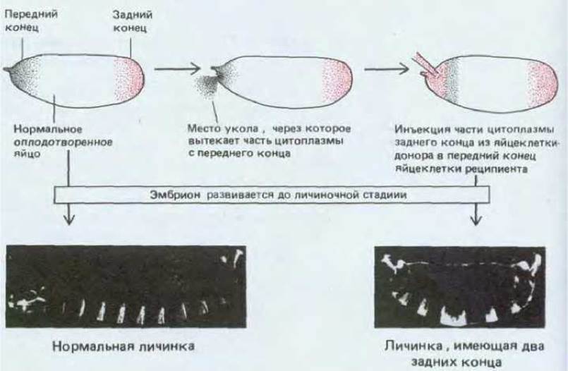
Fig. 16-59. Localized determinants at the poles of the Drosophila egg determine its anteroposterior polarity. If a small patch of cytoplasm is removed from the anterior end of the egg and replaced with cytoplasm from the posterior end, an abnormal larva with two posterior ends is produced (right photo); a normal larva is shown on the left. The larvae are photographed in dark-field illumination. [H.J. Frohnhofer, R. Lehmann, C. Nüsslein-Volhard, J. Embryol. Exp. Morphol., 97 (suppl.): 169–179, 1986.]
The bicoid and oskar mutations result in the absence of the corresponding gene products. These defects can be rescued by injecting normal cytoplasm into the mutant embryos. bicoid mutants develop relatively normally after the injection of anterior cytoplasm from normal embryos into the anterior pole of the mutant egg, whereas oskar mutants are rescued by the injection of posterior cytoplasm from a normal embryo into the prospective abdominal region of the mutant. In both cases, the normal gene product is localized at one end of the egg and acts as a localized source of a signal, controlling the generation of positional values along the anteroposterior axis.
Molecular genetic experiments have clarified this situation. Using in situ Hybridization with cloned bicoid cDNA probes, it was shown that mRNA corresponding to the bicoid gene is concentrated at the anterior pole of the egg. Initially, this mRNA is synthesized in the ovary by nurse cells connected to the oocyte (see Section 16.4.1, Fig. 16-39). As the bicoid mRNA moves through cytoplasmic bridges into the oocyte, it anchors to specific Components of the cytoplasm (presumably Cytoskeleton elements) at the oocyte's anterior end. Translation begins only after egg deposition. As a result, a bicoid protein concentration gradient is established, with its maximum at the anterior pole of the embryo. This concentration gradient can be altered genetically by creating mutants carrying multiple copies of the normal bicoid gene: increasing the maternal gene dose raises the protein concentration in the egg. Consequently, the embryonic segments shift toward the posterior end, even though their positions are dictated by the positional information provided by the bicoid protein concentration at any given site (Fig. 16-60). These considerations lead to the Classification of this protein as a morphogen (see Section 16.4.2).
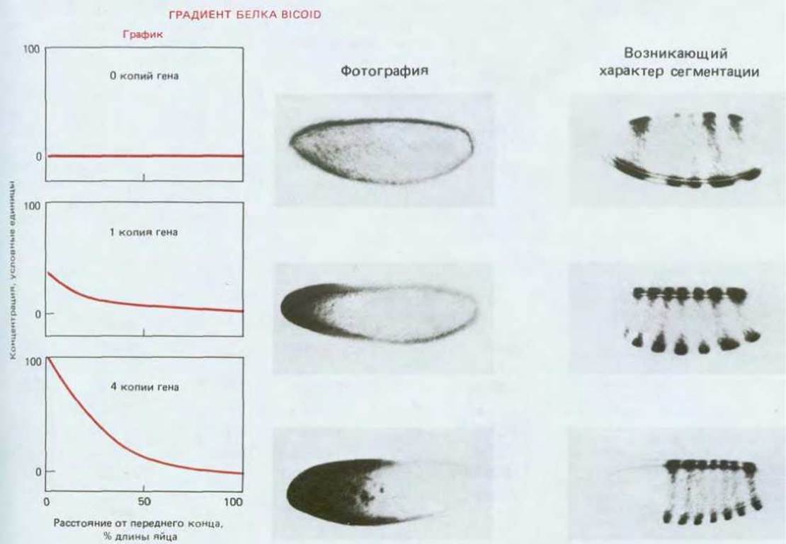
Fig. 16-60. The bicoid protein gradient in the Drosophila oocyte and its effect on segment pattern. The gradient is revealed by immunostaining with Antibodies against the bicoid protein; the segmentation pattern is visualized by antibody staining against the product of the pair-rule gene even-skipped (see Section 16.5.5). Three embryos are compared: one lacking the bicoid gene, one with a single copy of the gene, and one with 4 copies. In the absence of the bicoid gene, anterior segments fail to form; as the gene dosage increases, they form progressively further from the anterior pole, as if their positions were determined by the local bicoid protein concentration. The variation in this concentration, inferred from staining intensity, can be plotted graphically. Despite significant differences in the layout of segment primordia between embryos with one and four gene copies, both develop into completely normal larvae and adults. The mechanisms underlying this regulation are discussed in Section 16.4.9.
16.5.5. The Action of Three Classes of Segmentation Genes Subdivides the Embryo into Segments [49]
The products of egg-polarity genes provide universal graded signals that direct the ESTABLISHMENT OF THE segmental pattern. This process depends on the action of segmentation genes, which comprise a group of roughly 20 genes. Mutations in these genes can alter the number of segments or their fundamental internal properties without disrupting the overall polarity of the egg. Compared to egg-polarity genes, segmentation genes act at later developmental stages. Consequently, the embryonic phenotype is determined fully or partially by the embryonic genotype rather than solely by the maternal one. Thus, these genes are classified as zygotic-effect genes.
Most segmentation Gene Mutations are lethal; their effects are not seen in adult flies because the mutants die before reaching advanced developmental stages. However, such lethal mutations can propagate in a recessive state (as is typically the case). Therefore, heterozygotes carrying two gene copies—one normal and one mutant—remain viable. When heterozygous parents are crossed, one-fourth of the offspring are homozygotes carrying two copies of the mutant gene. These progeny die prematurely at late embryonic or early larval stages, but they live long enough for the altered phenotype to manifest. Nearly all segmentation genes were discovered by treating flies with mutagens and subsequently screening tens of thousands of dying larvae obtained from crosses of mutant strains.
Segmentation genes are subdivided into three classes (Fig. 16-61). Acting first are at least three gap genes, whose products establish the coarsest subdivision of the embryo. Mutations in any gap gene result in the deletion of large blocks of contiguous segments, while mutations in different gap genes lead to distinct but partially overlapping defects. For example, a larva mutant for the Krüppel gene lacks 8 segments, from T1 to A5 inclusive (corresponding to parasegments P3–P10).

Fig. 16-61. Phenotypic expression of mutations affecting the Three types of segmentation genes. In each case, the regions highlighted in color in normal larvae (left) are deleted in the mutant or replaced by mirror-image duplications of unaffected areas. Dominant mutations are conventionally denoted by uppercase letters, and recessive ones by lowercase letters. Several mutations affecting Drosophila body structure are classed as dominant because of their pronounced effect on the heterozygote phenotype, although their primary characteristic (lethal effect) is recessive, manifesting only in homozygotes. (C. Nüsslein-Volhard, E. Wieschaus, Nature, 287: 795–801, 1980; with minor modifications.)
The next group of segmentation genes to act comprises the 8 pair-rule genes. Mutations in these genes cause a series of deletions affecting alternating segments, leaving only half of the segments intact. This pair-segmental periodicity is characteristic of all pair-rule mutants, though they differ in the exact localization of the mutations within segment or parasegment boundaries. For instance, one pair-rule mutant called even-skipped loses all even-numbered parasegments, whereas another, fushi tarazu (ftz), loses all odd-numbered parasegments. A mutation in this same class, named hairy, results in the loss of a series of identically sized regions that do not coincide with parasegments. Finally, there are about 10 segment-polarity genes. Mutations in these genes lead to the loss of a portion of a segment and its replacement by a mirror-image copy of the whole segment or part of it. For example, in gooseberry mutants, the posterior half of each segment (i.e., the anterior half of each parasegment) is replaced by a nearly mirror-reversed anterior half of the corresponding segment (see Fig. 16-61).
Analysis of the phenotypes of various segmentation gene mutants suggests that these genes form a coordinated system that subdivides the developing embryo into increasingly finer domains distinguished by their patterns of gene expression. Here, too, Molecular Genetics makes it possible to investigate the mechanisms underlying this system.
16.5.6. Localized Expression of Segmentation Genes Is Regulated by a Hierarchical System of Positional Signals [44, 50]
Several genes from each segmentation gene class have been cloned, and the corresponding probes used to localize transcripts in normal embryos via in situ hybridization (see Section 4.6.11). We previously discussed how this method demonstrated that bicoid transcripts serve as a source of positional signal: the transcripts are localized at one pole of the egg, even though the phenotypic effects of the mutation extend over most of the embryo. Similarly, it has been shown that certain segmentation genes (namely, gap genes) participate directly or indirectly in forming positional signals that control local developmental patterns. For example, mutants defective in the gap gene Krüppel exhibit a severe anomaly extending across the region where Krüppel transcripts are detected in the normal embryo, as well as several segments beyond it (Fig. 16-62). The Krüppel gene has been sequenced and found to be homologous (as is another gap gene, hunchback) to a family of genes known to encode DNA-binding regulatory proteins in vertebrates, including the Xenopus transcription factor TFIIIA (see Section 9.1.9). By analogy with bicoid, it is tempting to propose that the Krüppel protein acts as a diffusing morphogen spreading from the Krüppel transcription site, although the observed protein spread is not as extensive as this hypothesis requires.
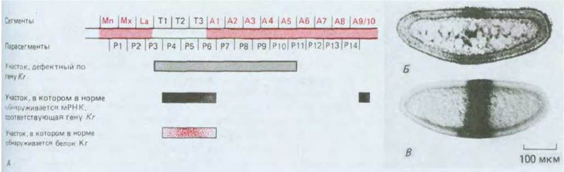
Fig. 16-62. Spatial domains of action of the gap gene Krüppel mapped on the Drosophila blastoderm. A. The diagram shows how the defect caused by the absence of a functional Krüppel product extends beyond the region where Krüppel transcripts are normally localized. B. Normal distribution of Krüppel transcripts as revealed by in situ hybridization at the blastoderm stage. C. Normal distribution of Krüppel protein visualized by antibody staining at the same stage. At other stages, the protein may have a broader distribution. The phenotype of a mutant lacking a functional Krüppel product is shown in Fig. 16-61A. (B — from H. Jäckle, D. Tautz, R. Schuh, E. Seifert, F. Lehmann, Nature, 324: 668–670, 1986; C — from U. Gaul, E. Seifert, R. Schuh, H. Jäckle, Cell, 50: 639–647, 1987, copyright Cell Press.)

Fig. 16-63. In situ hybridization of a radioactive DNA probe for the ftz gene to Drosophila blastoderm reveals seven distinct bands among the gene's transcription products, corresponding to the phenotypic defects observed in ftz mutants. In the autoradiograph, the ftz expression bands appear on a longitudinal section as black dots formed by silver grains. (Courtesy of Philip Ingham.)
Some pair-rule genes participate in short-range signaling, affecting cells adjacent to the sites of gene transcription; other genes, by contrast, exert developmental effects exclusively within the areas where they are transcribed. For example, transcripts of the normal ftz gene at the blastoderm stage appear as seven annular stripes reminiscent of zebra stripes (Fig. 16-63), marking the width and position of the precursors of even-numbered parasegments, which are lost in ftz mutants.
Summarizing these observations, one can suggest that egg-polarity gene products provide embryos with universal positional signals that prompt specific gap genes to express in precise locations. In turn, gap gene products represent a second tier of positional signals acting more locally to regulate finer details of the embryo's spatial organization, while also influencing the expression of yet another group of genes, including the pair-rule genes. Thus, the universal gradients established by egg-polarity genes take part in shaping finer details of spatial organization through the progressive subdivision governed by a hierarchy of sequential positional controls. This is an exceptionally reliable strategy: because positional signals do not dictate fine details, individual responding nuclei do not need to react with extreme precision to minimal shifts in the magnitude of such signals.
16.5.7. Products of One Segmentation Gene Control the Expression of Another [44, 50, 51]
According to the scheme presented above, the hierarchy of positional signals should correspond to the hierarchy of regulatory interactions between genes governing the establishment of spatial organization. This proposition can be tested by examining The Effect of a mutation in one gene on the expression of another. It turned out that the genes involved in spatial patterning along the anteroposterior axis form a hierarchical pyramid comprising five main tiers, with the products of each tier regulating the expression of genes in the tiers below. At the apex of this pyramid lie the egg-polarity genes, followed by gap genes, pair-rule genes, segment-polarity genes, and finally, homeotic selector genes.

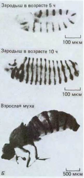
Fig. 16-64. A. Transcription patterns of four of the eight known pair-rule genes and one segment-polarity gene, engrailed, in the Drosophila blastoderm. Although each pair-rule gene on its own defines only a simple two-segment periodicity, the combined activity of the entire set of pair-rule genes leads to the specification of adjacent and overlapping structures, driving a much finer and more complex subdivision of the entire blastoderm into single-cell-wide stripes where the engrailed gene is expressed. B. Expression patterns of the engrailed gene in a 5-hour embryo (germ-band elongation stage), a 10-hour embryo, and an adult fly with its wings removed in this preparation. This expression pattern is detected using antibodies against the engrailed protein (for the 5-hour embryo) or (for the other two specimens) by generating a transgenic fly line carrying regulatory sequences of the engrailed gene fused to the coding sequence of a reporter enzyme, whose presence is easily detected histochemically by the formation of a colored product via the enzyme-catalyzed reaction. Note that the distribution of the engrailed protein persists throughout the animal's life once established. (A — after M. Akam, Development, 101, 1–22, 1987; B — courtesy of Tom Kornberg.)
Mutant embryos lacking normal Krüppel gene products can be obtained, and the expression of the normal ftz gene can be assessed via in situ hybridization using a cloned probe complementary to the latter. The characteristic ftz stripes fail to form precisely in those regions of the blastoderm affected by the Krüppel mutation. Consequently, the Krüppel gene product directly or indirectly regulates ftz expression. At the same time, the distribution of the normal Krüppel product remains unaffected in ftz mutants: the ftz product does not regulate the expression of the Krüppel gene.
Interactions between GENES OF THE same tier of the hierarchical pyramid are occasionally observed; unlike the interaction between Krüppel and ftz, some of these interactions can be reciprocal (i.e., cross-regulatory). For example, the gap genes Krüppel and hunchback mutually inhibit each other, preventing their products from being expressed simultaneously in embryonic nuclei. Consequently, under normal conditions, they are expressed in adjacent regions of the blastoderm with a sharp boundary between the anteriorly located hunchback expression domain and the posteriorly located Krüppel domain. However, in the absence of one product from this pair, the domain of the other expands beyond the normal demarcation line. The mutual inhibition exhibited by these two genes, which forces each Cell Nucleus to choose between alternative developmental fates, can explain how a set of strictly defined, non-overlapping expression domains arises in response to a continuous morphogen gradient.
The regular periodic pattern of pair-rule gene expression is likely established in a similar manner. Here, the graded positional signals determined by gap gene expression provide a rough developmental direction, while interactions among pair-rule genes (such as hairy and runt) lead to a stricter and more precise refinement. Thus, the expression domains of various pair-rule genes form a precisely reproducible distribution of mutual exclusions and overlaps within each two-segment module of the normal blastoderm embryo (Fig. 16-64). Different bands of blastoderm cells encircling the embryo are characterized by distinct combinations of pair-rule gene expression, which, organized hierarchically, descend to the finest possible level of resolution — single-cell width, corresponding to approximately 1/4 the width of a future segment or parasegment.
16.5.8. Egg-Polarity, Gap, and Pair-Rule Genes Establish Transient Spatial Organization; Segment-Polarity and Homeotic Selector Genes Provide Permanent Record [44, 52]
The events described in the previous section take place during the first few hours following fertilization. Gap genes and pair-rule genes are activated sequentially, and their encoded mRNAs (visualized via in situ hybridization) generate an averaged and heavily coarse-grained pattern (compared to the final outcome). The newly synthesized products themselves regulate gene expression until, through a series of mutual adjustments, the initial distribution of gene products resolves into a sharply defined system of repeating stripes. Yet even this system is unstable and transient. As development proceeds from the gastrula stage onward, the regular segmental arrangement of gap and pair-rule gene products fades away. However, their action has given rise to a temporary set of positional values—that is, molecular addresses of the blastoderm cells—that will maintain the segmental ORGANIZATION OF THE larva and adult animal. These positional values are recorded through the sustained activation of segment-polarity genes and homeotic selector genes.
16.5.9. Segment-Polarity Genes Control Major Subdivisions of Each Parasegment [44, 53]
A similar pattern of segment-polarity gene expression is observed within specific regions of all parasegments. A prime example is the engrailed gene. The presence of its mRNA can be demonstrated by in situ hybridization at the cellular blastoderm stage, where this mRNA forms a series of 14 single-cell-wide stripes localized at the anterior margins of future parasegments. The appearance of these stripes is closely linked to the expression of stripes corresponding to pair-rule genes (see Fig. 16-64); by observing disruptions in engrailed stripe distribution in pair-rule mutants, the rules governing engrailed activation can be formulated. Key roles are presumably played by specific combinations of pair-rule gene products: in even-numbered parasegments, engrailed is expressed only following the expression of ftz and odd-paired, whereas in odd-numbered parasegments, it follows the combination of even-skipped and paired. Other combinations of pair-rule products activate or repress different segment-polarity genes: Transcription of the wingless gene is turned on in single-cell-wide stripes located in the most posterior regions of the parasegments. Thus, each future parasegment is subdivided into at least three distinct regions already at the cellular blastoderm stage. These chemical differences are maintained and reinforced by the ongoing transcription of at least some segment-polarity genes even after pair-rule gene products have largely disappeared (see Fig. 16-61B). Some of the segment-polarity genes expressed in this manner (including, for instance, wingless) produce factors that subsequently act as local morphogens within segments, regulating finer details of the internal spatial organization and growth of the parasegment.
Pair-rule gene products not only regulate The activity of segment-polarity genes but also interact with gap gene products (and evidently egg-polarity genes), eliciting precisely localized differential activation of the genes that form the fifth and final tier of the hierarchical pyramid—the homeotic selector genes, whose activity maintains permanent distinctions between different parasegments. Next, we will examine the manifestations of selector gene activity in greater detail and discuss their role in underlying cellular memory mechanisms.
16.5.10. Homeotic Selector Genes of the Bithorax and Antennapedia Complexes Determine Parasegmental Differences [54]
Numerous homeotic mutations have been studied in Drosophila, each leading to the replacement of one body part by another; for instance, wings developing in place of eyes, legs in place of antennae, and so forth (see Fig. 16-53). Particularly crucial sets of mutations occur within two gene clusters known as the bithorax complex and the Antennapedia complex. Each complex contains several genes with distinct functions: the bithorax complex genes control differences among abdominal and thoracic body segments, while the Antennapedia complex genes control differences between thoracic and head segments. The genes of these two complexes are termed homeotic selector genes because they execute choices between determination states corresponding to different yet homologous, or homeomorphological, structures. Each homeotic selector gene is restricted in its action to a specific domain, or body region, which undergoes transformation upon mutation of that gene. Typically, such domains possess sharply defined boundaries that roughly correspond to half a "segment" — not coinciding with morphological segment boundaries, but rather aligning with parasegment boundaries (Fig. 16-56).
Homeotic selector genes were identified, as shown in Section 16.5, through the discovery of mutant adult flies; many mutations in this group are recessive lethals. Such mutations cause the death of the individual almost immediately after hatching. Thus, as with recessive lethal mutations of segmentation genes (see Section 16.5.5), the phenotypic effects of these genes are never manifested in adult flies but can be observed in embryos or early larvae. Consequently, analysis of these early stages provides the clearest and, in some respects, most complete picture regarding the role of homeotic selector genes.

Fig. 16-65. Normal Drosophila embryo (A) and mutant embryo (B) lacking the majority of the bithorax complex genes. In the mutants, all parasegments posterior to P5 resemble P5. [Courtesy of Gary Struhl; A — reprinted (with permission) from Nature, 293, 36–41, Copyright 1981, Macmillan Journals Limited.]
Larvae defective in all genes of the bithorax complex exhibit a relatively simple structure: the head and anterior Thorax up to parasegment P4 are normal, but all remaining 10 parasegments acquire The properties of parasegment P4. Partial deletions in the bithorax complex lead to less severe transformations: for example, P5 and all more anterior parasegments may appear normal, whereas parasegments from P5 posteriorly are transformed into P5 (Fig. 16-65). These observations, alongside analogous findings regarding the Antennapedia complex, illustrate the essential role of homeotic selector genes in specifying differences among parasegments: in the absence of these genes, such parasegmental differentiation fails to occur.
16.5.11. Homeotic Selector Genes Encode a System of Molecular Addresses [44, 55]
Like segmentation genes, homeotic selector genes are first activated at the blastoderm stage. Because the entire DNA of the Antennapedia and bithorax complexes has been cloned, the transcriptional sequence of all homeotic selector genes can now be elucidated using in situ hybridization and available DNA probes. The Conclusions drawn from these studies are striking: at first glance, each homeotic selector gene is normally expressed only in regions that develop abnormally — that is, as if they were out of place when the gene is mutated or absent. This apparent lack of logic allows us to view selector gene products as molecular addresses. Each parasegment possesses its own unique set of such addresses, and if the addresses are altered, the parasegment behaves as though it were located elsewhere. The activation of homeotic selector genes is regulated by segmentation genes; consequently, the expression pattern of homeotic selector genes coincides with the parasegmental boundaries defined by pair-rule and segment-polarity gene products. Thus, the combination of a specific homeotic selector gene product (or a set of such products) with particular segment-polarity gene products reliably specifies a unique address possessed exclusively by cells in one particular region of a given segment.
16.5.12. Products of homeotic selector genes are involved in regulating the expression of these genes [56]
Just as with segmentation genes, the expression pattern of homeotic selector genes undergoes complex adjustments from the moment transcripts first appear in the blastoderm. From a certain point onward within each parasegment, this pattern acquires strictly defined features, with the final expression pattern becoming established only after gastrulation is complete. Figure 16-66A illustrates the expression pattern at the germ-band elongation stage, approximately 5 hours after fertilization (see Figure 16-55).
This pattern is partially modified as a result of interactions among several homeotic selector genes. When homeotic selector genes are arranged in order—starting with genes expressed at the anterior end and ending with those at the posterior end—each gene in the series is repressed by the products of its subsequent members. Thus, in the absence of downstream gene products, any given gene in this series will exhibit a high level of expression both in its normal domain and in the parasegmental domains located posterior to it. This arrangement of genes, which corresponds to their spatial expression and is controlled by mutual interactions, is reflected in the sequence in which they are located along the chromosome in each of the two gene complexes (Figure 16-66B). This topic is discussed in Section 10.3.19.
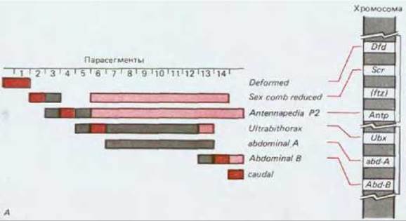

Fig. 16-66. (A) Expression patterns of genes in the Antennapedia and bithorax complexes in relation to their chromosomal localization. Note that The sequence of genes in each chromosomal complex corresponds to the spatial sequence in which these genes are expressed. (B) Transcription pattern of homeotic selector genes 5 hours after fertilization. All of these genes, except for caudal, are located within the Antennapedia and bithorax complexes, and at this stage all of them appear to be expressed in parasegmental domains. Most of these genes are actively expressed in at least one parasegment (highlighted in the darkest shade) and at lower levels in some neighboring parasegments: where transcript presence is required for the normal phenotype to manifest (lighter shade) and where transcripts are not needed (lightest shade). (B, adapted from M. Akam, Development, 101:1–22, 1987; modified.)
16.5.13. The adult fly develops from a set of imaginal discs that have memorized their positional information [57]
The expression pattern of homeotic selector genes is established in the Drosophila embryo and determines the body plan of not only the larva, but also the adult fly. To fully appreciate the role of these genes, one must understand how the adult organism, or imago, ultimately develops.

Fig. 16-67. Imaginal discs in a Drosophila larva (schematic representation) and the adult structures derived from them. Of the three pairs of leg discs, only one is shown. [J. W. Fristrom et al., in Problems in Biology: RNA in Development (E. W. Hanley, ed.), p. 382, Salt Lake City: University of Utah Press, 1969.]
The adult fly is formed primarily from undifferentiated, so-called imaginal cells gathered into clusters located laterally in each of the larval body segments. The source of imaginal cells is the embryonic epithelium that covers the entire body. These cells maintain their connection with the epidermis and participate mainly in forming the epidermal structures of the adult organism. The imaginal cells that will give rise to the head, thorax, and genitalia are organized into imaginal discs, whereas the abdominal imaginal cells are grouped into clusters called abdominal histoblast nests. Disc cells have been investigated in the greatest detail. A total of 19 discs are known, one of which lies on the body midline, while the remaining 18 lie in pairs laterally along the larva (Fig. 16-67). The discs are epithelial sacs shaped like crumpled and flattened spheres; during metamorphosis, they unfold and differentiate. One pair of discs gives rise to the eyes and antennae, another to the wings and part of the thorax, a third to the first pair of legs, and so on. Externally, the cells of different imaginal discs are indistinguishable, and during differentiation, they produce a similar set of differentiated cell types. However, transplantation experiments show that they are actually regionally determined and non-equivalent.
If an imaginal disc is transplanted from one site to another within a larva and allowed to develop until the onset of metamorphosis, the transplanted disc will differentiate autonomously into a structure appropriate to its origin, regardless of its new location. It follows that, during differentiation, imaginal disc cells are guided by a memory of their original location. Using more sophisticated transplantation techniques, in which imaginal disc cells proliferate for a prolonged period before differentiation, it can be demonstrated that this state of cellular memory is inherited (with a few exceptions to this general rule) over an indefinitely large number of cell generations (Fig. 16-68).
Homeotic selector genes are an essential component of cellular memory mechanisms. If they are eliminated from imaginal disc cells at any stage of the extended period leading to differentiation during metamorphosis, the cells begin to differentiate into different structures, as if they belonged to other body segments. This phenomenon can be demonstrated using X-ray-induced mitotic recombination. This approach can be viewed as the genetic surgery of individual cells, used to generate clones of mutant cells with a specific genotype at any stage of development. These issues will be discussed in the next section.

Fig. 16-68. Experiments to determine the state of determination of imaginal disc cells. The method involves implanting cells into larvae just prior to metamorphosis; the cells differentiate to form specific adult structures that, following metamorphosis, lie within the body cavity of the host fly and are not integrated with its tissues. Cells can be tested immediately after extraction of the discs or after implantation into the abdomen of an adult fly, which serves in this case as a natural culture chamber. The hormonal environment within adult flies allows these cells—which have thus escaped metamorphosis—to proliferate without differentiating (prior to The Cell determination assay). In both cases, following implantation back into a larva, the cells typically differentiate and form structures corresponding to The Fate of the original disc.
16.5.14. Homeotic selector genes play a vital role in the retention of positional information by imaginal disc cells [58]
A normal somatic cell contains two homologous sets of Chromosomes. One homolog of each pair is of paternal origin, and the other is maternal. Normally, DNA exchange via Crossing Over between homologous paternal and maternal chromosomes occurs only during Meiosis in germ Cell Formation. Occasionally, however, crossing over takes place during the division of ordinary somatic cells. Although mitotic recombination is normally a rather rare event, it can be induced in insects by X-ray irradiation (likely as a side effect of radiation damage to chromosomes). As shown in Fig. 16-69, if the maternal and paternal chromosomes carry different alleles of a gene, the cell is heterozygous. Through a single mitotic recombination event, two homozygous daughter cells can be produced that differ genetically both from the parental cells and from each other: one inherits two copies of the paternal allele, and the other two copies of the maternal allele. Subsequently, each daughter cell reproduces in the usual manner, generating a pair of homozygous clones surrounded by heterozygous tissue (Fig. 16-70). If, for instance, all Cells of the organism are initially heterozygous for a mutation that causes yellow pigmentation instead of normal brown, but this mutation is recessive (so that heterozygous cells have a brown phenotype), mitotic recombination can give rise to a clone of homozygous yellow cells against a brown Background.

Fig. 16-69. Normal mitosis (A) and mitosis accompanied by recombination (B). The diagram illustrates the fate of a single pair of homologous chromosomes, one of which is of paternal origin (shaded, with a black circle for the centromere) and the other maternal (with a white circle for the centromere). These chromosomes contain a pigmentation gene (or other marker gene) with the wild-type allele A (white square on the paternal chromosome) and a recessive mutant allele a (red square on the maternal chromosome). Homozygous A/A and heterozygous A/a cells exhibit the normal phenotype, whereas homozygous a/a cells exhibit an altered phenotype. Recombination via DNA exchange between the paternal and maternal chromosomes results in a pair of daughter cells, one of which is homozygous A/A (normal phenotype) and the other homozygous a/a (mutant phenotype). Mitotic recombination is a rare, stochastic event.

Fig. 16-70. Mitotic recombination can be used to generate a clone of genetically marked mutant cells in the Drosophila wing. The earlier recombination occurs, the larger the potential clone will be.
Of course, it is impossible to control precisely which cells will undergo mitotic recombination, nor can one predict with absolute certainty the exact chromosomal site where crossing over will take place. However, experimenters can exercise considerable control over the timing of such events (by irradiating cells at a strictly defined time) and, more importantly, over the initial genotype. First, one can cross homozygotes in which a mutation of the homeotic selector gene of interest is linked to a marker mutation (such as one causing abnormal pigmentation). In this case, the marker makes it possible to identify homozygous clones generated by mitotic recombination based on their abnormal coloration. Such marked clones can be reliably considered homozygous for the mutation under study.
The primary effects of homeotic selector gene mutations are mostly recessive: only homozygous mutant organisms display homeotic transformation. By utilizing the mitotic recombination technique described above, one can generate a clone of cells in an imaginal disc marked for a homeotic gene and observe their behavior against a heterozygous, phenotypically normal background. This is how it was demonstrated that marked cells (and only they) are capable of homeotic transformation (thereby proving that they lie within the domain of action of the homeotic selector gene); from this, one can conclude that a recombination event took place during early development. For example, a two-day-old larva heterozygous for a mutation disrupting the function of the Ultrabithorax (Ubx) gene from the bithorax complex can be exposed to X-rays. As a result, isolated clones of homozygous Ubx/Ubx cells will be produced within the affected imaginal discs. If these clones are located on the haltere disc, wild-type tissue patches will arise within the haltere. Such observations indicate that positional memory in each cell is maintained by the continuous activity of the normal homeotic selector gene, and that this memory operates cell-autonomously.
16.5.15. Homeotic selector genes and segment polarity genes define body compartments [59]
The memorized differences controlled by homeotic selector genes are discrete: GENE EXPRESSION IN cells of adjacent paraseptal domains exhibits sharp distinctions. The same holds true for at least one segment polarity gene—engrailed (see Fig. 16-64B)—whose differential expression corresponds to a clear-cut distinction between cells of the posterior and anterior regions of the parasegment (corresponding to cells of the anterior and posterior compartments of the segment). Thus, through the differential expression of these two classes of genes, the body is subdivided into a series of distinct regions whose cells are characterized by different states of determination.
At the boundary between two such compartments, cells do not mix, as though the selective adhesion of cells sharing the same molecular address enables them to break away from cells bearing a different one. Thus, for example, if genetic recombination produces a clone of marked yet normal cells in the wing, this clone remains strictly on one side of the sharp boundary separating the two parasegments that form the wing. Body subdivisions (in the wing or any other appendage) formed in this manner are called compartments (Fig. 16-71). Although compartment boundaries generally do not coincide with the borders of normal organs, they match the limits of domains within which homeotic selector genes operate, which can be observed by the expression of the engrailed gene (see Fig. 16-71, B). In the corresponding mutants, the tissue on one side of the compartment boundary is transformed, while normal tissue lies on the other side (Fig. 16-72).

Fig. 16-71. A. The shape of marked clones in a Drosophila wing indicates the existence of compartment boundaries. At such a boundary, the edge of each patch appears straight. Even if a marked clone is genetically altered in a way that allows it to grow faster than the rest of the wing and become very large, it still does not cross the compartment boundaries (far right drawing). Note that this boundary does not coincide with the central vein of the wing. B. The expression pattern of the engrailed gene in the wing, revealed by the same method as in Fig. 16-64, B. The compartment boundaries coincide with the borders of engrailed gene expression. (A - after F.H.C. Crick, P.A. Lawrence, Science, 189, 340-347, 1975; B - courtesy of Tom Kornberg.)
Compartment boundaries are, by definition, the borders between two populations of cells with different states of determination that cannot intermingle. Although the state of determination is normally irreversible, each compartment must nevertheless function as a self-sustaining unit. It cannot recruit cells from a neighboring compartment or share its own surplus of cells with a neighbor. However, it is capable of regulating its internal organization through adjustments that do not violate these constraints. Each compartment possesses its own detailed internal structure, which arises within the imaginal disc as a result of cell-cell interactions at relatively late Selection/3.html">Stages of development. The Molecular Basis of these local pattern-forming mechanisms remains largely unstudied, although, as mentioned in Section 16.5.9, the product of the segment-polarity gene wingless may possibly be involved. It can be demonstrated, however, that persistent intercellular interactions dictate not only spatial organization but also the growth of imaginal disc parts: this system obeys the intercalation rule (see Section 16.4.9). During The regulation of growth and pattern formation, each compartment behaves as a more or less independent, integrated entity. This was demonstrated in experiments where mitotic recombination was used to create labeled cell clones with a genotype enabling them to proliferate faster than other clones of the imaginal disc. During growth, these clones occupy a disproportionately large part of the compartment in which they reside, yet they are unable to disrupt compartment boundaries; they cannot increase the size of the compartment or alter its spatial organization (see Fig. 16-71).
16.5.16. Auto-regulation of homeotic selector genes contributes to cellular memory mechanisms [60]
Having taken an excursion into the future—that is, having examined later stages of Drosophila development—let us return to the past to discuss the important properties of homeotic selector genes from a molecular perspective. First and foremost, we must consider how these mechanisms provide each cell with a memory of which segment it resides in. One way to achieve this effect is through a positive feedback loop in which a gene product stimulates its own transcription (see Section 16.2.10). Evidence indicates that homeotic selector genes indeed possess this self-maintaining property. For example, cultured cells can be transfected with an artificially constructed plasmid vector containing either the protein-coding sequence of a homeotic selector gene linked to some other strong promoter, or the regulatory sequence of that same gene linked to the sequence of another gene encoding a different protein (such as an easily detectable enzyme). In the first case, one can ensure the required level of Synthesis of the protein encoded by the homeotic selector gene, while In the second, one can test how effectively the regulatory function of this protein operates (determined by the synthesis of the reporter enzyme in cells co-transfected with both types of Plasmids). Such experiments have shown that the Ultrabithorax protein can exert a feedback effect on the Ultrabithorax regulatory sequence, stimulating the expression of the corresponding gene (Fig. 16-73). Furthermore, it can also influence the Antennapedia regulatory sequence, controlling the synthesis of its corresponding protein, and the ftz protein possibly stimulates both Ultrabithorax and Antennapedia. Of course, the conditions established in cell culture as a model do not replicate those arising in the embryo; nevertheless, these observations demonstrate that homeotic selector genes and their relatives can not only stimulate or inhibit one another, but also self-stimulate.

Fig. 16-72. Haltere of a bithorax mutant (B); for comparison, photographs of a normal wing and a normal haltere are shown (A). The anterior compartment of the mutant haltere is transformed into the anterior compartment of a wing. (A - courtesy of Peter Lawrence; B - from F.H.C. Crick, P.A. Lawrence, Science, 189, 340-347, 1975. Copyright, 1975 by the American Association for the Advancement of Science.)
Such positive feedback is likely important for the initial establishment of the gene's activation state, whereas other mechanisms may be employed to maintain this state over a prolonged period. Nonetheless, we are now beginning to understand how homeotic selector gene products can simultaneously serve as molecular addresses, archival records, and tools for regulating the activity of other genes.

Fig. 16-73. Experiments utilizing cells transfected with engineered plasmids bearing the Ultrabithorax (Ubx) and fushi tarazu (ftz) genes demonstrate that the ftz (B) and Ubx (A) proteins can act on the regulatory region of the Ubx gene, stimulating its expression. Ubx auto-stimulation (A) establishes a positive feedback loop that ensures self-sustaining Ubx expression in vivo. A similar effect is observed for the Antennapedia gene. However, the situation depicted here is vastly simplified. In the actual embryo, there are presumably numerous regulatory proteins that combine and/or compete to control the expression of these genes.

Fig. 16-74. A mutant embryo defective in the extra sex combs (esc) gene, obtained from a mother who also lacks this gene. All segments are transformed and resemble the terminal abdominal segment (compare with Fig. 16-65). In this mutant, the expression pattern of homeotic selector genes (initially nearly normal) becomes unstable, soon resulting in the ectopic activation of all these genes along the entire body axis. (G. Struhl, Nature, 293, 36-41, 1981.)
16.5.17. The expression of homeotic selector genes is regulated by differential splicing as well as transcriptional control [56, 61]
In a broad sense, The system of homeotic selector genes likely possesses a simple, rational organization and function. However, many complexities exist in the details. For instance, the mere interaction of homeotic selector gene products with other genes appears insufficient to stabilize the initial expression pattern. An additional set of genes is required, upon whose inactivation via mutation homeotic selector genes become independently switched on in all cells of the embryo (Fig. 16-74). The persistent differences between parasegments may fundamentally rely on heritable modifications of the regulatory regions of homeotic selector genes that have somehow undergone imprinting at the level of Chromatin Structure (see Section 10.3.10). Moreover, although the bithorax complex dictates the identity of nine parasegments, it contains a mere three genes: Ultrabithorax, abdominal-A, and abdominal-B. These genes, much like those of the Antennapedia complex, are exceptionally large and characterized by highly intricate regulatory sequences. Consequently, each gene produces a set of alternatively spliced transcripts. For instance, the Ultrabithorax gene spans approximately 75,000 nucleotide pairs, and its corresponding transcripts undergo differential splicing depending on the developmental stage and the cell's position within the embryo. Mutations localized in different regions within the gene can affect distinct parasegments or specific parts thereof. Thus, for example, mutations localized in one region of the Ultrabithorax gene transform the anterior half of the haltere into the anterior half of the wing (see Fig. 16-72), whereas mutations in another region transform the posterior part of the haltere into the posterior part of the wing. Because these mutations reside in different sites and affect different body parts, they were once thought to correspond to different genes and were consequently given separate names: bithorax and postbithorax. It has now become clear that many mutations in homeotic selector genes affecting individual parasegments or parts of parasegments are not mutations of separate genes, but rather map to different regions of the same large, complex gene (see Section 10.3.19).
16.5.18. Many pattern-forming genes contain conserved homeobox sequences that encode a portion of a DNA-binding protein [62]
A striking outcome of the cloning of homeotic selector genes was the discovery of a sequence of roughly 180 nucleotide pairs, termed the homeobox, which is contained with minor variations in virtually all genes of this type as well as in certain other genes. For example, a pair-rule gene (ftz) contains a nucleotide sequence 77% identical to the Antennapedia homeobox, and the match between the Amino acid sequences encoded by these genes is even higher, reaching 83%. In Drosophila, at least 16 genes contain a homeobox, although the degree of Homology varies widely, and nearly all homeobox-containing genes are key members of the roughly 50 genes controlling spatial organization along the anteroposterior body axis. These include egg-polarity genes (bicoid), pair-rule genes (ftz, even-skipped, paired), segment-polarity genes (gooseberry, engrailed), and virtually all homeotic selector genes from the Antennapedia and bithorax complexes. At least one gene (zerknüllt), involved in dorsoventral patterning, also contains a homeobox. These observations imply that the presence of a homeobox is a hallmark of pattern-controlling genes.
Homeobox-containing genes typically encode proteins localized to cell nuclei, suggesting their direct involvement in controlling gene expression. Furthermore, the Amino Acid Sequence forming the homeobox presumably enables these Drosophila proteins to bind to specific DNA sites that function as enhancers and silencers (Section 10.2.7) of gene expression, including the expression of other homeobox-containing genes.
The existence of specific regulatory sequences in DNA recognized by proteins containing a homeobox-domain suggests that they are also a defining feature of homeobox-containing pattern-forming genes. It is easy to envision how such genes, with extremely minor differences in their regulatory and protein-coding sequences, could combine to form a complex network in which the products of one gene control the expression of another—a mechanism discussed when we examined pattern formation along the anteroposterior axis of the Drosophila embryo. In such a system, cellular memory mechanisms and determination can be explained quite simply by autoregulation of gene expression, similar to the autoregulation described for the Ultrabithorax genes (see Fig. 16-73).
16.5.19. The homeobox exhibits a high degree of evolutionary conservation [62, 63]
The remarkable homology among homeobox-containing genes suggests that they arose through tandem duplication and divergence. The Emergence of genes via duplication and divergence is also supported by the clustered arrangement of many of these genes within the Drosophila genome. The Antennapedia complex, for instance, includes the ftz, bicoid, and zerknüllt genes, alongside a set of homeotic selector genes. The formation of homeobox gene clusters may reflect their evolutionary history rather than a physiological necessity; flies in which genetic rearrangements have excised the Ultrabithorax gene from the bithorax complex display no anatomical abnormalities.
It appears likely that as body plans grew more complex, series of modified homeobox-containing genes were added to The Genome. Drosophila mutants lacking specific sets of homeobox genes exhibit phenotypic traits potentially reminiscent of ancestral structures. For instance, by deleting certain homeotic selector genes, one can produce an animal with an array of numerous identical segments akin to a centipede; if one could step back yet another step in evolution by removing segment-polarity and pair-rule genes, one might obtain a hypothetical ancestor organized like an unsegmented nematode, while retaining a distinction between head and tail. Based on such a sequence of events, the egg-polarity gene bicoid can arguably be considered an ancestral representative of the homeobox gene family. Naturally, this remains largely a product of our imagination, yet the involvement of such an ancestral homeobox gene in body patterning is plausible, given that virtually all its Drosophila descendants perform this exact function.
The Discovery of the homeobox raises questions of broader evolutionary significance. Homeobox-containing genes have been discovered not only in insects and other Arthropods, but also in the nematode C. elegans, Annelids (leeches and earthworms), sea urchins, primitive Chordates, and vertebrates (including frogs, chicks, mice, and humans). The homeobox sequence displays an astonishing degree of conservation at the Protein Structure level; one of the proteins corresponding to a Xenopus homeobox gene possesses a sequence in which 59 out of 60 Amino Acids are identical to the homeodomain of the Drosophila Antennapedia protein, despite 500 million years of independent evolution. Such a high degree of conservation suggests that the mechanisms controlling the basic body plan in insects and vertebrates may share a fundamental similarity.
16.5.20. Mechanisms of regional cellular determination in vertebrates and insects may be similar [63, 64]
When describing the STRUCTURE OF THE early Drosophila embryo (Section 16.5.2), we noted similarities to vertebrates. Animals of both classes pass through a gastrulation stage during development, resulting in the formation of distinct germ layers; the bodies of representatives from both groups are subdivided into segments oriented more or less perpendicularly to the germ layer primordia. Vertebrate embryonic somites and Drosophila embryonic segments are superficially similar, but an analysis of their behavior reveals that they possess distinct positional values, which dictate their (the somites') subsequent differentiation. As with Drosophila imaginal discs, such positional determination can be demonstrated through transplantation experiments. For example, newly formed somites in early chick embryos can be grafted to a new location. At this stage, thoracic and cervical somites are externally nearly indistinguishable, yet thoracic somites transplanted into the neck region give rise to thoracic vertebrae bearing Ribs, whereas neck somites transplanted into the thoracic region give rise to characteristic cervical vertebrae. The similarities do not end there. It is known that each insect body segment is divided into anterior and posterior compartments that differ in their expression of the engrailed gene. Similarly, the anterior and posterior halves of chick embryo somites differ sharply in their chemical nature (Fig. 16-75), as manifested by cell Surface Properties detectable with certain Lectins, as well as by their influence on the growth trajectory of nerve fibers.
The discovery of the homeobox made it possible to test whether such analogies reflect the distribution of specific molecules. To date, it has been shown that The nucleotide sequences of certain vertebrate homeobox-containing genes are homologous to the corresponding Drosophila genes, and this homology extends beyond the homeobox itself; using in situ hybridization, these vertebrate genes have been shown to be expressed in strictly defined regions of the vertebrate embryo (Fig. 16-76). These findings suggest that the molecular mechanisms governing pattern formation in vertebrates and insects are fundamentally very similar, although the extent of this similarity cannot yet be fully assessed. For instance, in vertebrates, spatial organization is established primarily through cell-cell interactions during cleavage stages. There is no equivalent here to the processes that take place in the syncytial insect embryo. Moreover, many phylogeneticists believe that body segmentation in these animal groups evolved independently. Unfortunately, it must be noted that vertebrates exhibit nothing remotely resembling the homeotic mutations seen in Drosophila. If this is the case, it remains doubtful whether studies on Drosophila will contribute significantly to uncovering a universal relationship between genes and body plan organization across all organisms.

Fig. 16-75. A. A series of somites (anterior end of the body to the left, posterior to the right) of a 2-day-old chicken embryo, immunostained with antibodies recognizing neural crest cells: the latter migrate exclusively through the rostral (anterior) half of each somite because the cells comprising this half differ significantly from those in the caudal (posterior) half (conditions are favorable for cell migration in the former case, but not in the latter). B. A region of an older chicken embryo stained to reveal spinal nerve roots as they pass through the somites. Like neural crest cells, the nerve roots migrate exclusively through the anterior half of the somite. Tissue-grafting experiments demonstrate that this segmental organization of the Peripheral Nervous system is determined by initial differences in determination between the anterior and posterior somite cells (reminiscent of the expression pattern of the engrailed gene in Drosophila). (A - from M. Rickman, J.W. Fawcett, R.J. Keynes, J. Embryol. Exp. Morphol., 90, 437-455, 1985; B - courtesy of Claudio Stern.)


Fig. 16-76. A. Expression patterns of two mouse homeobox-containing genes, Hox 2.1 and Hox 3.1, revealed by in situ hybridization in adjacent longitudinal sections through an entire 13.5-day mouse embryo. High transcript concentrations correspond to dense silver grain clusters on autoradiographs. These two genes are expressed in distinct, partially overlapping domains along the anteroposterior axis in the Brain AND SPINAL cord, as well as in several other tissues. B. Expression pattern of a chicken homeobox-containing gene (chicken en) that shares particularly close homology with the Drosophila engrailed gene. In this case, antibodies were used to visualize the distribution of the protein encoded by this gene in a 2-day chicken embryo. Unlike its Drosophila counterpart, which forms a series of segmental stripes, the en protein in the chick localizes as a discrete cluster in the Midbrain and Hindbrain. (A - from P.W.H. Holland, B.L.M. Hogan, Development, 102, 159-174, 1988; B - courtesy of Tom Kornberg.)
Summary
The body of Drosophila, like that of other insects, consists of a set of repeating, modified fundamental modules (segments) with specialized, non-segmented structures at the anterior and posterior extremities. Major subdivisions of each segment are specified by the expression of a distinct set of regulatory genes that assign molecular addresses to these subregions. Pattern formation is initiated by the establishment of egg Asymmetry. Two classes of maternal-effect genes, also known as egg-polarity genes, can be distinguished here. One group of these genes controls the dorsoventral distribution of germ layers and major tissue types, while the other governs the anteroposterior (head-to-tail) arrangement of segments. An example of the latter class is the bicoid gene, whose mRNA becomes localized at the anterior pole of the egg. Because the early embryo is a syncytium, the bicoid gene product can readily diffuse through the Cytosol across the embryo, establishing a morphogen gradient that determines the overall organization of the anterior half of the embryo. This gradient triggers the ordered expression of a hierarchically organized gene network comprising gap genes, pair-rule genes, segment-polarity genes, and homeotic selector genes. Influenced by bicoid and mutual interactions, these latter genes begin to be expressed in discrete regions of the embryo, progressively leading to the partitioning of the body into repeating segments and their subparts.
The expression of gap and pair-rule genes is transient, but it imprints the expression of segment-polarity and homeotic selector genes; the expression of these latter genes is maintained, undergoing further refinement during subsequent development to provide cells with positional information. The cellular memory mechanism is sustained in part by positive feedback (whereby the protein products of homeotic selector genes stimulate the transcription of their own genes) and in part by heritable changes in chromatin structure. The necessity for certain forms of positional memory can be demonstrated in experiments using imaginal disc cells, which give rise to the adult external body structures; these cells retain the memory of their original developmental fates over an indefinite number of cell divisions. This behavior is driven by the continuous presence of homeotic selector genes within every individual cell of each imaginal disc. Compartment boundaries, which are presumably maintained through differential Cell Adhesion, segregate cells of differing differentiation states in accordance with the expression of these genes.
In the genome, homeotic selector genes are clustered into two major complexes: the Antennapedia complex and the bithorax complex. All of them contain a homeobox sequence that encodes a highly conserved domain of approximately 60 amino acids. This nucleotide sequence is also present in certain egg-polarity genes, pair-rule genes, and segment-polarity genes, pointing to a common evolutionary origin from an ancestral gene that participated in the regulation of pattern formation. Homeobox-containing genes have been discovered in various animals, including nematodes and mammals, suggesting that pattern formation in these organisms relies on molecular mechanisms fundamentally similar to those operating in Drosophila.
Last update: 12/08/2026
Editorial and Educational Adaptation: This material has been compiled based on the primary/original source text. The project team performed an editorial review, corrected technical inaccuracies, structured sections, and adapted the content for an educational format.
What was processed:
- elimination of formatting defects (OCR errors, structural breaks, corrupted characters);
- editorial organization of content;
- standardization of terminology in accordance with academic sources;
- verification of factual statements against the original source text.
All mentions of the author, publication year, and origin of the primary text have been preserved in accordance with the source.