Pharmacognosy with the Basics of Plant Biochemistry - Kovalyov V. M. 2004
Special Part
Peptides and Proteins
Lectins
Lectins (from Latin legere meaning to choose) are Proteins or Glycoproteins capable of binding sugars, thereby enabling Cell agglutination and the precipitation of Glycoconjugates.
Lectins contain at least two binding sites that interact with free mono- and Oligosaccharides, as well as with sugar residues within Polysaccharides, glycoproteins, and Glycolipids. In their simplest form, the interaction between lectins and CARBOHYDRATES manifests as the agglutination of particles and Cells, such as erythrocytes, or the precipitation of polysaccharides and glycoproteins.
The discovery of lectins was prompted by the toxicity of castor oil (Oleum Ricini), a puzzle that troubled many pharmacologists and toxicologists in the late 19th century. The Study of lectins began with the pioneering work of P. H. Stillmark, who established that ricin—a toxic lectin found in castor seeds—induces erythrocyte agglutination and hemolysis. This milestone in The history of science is considered the birth of a new field: lectinology.
The modern era of lectin research began after 1945, when W. Boyd discovered Blood group-specific agglutinins. In our country, Ukrainian scientists M. D. Lutsyk and Ye. M. Panasiuk made significant contributions to this research. Today, the core of lectin studies is centered at the Institute of Molecular Biology and Genetics of the National Academy of Sciences of Ukraine. Research on lectins focuses on obtaining pure preparations and determining their carbohydrate Specificity. By 1965, only three crystalline lectins were known (phytohemagglutinin, concanavalin A, and ricin); today, their number exceeds 100.
Structure and Classification of Lectins
The non-protein components of lectins include carbohydrates and divalent Metal Ions such as Ca2+, Mn2+, Zn2+, Mg2+, Co2+, Ni2+, Cd2+, and others.
For most lectins, metal ions determine the specificity of their carbohydrate interaction. Removing these metals from the molecule leads to a decrease or even complete loss of biological activity. However, for certain lectins, metals are not essential components, as seen in wheat germ agglutinins and animal lectins.
The carbohydrate content in various lectins varies widely—from 3 to 80%—though it typically ranges between 3-10%. N-acetylglucosamine and mannose are generally the primary Monosaccharides forming the main chain. The carbohydrate component is not always crucial for a lectin's biological activity; among known lectins, concanavalin A and pea lectin contain no sugars at all.
The Amino Acid Composition of lectins from evolutionarily distant organisms varies significantly, making it inappropriate to speak of a single, characteristic Primary Structure.
With very few exceptions, the molecules of known lectins are built from multiple polypeptide chains, or subunits, meaning they possess a quaternary structure. These subunits may be identical or different. Among plant lectins, di- and tetrameric forms are observed, whereas animal lectins are most often higher-order polymers. Polypeptide chains in lectin molecules can be linked by both non-covalent bonds (hydrophobic, hydrogen, and ionic bonds) and disulfide bridges. Various combinations of polypeptide chains give rise to lectin isoforms, which may exhibit differing carbohydrate specificities.
One of the earliest classifications of lectins, which remains in use today, was proposed by H. J. Möckel.
According to this system, the specificity of lectin-carbohydrate interactions is determined by THE POSITION OF the hydroxyl group at C3 and C4, as well as the D- or L-configuration of the sugar. Based on this, all lectins are divided into four groups specific to the following sugars:
1) 3,4-OH cis, L-form (L-fucose, L-galactose);
2) 3,4-OH cis, D-form (D-galactose);
3) 3,4-OH trans, D-form (D-glucose, D-manose);
4) 3,4-OH trans, L-form (L-glucose, L-gulose).
Types of carbohydrates depending on the hydroxyl position and the pyranose ring conformation:
Class="center">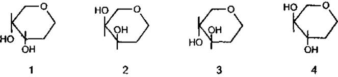
Lectins that interact with carbohydrates of the fourth group are currently unknown. Another classification divides lectins into 9 groups based on their specific affinity for monosaccharides such as L-fucose, D-galactose, D-glucose, D-mannose, N-acetyl-D-glucosamine, N-acetyl-D-galactosamine, N-acetyl-D-galactosamine combined with galactose, N-acetyl-D-glucosamine combined with D-glucose, and N-acetylneuraminic acids.
These classifications are somewhat conventional because they do not account for the fine specificity of lectins toward oligosaccharides. Commercial suppliers often provide a practical classification of lectins that includes data on specific oligosaccharides.
A more robust approach is a combined classification that divides lectins into three groups.
Based on carbohydrate specificity: lectins reacting with acidic sugars, and lectins reacting with neutral sugars.
Based on structural and chemical carbohydrate recognition:
lectins reactive only toward terminal residues; lectins reactive toward terminal di-, tri-, and tetrasaccharides; and lectins reactive toward internal oligosaccharides of the chain backbone.
Based on functional activity:
simple lectins, which may or may not agglutinate; mitogenic lectins; Toxic Lectins.
To date, there is no single unified classification of lectins, so various systems are used depending on the research objectives.
Distribution and Biological Role of Lectins
Lectins are characteristic of organisms at any level of Organization—from Viruses and Bacteria to mammals. The first purified microbial lectins were obtained in the late 1970s, specifically toxins from the bacterium Pseudomonas aeruginosa and Fungi of the genus Streptomyces. The structure of Influenza virus hemagglutinin has been studied in detail. Pectins have been found in Lichens, while data on fern and pine lectins are currently absent. The highest Abundance of lectins is found in flowering plants, with several thousand species investigated. Lectins have also been detected in many invertebrate and vertebrate animals. Lectin-like proteins are found On the surface of platelets and in human Liver, lung, Heart, placental, and Spleen Tissues.
The presence of lectins in living organisms across various evolutionary levels indicates their significant biological importance, although the universal function of these proteins remains not fully elucidated. The most well-supported hypotheses regarding The Role of lectins suggest their involvement:
as factors in the "recognition" of molecules and cells during intercellular interactions;
in the clearance of damaged glycoproteins and cells from the mammalian bloodstream;
in cell aggregation in lower plants and animals.
Undoubtedly, the hypothesis regarding the participation of lectins in the transport, accumulation, and immobilization of carbohydrates is of considerable interest.
In plants, the protective function of lectins is manifested in deterring herbivores and inhibiting the growth of infectious bacteria and fungi.
Research indicates that lectins may ensure the specificity of pollen-pistil interactions during plant Fertilization.
It is hypothesized that plant seed lectins stimulate the GROWTH AND DEVELOPMENT of embryos through their mitogenic activity.
Methods FOR ISOLATION and Study of Lectins
Extraction of lectins from raw Materials is typically carried out using a 0.9% sodium chloride solution. If the raw material contains a significant amount of Lipids, it is preliminarily defatted with petroleum ether. The extract is clarified by ultracentrifugation. Lectins are concentrated by precipitation from the extract using salts (such as ammonium sulfate) or organic Solvents (acetone, ethanol). The resulting lectin concentrate is further purified using affinity techniques or other methods. Ion-exchange chromatography, Gel filtration, and Electrophoresis are employed for the final purification of lectins, as well as for separating mixtures of isoforms.
The most rational approach for obtaining pure lectins is Affinity Chromatography. The Essence of the method lies in the reversible interaction of lectins with carbohydrate residues that are part of the sorbents. Both natural sorbents—such as Sephadex (dextrin-based), Sepharose (agarose-based), and Chitin—and artificial ones are utilized. Lectin desorption is performed using a solution of a specific carbohydrate,
which blocks lectin activity, or by lowering the pH to 3.0, leading to the dissociation of the lectin-Ligand complex.
The study of lectins is based on the specificity of their interaction with carbohydrates. To detect lectins, the hemagglutination assay is used in various formats and modifications. The results are recorded either subjectively (visually) or objectively (spectrophotometrically). The principle is that a suspension of erythrocytes is added to a series of consecutive dilutions of lectin, and after incubation, agglutination is noted. The lectin titer is expressed as the highest dilution of the solution that still produces agglutination. To increase the sensitivity of the reaction, erythrocytes can be treated with Proteolytic Enzymes.
In addition to the agglutination assay, the precipitation reaction with glycoproteins and polysaccharides is used to detect lectins, although it is more selective and suitable for determining the carbohydrate specificity of lectins. In general, the carbohydrate affinity of lectins is revealed through the method of inhibiting lectin activity with a corresponding sugar. A precipitation or hemagglutination assay is used as the test system. A negative effect in these assays indicates the interaction of the lectin with a specific carbohydrate. A quantitative characterization of carbohydrate specificity is provided by the minimum concentration of the carbohydrate that inhibits lectin activity.
Applications and Biological Activity of Lectins
The specificity of lectin-carbohydrate interactions underlies their practical use as Reagents:
in investigating the Structure and function of cell membranes under both normal and pathological conditions (e.g., malignantly transformed cells);
in studying The Effect of lectin-membrane interactions on cellular METABOLISM, including the mitogenic and anti-mitogenic effects of lectins on T- and B-lymphocytes;
for rapid blood typing;
for the purification of glycoproteins via affinity chromatography on Immobilized Lectins;
for the identification of bacteria and viruses.
In addition, lectins are used in forensic medicine for the identification of BIOLOGICAL OBJECTS AND physical evidence.
The antitumor activity of certain toxic lectins capable of blocking Protein Synthesis has been established, particularly in tumor cells, which are more sensitive to their action than normal cells. Such lectins include ricin, abrin, diphtheria toxin, death cap lectin, mistletoe lectin, and others.
Some lectins, such as concanavalin A, exhibit immunosuppressive effects, which have found application in organ transplantation. A lectin from the hemolymph of the beetle Allomyria dichotoma has been proposed as a chemotherapeutic agent. It exerts a mitogenic effect on T-lymphocytes, stimulates The production of interleukin-2, and activates natural killer cells.
The Use of lectins for Diagnostics on living organisms, as well as Pharmaceuticals, is limited by their high toxicity, accumulation in the body, narrow therapeutic index, and the difficulty of determining the concentration of these substances in the blood.
Last update: 06/08/2026
Editorial and Educational Adaptation: This material has been compiled based on the primary/original source text. The project team performed an editorial review, corrected technical inaccuracies, structured sections, and adapted the content for an educational format.
What was processed:
- elimination of formatting defects (OCR errors, structural breaks, corrupted characters);
- editorial organization of content;
- standardization of terminology in accordance with academic sources;
- verification of factual statements against the original source text.
All mentions of the author, publication year, and origin of the primary text have been preserved in accordance with the source.