LEHNINGER PRINCIPLES OF BIOCHEMISTRY - VOL. 1. THE FOUNDATIONS OF BIOCHEMISTRY: STRUCTURE AND CATALYSIS - 2011
PART I. STRUCTURE AND CATALYSIS
7. CARBOHYDRATES AND GLYCOBIOLOGY
7.3. Glycoconjugates: Proteoglycans, Glycoproteins, and Glycolipids
Oligo- and Polysaccharides not only play critical roles in cellular energy storage (starch, Glycogen) and serve as structural Materials (Cellulose, Chitin, peptidoglycans), but also participate in information transfer. Some sugars act as mediators of specific Cell-cell interactions and cell-matrix interactions; others serve as directional tags that specify the intracellular trafficking and localization of certain Organelles, or signal for degradation if a protein is misfolded or present in excess. In addition, they can act as recognition sites for extracellular signaling molecules (e.g., growth factors) or extracellular parasites (Bacteria or Viruses). The surface of nearly every Introduction/5.html">Eukaryotic Cell features specific oligosaccharide chains covalently attached to the exterior of The Plasma Membrane, forming a carbohydrate layer known as the glycocalyx that is several nanometers thick. This layer encodes a wealth of information about The Cell for its microenvironment. Such Oligosaccharides play a primary role in Cell Recognition, adhesion, and migration during development, Blood clotting, immune responses, wound healing, and a multitude of other processes. In most cases, these informational CARBOHYDRATES are covalently linked to Proteins or Lipids to form biologically active glycoconjugates.
Proteoglycans are macromolecules of the cell surface or Extracellular matrix in which one or more glycosaminoglycan chains are covalently joined to a membrane or secreted protein. The glycosaminoglycan chain can also attach to extracellular proteins through Electrostatic Interactions with the negatively charged groups of the polysaccharide. Proteoglycans are a major component of the extracellular matrix.
Glycoproteins are proteins covalently linked to one or more oligosaccharide chains of varying complexity. These molecules are typically found on the outer surface of the plasma membrane, in the extracellular matrix, and in the blood. Intracellularly, they reside in organelles such as the Golgi apparatus, secretory granules, and Lysosomes. The oligosaccharides of glycoproteins are structurally more diverse than the glycosaminoglycans of proteoglycans. Glycoprotein oligosaccharides carry extensive biological information, forming highly specific recognition sites and high-affinity binding centers for carbohydrate-binding proteins known as Lectins. Certain nuclear and cytoplasmic proteins may also be glycosylated.
Glycolipids are membrane Sphingolipids in which the hydrophilic HEAD group is an oligosaccharide that serves, much like in glycoproteins, as a specific recognition site for lectins. Glycolipids are particularly abundant in the Brain and Nervous Tissue, where they participate in Nerve Impulse Conduction and myelin sheath formation. Furthermore, glycolipids are involved in cellular signal Transduction.
Proteoglycans: Macromolecules of the Cell Surface and Extracellular Matrix Containing Glycosaminoglycans
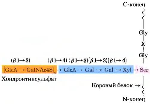
Mammalian Cells synthesize approximately 40 distinct types of proteoglycans. These molecules influence numerous cellular processes, such as growth factor activation and adhesion. A typical proteoglycan consists of a core protein to which one or more glycosaminoglycan chains are covalently attached. Attachment to the protein typically occurs at a Serine residue via a trisaccharide bridge (Fig. 7-24). The Ser residue involved in this linkage is most frequently found within the consensus sequence -Ser-Gly-X-Gly- (where X represents any amino acid residue), although not every protein containing this sequence is conjugated to a glycosaminoglycan. Many proteoglycans are secreted into the extracellular matrix, whereas others function as integral Membrane Proteins (Fig. 11-6). For example, the extracellular matrix separating organized groups of cells contains a family of core proteins with molecular weights ranging from 20,000 to 40,000, each covalently linked to multiple heparan sulfate chains. There are two major families of membrane-bound heparan sulfate proteoglycans. Syndecans possess a single transmembrane domain and an extracellular domain linked to three heparan sulfate chains and two chondroitin sulfate chains (Fig. 7-25a). Glypicans are anchored to the membrane via a covalently attached glycosylphosphatidylinositol (GPI) membrane lipid derivative (Chapter 11). Both syndecans and glypicans can be shed into the extracellular space. An extracellular matrix protease cleaves the polypeptide chain close to the membrane surface, releasing the syndecan ectodomains (the domains projecting outward from the plasma membrane), whereas a phospholipase cleaves the linkage to the Membrane Lipids, releasing glypicans. Numerous chondroitin sulfate and dermatan sulfate proteoglycans also exist; some are membrane-associated, while others are secreted into the intercellular space.
Class="center">Fig. 7-24 Structure of a proteoglycan. A typical trisaccharide (highlighted in blue) links a glycosaminoglycan (here, chondroitin sulfate, highlighted in orange) to a serine residue (pink) in the core protein. The xylose residue at the reducing end of this linker sequence is attached via its anomeric carbon to the hydroxyl group of the Ser residue.
Glycosaminoglycan chains can bind to various extracellular ligands, thereby modulating Ligand-receptor interactions at the cell surface. Detailed structural analysis of heparan sulfate has revealed a defined, non-random heterogeneity; specific segments (typically 3 to 8 disaccharide units in length) differ from adjacent regions in their sequence and binding Specificity for particular proteins. Highly sulfated domains (NS domains) alternate with regions of unmodified N-acetylglucosamine GlcNAc and glucuronic acid GlcA residues (N-acetylated or NA domains) (Fig. 7-25b). The precise sulfation pattern of an NS domain depends on the specific proteoglycan; considering the combinatorial possibilities of modifications in the GlcNAc-IdoA dimer, at least 32 distinct disaccharide variants can theoretically exist. Moreover, certain core proteins synthesized in different cell types may exhibit distinct heparan sulfate structures.
Fig. 7-25 Structure of integral membrane proteoglycans, (a) Schematic representation of a syndecan and a glypican in the plasma membrane. Syndecan is anchored in the membrane through hydrophobic interactions between a stretch of nonpolar amino acid residues and the plasma membrane lipids; it can be released by proteolytic Cleavage near the membrane surface. In a typical syndecan, the amino-terminal domain of the protein on the extracellular membrane surface is covalently linked (via trisaccharide bridges; see Fig. 7-24) to three heparan sulfate chains and two chondroitin sulfate chains. Glypicans are anchored to the membrane via covalent linkages to membrane lipids (a GPI anchor, see Chapter 11) and are shed when this protein-lipid bond is cleaved by a phospholipase. All glypicans contain 14 conserved Cys residues that form Disulfide Bonds to stabilize the protein core, along with two or three glycosaminoglycan chains attached near the C-terminus close to the membrane surface, (b) In the heparan sulfate chain, regions rich in sulfated residues (S domains, shown in green) alternate with regions of unmodified GlcNAc and GlcA residues (NA domains, shown in gray). The structure of a single S domain, characterized by a high density of modified residues, is shown in detail: GlcNAc is sulfated at C-6, and IdoA is sulfated at C-2. The exact positioning of sulfated groups within an S domain varies According to the proteoglycan type.

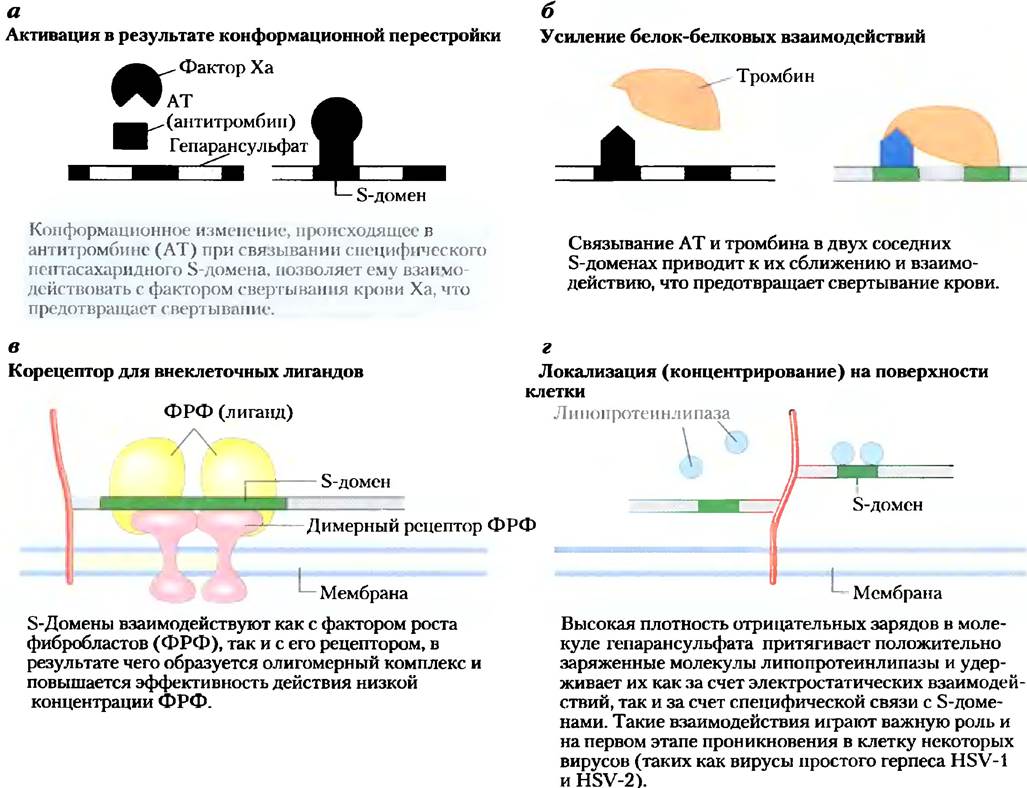
NS domains bind extracellular proteins and signaling molecules in a specific manner, thereby modulating their activity. This modulation can occur through Conformational Changes in the protein molecule induced by binding (Fig. 7-26a), or because adjacent heparan sulfate domains can bind distinct proteins, bringing them into close proximity and enhancing Protein-Protein Interactions (Fig. 7-26b). A third common mechanism involves the binding of extracellular signaling molecules (such as growth factors) to heparan sulfate, which increases their local concentration and facilitates interaction with
their corresponding cell-surface receptors. In such cases, heparan sulfate acts as a coreceptor (Fig. 7-26c). For instance, fibroblast growth factor (FGF), an extracellular signaling protein that stimulates Cell Division, first binds to cell-surface syndecan-associated heparan sulfate. Syndecan then presents FGF to its plasma membrane receptor, allowing FGF to productively interact with the receptor and stimulate cell proliferation. Finally, through yet another mechanism, NS domains interact via electrostatic or other forces with various soluble extracellular molecules, thereby maintaining a high local concentration near the cell surface (Fig. 7-26d). The vital importance of the proper structure of sulfated heparan sulfate domains has been demonstrated in studies with "knockout" mice lacking the enzyme that sulfates the C-2 hydroxyl group of the IdoA residue. These animals lack Kidneys and exhibit severe congenital skeletal and ocular developmental abnormalities. Other studies indicate that membrane proteoglycans play a crucial role in hepatic lipoprotein clearance. Furthermore, accumulating evidence suggests that axon outgrowth pathways, and consequently neural wiring, are guided to some extent by proteoglycans (heparan sulfate and chondroitin sulfate), which provide directional guidance cues for extending axons.
Fig. 7-26 Four types of interactions between heparan sulfate NS domains and proteins.
Some proteoglycans are capable of forming aggregates—massive supramolecular complexes consisting of numerous core proteins bound to a single, elongated hyaluronan molecule. The Ser residue in the core protein aggrecan ($M_r$ ≈ 250,000) is linked via trisaccharide bridges to numerous chondroitin sulfate and keratan sulfate chains, resulting in an aggrecan monomer with a Molecular Weight of approximately 2 · 106. When hundreds of such decorated core proteins associate with a single extended hyaluronan molecule (Fig. 7-27), the resulting proteoglycan aggregate, with a molecular weight exceeding 2 · 108 Da, together with its associated Hydration shell, occupies a volume roughly equivalent to that of an entire bacterial cell! Aggrecan binds tightly to Collagen in the extracellular matrix of Cartilage, which is essential for the development and mechanical resilience of this Connective Tissue.
Fig. 7-27 Proteoglycan aggregates in the extracellular matrix. Schematic model of a proteoglycan aggregate featuring numerous aggrecan molecules. A single, very long hyaluronan molecule is noncovalently associated with approximately 100 aggrecan core protein molecules. Each aggrecan molecule contains multiple covalently attached chondroitin sulfate and keratan sulfate chains. The interaction between these core proteins and hyaluronan is stabilized by link proteins located at each junction between the hyaluronan chain and a core protein. The inset atomic force micrograph shows an individual aggrecan molecule (see Box 11-1).

Massive extracellular proteoglycans are intertwined with fibrous matrix proteins such as collagen, Elastin, and Fibronectin, forming an expansive network that provides both tensile strength and elasticity to the extracellular matrix. Several of these matrix proteins possess binding sites for multiple matrix components. For instance, fibronectin contains distinct domains for binding fibrin, heparan sulfate, collagen, and a family of plasma membrane proteins called Integrins, which function to transmit signals between the cell interior and the extracellular matrix (see Fig. 12-28). The overall architecture of cell-matrix interactions (Fig. 7-28) is characterized by a complex network of connections among cellular and extracellular molecules. These interactions are critical not only for anchoring cells within the matrix, but also for providing pathways for cell migration during tissue development and for relaying information (via integrins) into and out of the cell across the plasma membrane.
Fig. 7-28 Interaction of a cell with the extracellular matrix. Cells anchor to extracellular matrix proteoglycans via membrane-spanning integrin proteins and extracellular linker proteins (such as fibronectin) that contain binding sites for both integrins and proteoglycans. Note the intimate contact between collagen fibrils, fibronectin, and proteoglycans.

Glycoproteins Contain Covalently Bound Oligosaccharides
Glycoproteins are protein-carbohydrate conjugates in which the carbohydrate chains are shorter and more structurally diverse than the glycosaminoglycan chains of proteoglycans. The carbohydrate is attached via its anomeric carbon atom to the OH group of a Ser or Thr residue (O-linked glycosidic bond) or to the amide nitrogen of an Asn residue (N-linked glycosidic bond) (Fig. 7-29). Some glycoproteins contain only a single oligosaccharide chain, whereas many have several. The carbohydrate content can range from 1% to 70% of the glycoprotein's mass. Mucins are secreted or membrane-bound glycoproteins that can contain a high density of O-linked oligosaccharide chains. Mucins are present in most secretions, imparting the characteristic slipperiness of mucus. About half of all mammalian proteins are glycosylated, and roughly 1% of mammalian genes encode Enzymes involved in the synthesis and attachment of these oligosaccharide chains. O-linked sequences are typically rich in Gly, Val, and Pro residues. Conversely, the attachment of N-linked chains depends on the presence of the consensus sequence N-{P}-[ST] (Methods for depicting consensus sequences were discussed in Box 3-3). As with proteoglycans, not all potential attachment sites are necessarily occupied in any given molecule.
Fig. 7-29. Types of oligosaccharide linkages in glycoproteins, (a) Attachment of an oligosaccharide via an O-glycosidic bond occurs by linking to the hydroxyl group of a Ser or Thr residue (highlighted in pink); in this example, GalNAc is located at the reducing end of the oligosaccharide chain. Examples of one simple and one complex oligosaccharide chain are shown. (b) In oligosaccharides attached via an N-glycosidic bond, linkage is established with the amide nitrogen of an Asn residue; here, the terminal residue is GlcNAc. Examples of three common types of N-linked oligosaccharide chains are provided. Complete elucidation of an oligosaccharide structure requires determining the position and stereochemistry (α/β) of each glycosidic bond.

One class of glycoproteins found in the Cytoplasm and nucleus is unique in that the glycosylated sites of the protein contain only single N-acetylglucosamine residues linked via an O-glycosidic bond to the hydroxyl group of a serine side chain. This modification is reversible and frequently targets the same serine residues that undergo phosphorylation during certain Stages of Protein function. These two modifications are mutually exclusive, and this type of glycosylation may play a critical role in regulating protein activity. We will discuss this further in Chapter 12 in the context of protein phosphorylation.
As we will see in Chapter 11, the outer surface of the plasma membrane is densely populated with membrane glycoproteins bearing covalently attached oligosaccharides of varying complexity. One of the best-characterized membrane glycoproteins is Glycophorin A of The erythrocyte membrane (Fig. 11-7). Approximately 60% of its mass consists of carbohydrates organized into 16 oligosaccharide chains, each containing 60 to 70 monosaccharide residues, which are covalently linked to amino acid residues near the N-terminus of the polypeptide chain. Fifteen of these oligosaccharide chains are attached via O-glycosidic bonds to Ser or Thr residues, and one via an N-glycosidic bond to an Asn residue.
Glycomics is the systematic characterization of all carbohydrate-containing components of a given cell or tissue, including those conjugated to proteins and lipids. Glycomics also investigates which proteins bind specific glycoproteins and identifies the specific amino acid in the sequence to which each oligosaccharide is attached. This challenging yet rewarding field of study allows researchers to examine normal glycosylation patterns and how they are altered in genetic diseases and Cancer. Modern methods for characterizing the complete cellular Complement of carbohydrates rely heavily on mass spectrometry (see Fig. 7-37). To date, the structures of numerous O- and N-linked oligosaccharides from various glycoproteins have been elucidated, with several representative examples shown in Fig. 7-29. In Chapter 27, we will examine the mechanisms that enable specific proteins to recognize and bind particular oligosaccharide sequences.
Many proteins secreted by Eukaryotic cells, including most blood proteins, are glycoproteins. For example, IMMUNOGLOBULINS (Antibodies) and certain Hormones—such as follicle-stimulating hormone, luteinizing hormone, and thyroid-stimulating hormone—are glycoproteins. Many milk proteins, such as lactalbumin, as well as several pancreatic proteins (e.g., Ribonuclease), are also glycosylated, as are the vast majority of lysosomal proteins.
The precise "biological advantages" conferred by protein glycosylation remain incompletely understood. Hydrophilic carbohydrate clusters alter the polarity and solubility of the proteins to which they are attached. Oligosaccharide chains added to Newly synthesized proteins in the Endoplasmic reticulum and finalized in the Golgi apparatus serve as sorting signals and participate in protein quality control by tagging misfolded proteins for degradation (Fig. 27-39). When numerous negatively charged oligosaccharide chains cluster in a specific region of a protein, mutual electrostatic repulsion drives The formation of an extended, rod-like conformation in that region. The large bulk and negative charge of oligosaccharide chains also protect certain proteins from proteolytic attack. Beyond these global effects on Protein Structure, oligosaccharide chains mediate more specific biological interactions (Section 7.4). The crucial importance of normal protein glycosylation is underscored by the fact that at least 18 different inherited human congenital disorders of glycosylation are associated with severe physical and mental developmental abnormalities, some of which are fatal.
Glycolipids and Lipopolysaccharides Are Membrane Components
Glycoproteins are not the only cellular molecules that feature complex oligosaccharide chains; several classes of lipids are also covalently linked to oligosaccharides. Gangliosides are membrane lipids of eukaryotic cells whose polar head groups—which project from the outer surface of the plasma membrane—consist of a complex oligosaccharide containing sialic acid (Fig. 7-9) and other monosaccharide residues. Some ganglioside oligosaccharide chains, such as those that determine human Blood Groups (Fig. 10-15), are identical to oligosaccharides found in certain glycoproteins, which therefore also contribute to blood group specificity. Like the oligosaccharide chains of glycoproteins, the oligosaccharides of membrane lipids are typically (and perhaps exclusively) located on the outer face of the plasma membrane.
Lipopolysaccharides are a defining structural feature of the outer membrane of Gram-negative bacteria such as Escherichia coli and Salmonella typhimurium. These molecules are primary targets for antibodies elicited by The Immune System during a bacterial infection, and they serve as major determinants of the serotypes of bacterial strains (serotypes being strains distinguishable by their antigenic properties). The lipopolysaccharides of S. typhimurium contain six Fatty acids linked to two glucosamine residues, one of which has a complex oligosaccharide attached to it (Fig. 7-30). The outer membrane of E. coli contains structurally similar but distinct lipopolysaccharides. The lipopolysaccharides of certain bacteria are toxic to humans and animals; for instance, they are responsible for the drastic drop in blood pressure associated with septic Shock induced by Gram-negative bacterial infections. ■
Fig. 7-30. Bacterial lipopolysaccharides. Schematic representation of the outer membrane lipopolysaccharide structure from Salmonella typhimurium: Kdo is 3-deoxy-D-manno-octulosonic acid, Hep is L-glycero-D-manno-heptose, and AbeOAc is abequose (3,6-dideoxygalactose) acetylated at one of its hydroxyl groups. The lipid portion of the molecule (lipid A) contains six fatty acids. While lipopolysaccharide structures vary somewhat among different bacterial species, all share a conserved tripartite architecture comprising a lipid region (lipid A), a core oligosaccharide, and an O-specific chain that determines the bacterial serotype (its immunogenicity). The outer membranes of the Gram-negative bacteria S. typhimurium and E. coli are so densely packed with lipopolysaccharide molecules that virtually the entire cell surface is coated with O-specific chains.

Summary of Section 7.3 Glycoconjugates: Proteoglycans, Glycoproteins, and Glycolipids
■ Proteoglycans are glycoconjugates in which a core protein is covalently attached to one or more large glycan molecules, such as heparan sulfate, chondroitin sulfate, or keratan sulfate. Glycans account for the major fraction of the molecular mass. Proteoglycans are anchored to the outer surface of the plasma membrane via a transmembrane peptide or a covalently attached lipid, providing sites for Cell Adhesion, Molecular recognition, and signal transduction between cells or between cells and the extracellular matrix.
■ Glycoproteins contain covalently bound oligosaccharides that are smaller and more structurally complex (and thus carry more biological information) than glycosaminoglycans. Many cell-surface proteins, extracellular proteins, and the majority of secreted proteins are glycoproteins. Covalently attached oligosaccharides influence Protein folding and stability, provide intracellular sorting signals for newly synthesized proteins, and mediate specific recognition events with other proteins.
■ Glycomics is the comprehensive identification and quantification of the complete complement of carbohydrate-containing molecules in a cell or tissue, coupled with the functional analysis of all such molecules.
■ PLANT AND ANIMAL glycolipids and bacterial lipopolysaccharides are cell-surface components featuring covalently bound oligosaccharide chains that project from the outer surface of the cell.
Last update: 06/08/2026
Editorial and Educational Adaptation: This material has been compiled based on the primary/original source text. The project team performed an editorial review, corrected technical inaccuracies, structured sections, and adapted the content for an educational format.
What was processed:
- elimination of formatting defects (OCR errors, structural breaks, corrupted characters);
- editorial organization of content;
- standardization of terminology in accordance with academic sources;
- verification of factual statements against the original source text.
All mentions of the author, publication year, and origin of the primary text have been preserved in accordance with the source.