LEHNINGER PRINCIPLES OF BIOCHEMISTRY - VOLUME 1. THE FOUNDATIONS OF BIOCHEMISTRY: STRUCTURE AND CATALYSIS - 2011
PART I. STRUCTURE AND CATALYSIS
7. CARBOHYDRATES AND GLYCOBIOLOGY
7.2. Polysaccharides
Most CARBOHYDRATES found in nature occur as Polysaccharides, polymers of high molecular weight. Polysaccharides, also called glycans, differ from one another in the identity of their recurring monosaccharide units, in the length of their chains, in the types of bonds linking them, and in the degree of branching. Homopolysaccharides contain only a single type of monomeric unit; Heteropolysaccharides contain two or more different kinds (Fig. 7-13). Some homopolysaccharides, such as starch and Glycogen, serve as storage forms of Monosaccharides that are used as fuel. Other homopolysaccharides (Cellulose and Chitin, for example) serve as structural elements in Plant Cell Walls and animal exoskeletons. Heteropolysaccharides provide extracellular support for organisms of all kingdoms. For example, the rigid layer of the bacterial cell envelope (peptidoglycan) is composed in part of a heteropolysaccharide built from two alternating monosaccharide units. In animal Tissues, the extracellular space is occupied by several heteropolysaccharides that form a matrix that holds individual Cells together and provides protection, shape, and support to cells, tissues, and Organs.
Class="center">Fig. 7-13. Homo- and heteropolysaccharides. Polysaccharides may consist of one, two, or several types of monosaccharide units, joined in linear or branched chains of varying length.

Unlike Proteins, polysaccharides generally do not have a defined molecular weight. This difference reflects the mechanics of their synthesis. As we discuss in Chapter 27, proteins are synthesized on a template (Messenger RNA) of defined sequence and length, with Enzymes directing the process to follow the template instructions precisely. There is no template for Polysaccharide synthesis; rather, the program for synthesis resides in The properties of the enzymes that catalyze the polymerization of monomeric units, and there is no specific stop command to halt the process.
Some Homopolysaccharides Serve as Energy Stores
The most important Storage Polysaccharides are starch in PLANT CELLS AND glycogen in animal cells. Both occur intracellularly as large clusters or granules. Starch and glycogen molecules are heavily hydrated because they have many exposed hydroxyl groups available to hydrogen-bond with Water. Starch is produced by most plant cells (see Fig. 20-2) and is particularly abundant in tubers (such as potatoes) and seeds.
Starch contains Two Types of glucose polymer: amylose and amylopectin (Fig. 7-14). Amylose consists of long, unbranched chains of D-glucose residues connected by (α1 —> 4) linkages. Such chains vary in molecular weight from a few thousand to more than a million. Amylopectin also has
a high molecular weight (up to 100 million Da), but unlike amylose, it is highly branched. The glycosidic linkages joining successive glucose residues in amylopectin chains are (α1 —> 4); the branch points (occurring every 24 to 30 residues) are (α1—> 6) linkages.
Fig. 7-14. Glycogen and starch. (a) A short segment of amylose, a linear polymer of D-glucose residues joined by (α1 —> 4) glycosidic bonds. A single amylose chain may contain several thousand glucose residues. Amylopectin has the same Structure between branch points. Glycogen has the same general structure as amylopectin but is more extensively branched. (b) A branch point in amylopectin, showing an (α1 —> 6) glycosidic bond joining two glucose residues. (c) A segment of a starch granule, composed of amylose and amylopectin threads. The amylopectin threads (shown in red) form Double helices with each other or with amylose molecules (shown in blue). In The Cell, when starch is mobilized for energy, glucose residues are removed enzymatically from the nonreducing ends of outer branches. Glycogen has a similar architecture but is more compact and highly branched.

Glycogen is the principal storage polysaccharide of animal cells. Like amylopectin, glycogen is a polymer of (α1 —> 4)-linked glucose residues, with (α1—> 6)-linked branches. However, glycogen is more densely branched (every 8 to 12 residues) and more compact than starch. Glycogen is especially abundant in the Liver, where it may constitute as much as 7% of the wet weight, and is also present in Skeletal Muscle. In hepatocytes, glycogen is found in large granules, which in turn consist of clusters of smaller granules composed of single, highly branched glycogen molecules with average molecular weights of several million. Associated with these granules are the enzymes responsible for the Synthesis and degradation of glycogen.
Because each branch of a glycogen molecule ends in a nonreducing sugar residue, a glycogen molecule has many nonreducing ends but only one reducing end. When glycogen is mobilized as a fuel, glucose residues are removed one at a time from the nonreducing ends. The enzymes that mobilize glycogen act simultaneously on the many nonreducing ends, allowing the rapid release of glucose monomers.
Why doesn't the cell store glucose as the monomer instead of polymerizing it? Calculations reveal that the concentration of glycogen in a hepatocyte is equivalent to a 0.4 M glucose concentration. The actual concentration of the insoluble glycogen is about 0.01 µM; its contribution to the osmolarity of the Cytosol is therefore negligible. If the cytosol contained 0.4 M free glucose, the osmolarity would be dangerously high, driving an influx of water that would lyse the cell (Fig. 2-12). Furthermore, with an intracellular glucose concentration of 0.4 M and an extracellular concentration of ~5 mM (the glucose level in mammalian Blood), the free-energy change required for transport of glucose into the cell against such a steep concentration gradient would be prohibitively large.
Dextrans are bacterial and Yeast polysaccharides made up of (α1 —> 6)-linked D-glucose residues. All dextrans have branch points, typically involving (α1 —> 3) bonds, and some also have (α1—> 2) or (α1—> 4) branch points. Dextrans produced by Bacteria growing on the surfaces of Teeth contribute to The formation of dental plaque. Synthetic dextrans are used in various commercial products, including Sephadex, which is used in Column Chromatography as a gel-filtration medium (Fig. 3-17b). In these products, dextrans are chemically cross-linked to form insoluble Materials of varying porosity, allowing the Separation of macromolecules of different sizes.
Some Homopolysaccharides Serve Structural Roles
Cellulose is a tough, fibrous, water-insoluble substance found in the cell walls of plants, particularly in the stalks, stems, and all woody portions of the plant body. Cellulose is the primary constituent of wood; cotton is almost pure cellulose. Like amylose and the main chains of amylopectin and glycogen, cellulose is a linear, unbranched homopolysaccharide, consisting of 10,000 to 15,000 D-glucose units. However, There is a fundamental difference: the glycosidic linkages in cellulose have the β configuration (Fig. 7-15), whereas those in amylose, amylopectin, and glycogen have the α configuration. Glucose residues in cellulose are linked by (β1—> 4) glycosidic bonds, compared with (α1—> 4) bonds in amylose, starch, and glycogen. This seemingly small difference accounts for the vastly different structural and Physical Properties of cellulose and amylose.
Fig. 7-15. The structure of cellulose. (a) Two monomer units of a cellulose chain, D-glucose residues joined by a (β1—> 4) linkage. The units are shown in the chair conformation, but can rotate relative to each other. (b) Scale drawing of portions of two parallel cellulose chains, showing the conformation of the D-glucose residues and cross-chain hydrogen bonding. In the lower left hexose, all hydrogen atoms are shown; in the other three residues, hydrogen atoms attached to carbon atoms are omitted for clarity, as they do not participate in hydrogen bonding.

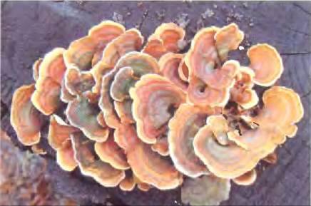
Dietary glycogen and starch are broken down by salivary and intestinal α-amylases, enzymes that hydrolyze (α1 —> 4) glycosidic bonds between glucose residues. Most animals cannot use cellulose as a food source because they lack an enzyme to hydrolyze (β1—> 4) linkages. Termites readily eat wood because their intestinal tract harbors a symbiotic microorganism, Trichonympha, which secretes cellulase, an enzyme that hydrolyzes the (β1—> 4) bonds. Wood-rotting Fungi and bacteria also produce cellulase (Fig. 7-16).
Fig. 7-16. Wood-rotting fungi. A shelf fungus growing on an oak stump. Wood-rotting fungi produce the enzyme cellulase, which hydrolyzes the (β1 —> 4) glycosidic bonds in cellulose, allowing the fungi to utilize the wood as a source of metabolizable sugars (glucose). Among vertebrates, only ruminants (cattle, sheep, goats, camels, giraffes) can use cellulose as a primary food source; their digestive tracts contain specialized compartments (such as the rumen) populated by bacteria and Protozoa that secrete cellulase.
Chitin is a linear homopolysaccharide composed of N-acetylglucosamine residues linked by β-glycosidic bonds (Fig. 7-17). Its only difference from cellulose is that in this polymer, the hydroxyl groups at the C-2 carbon atoms are replaced by acetylated amino groups. Chitin forms long fibers resembling those of cellulose and, much like cellulose, is indigestible by vertebrates. Chitin forms the structural basis of the tough exoskeleton in approximately one million arthropod species—including insects, lobsters, and crabs—and is arguably the second most abundant natural polysaccharide after cellulose, with roughly 1 billion tons produced in the biosphere annually!
Fig. 7-17. Chitin. (a) A short segment of a chitin molecule consisting of N-acetyl-D-glucosamine units joined by (β1—> 4)-glycosidic bonds. (b) The beetle Pelidnota punctata has a tough protective shell (exoskeleton) made of chitin.

The three-dimensional structure of homopolysaccharides is influenced by steric factors and hydrogen bonding
The three-dimensional Introduction/33.html">Structure of Polysaccharides is governed by the same principles that dictate the spatial conformation of polypeptide chains. Monomer units, whose strict structures are defined by covalent bonds, fold into three-dimensional macromolecular architectures stabilized by intra- or intermolecular weak interactions: Hydrogen Bonds, hydrophobic interactions, Van der Waals forces, and—in polymers containing charged monomer units—Electrostatic Interactions. Because polysaccharides are rich in hydroxyl groups, hydrogen bonding plays a particularly powerful role in shaping their structure. Glycogen, starch, and cellulose, as well as the oligosaccharide moieties of Glycoproteins and Glycolipids discussed later, are built from six-membered ring units. Such molecules can be visualized as a series of rigid pyranose rings linked by oxygen bridges (glycosidic bonds). In principle, this structure permits rotation about both C—O bonds connecting adjacent residues (Fig. 7-15a). However, much like in Polypeptides (Figs. 4-2, 4-8), rotation around each bond is constrained by steric factors arising from the substituents. The three-dimensional conformation of these molecules can be described by the dihedral angles φ and Ψ formed across the glycosidic bond (Fig. 7-18), analogously to the φ and Ψ angles of the peptide bond (Fig. 4-2).
Fig. 7-18. Conformation of glycosidic bonds in cellulose, amylose, and dextran. The polymers are depicted as rigid pyranose rings connected by glycosidic bonds, which theoretically allow free rotation. Note that in the dextran molecule, rotation is also possible around the bond connecting the C-5 and C-6 carbon atoms (torsion angle ω [omega]).

The specific shape and large size of the pyranose ring, along with its substituents, impose strict limitations on the possible values of the angles φ and Ψ; certain Conformations are vastly more stable than others, as demonstrated by energy surface plots as a function of φ and Ψ (Fig. 7-19).
Fig. 7-19. Ramachandran-like map of allowed conformations for oligosaccharide and polysaccharide chains. The torsion angles φ and Ψ (Fig. 7-18), which determine the relative spatial orientation of adjacent rings, can theoretically range from 0° to 360°. In reality, some angle values correspond to sterically forbidden conformations, whereas others permit the Formation of the maximum number of hydrogen bonds. (a) By plotting the energy (Σ) corresponding to each pair of φ and Ψ values and drawing isoenergetic contours (lines of equal energy) at 1 kcal/mol intervals, the preferred conformational regions can be identified. This map is analogous to the Ramachandran plot for Peptides (Figs. 4-3, 4-8). (b) Two conformational states of the disaccharide gal(β1—>3)gal, corresponding to the energy minimum and maximum on diagram (a), are marked with red and blue circles on the map. The red circle indicates the least favorable conformation, and the blue circle represents the most favorable one. The conformational states of the three polysaccharides shown in Fig. 7-18 were determined by X-ray crystallography: all of them fall within low-energy regions.

The most stable three-dimensional structure for (α1—> 4)-linked chains of starch and glycogen is a tightly coiled helix (Fig. 7-20) stabilized by intra-chain hydrogen bonds. In unbranched amylose, this structure is so highly ordered that the substance can be crystallized and its structure investigated by X-Ray Diffraction Analysis. On average, the plane of each residue in an amylose chain is rotated by 60° relative to the plane of the preceding residue, resulting in six residues per turn of the helix. The inner cavity of the amylose helix is of a size that perfectly accommodates complex iodide ions (I3– or I5–); the reaction with iodine produces an intense blue color and is widely used as a standard quantitative test for amylose.
Fig. 7-20. Structure of starch (amylose). (a) In its most stable state, the amylose polymer chain—consisting of residues rigidly locked in the chair conformation—forms a helix, distinguishing it from cellulose, which forms extended straight fibers (Fig. 7-16). (b) A space-filling model of a segment of the amylose helix. The presence of (α1—> 4) linkages endows amylose, amylopectin, and glycogen with a highly coiled, compact structure. This architecture forms The basis of dense storage granules of starch and glycogen found in many cells (Fig. 7-14).

In the most stable conformation of cellulose, each glucose residue in the chair conformation is rotated by 180° relative to its neighbor, resulting in extended, straight, uncoiled ribbons. All hydroxyl groups within the molecule are available to form hydrogen bonds with adjacent chains. The extensive network of intra- and intermolecular hydrogen bonds between neighboring cellulose chains imparts remarkable tensile strength to the fibers (Fig. 7-15b). Humans have utilized this property of cellulose for millennia in materials such as papyrus, paper, cardboard, rayon, insulation board, and countless other useful products. The water content in such materials is low because the extensive network of intermolecular hydrogen bonds fully satisfies the hydrogen-bonding capacity of the cellulose molecules.
Bacterial and algal Cell walls contain structural heteropolysaccharides
The rigid framework of bacterial cell walls (peptidoglycan) is composed of a heteropolymer of alternating N-acetylglucosamine and N-acetylmuramic acid residues joined by (β1—> 4) linkages (see Fig. 20-31). These linear polymers are packed tightly within The Cell wall and held together by short cross-links formed by various peptides, the exact nature of which depends on the bacterial species. These peptide cross-bridges knit the polysaccharide chains into a robust, mesh-like exoskeleton that encases the entire cell, preventing it from Swelling and lysing under the inward Osmotic Pressure of water. The enzyme Lysozyme kills bacteria by hydrolyzing the (β1—> 4)-glycosidic bonds between N-acetylglucosamine and N-acetylmuramic acid (see Fig. 6-24). Lysozyme is present in tears, where it plays a protective role by defending against bacterial infections. Certain bacterial Viruses (Bacteriophages) produce lysozyme to rupture the host cell wall and escape—a critical step in the viral life cycle. Penicillin and related Antibiotics destroy bacteria by interfering with the synthesis of these structural cross-links, leaving the cell wall unable to withstand osmotic lysis (see pp. 314–316).
The cell walls of certain red marine Algae contain Agar—a complex mixture of sulfated heteropolysaccharides composed of D-galactose and an L-galactose derivative in which the C-3 and C-6 carbon atoms are bridged by an ether bond (Fig. 7-21). The two principal components of agar are unbranched agarose (Mr ≈ 120,000) and branched agaropectin. Agarose possesses a remarkable gel-forming property that is widely exploited in biochemical laboratories. When an aqueous suspension of agarose is heated and subsequently cooled, its molecules form a double helix: two parallel-oriented molecules intertwine around each other, with each turn containing three sugar residues while water molecules occupy the central cavity. These Helical structures further aggregate to form a gel—a three-dimensional matrix that entraps large volumes of water. Agarose gels serve as inert media for the electrophoretic separation of Nucleic Acids, notably in DNA Sequencing (see Fig. 8-33). Agar is also used to prepare solid growth media for culturing bacterial colonies. Furthermore, agar forms the shells of capsules for various Vitamins and medications; it dissolves rapidly in The Stomach and is metabolically inert.
Fig. 7-21. Structure of agarose. The repeating units of the agarose molecule consist of D-galactose residues linked via a (β1—> 4) bond to 3,6-anhydro-L-galactose, in which an internal ether bridge connects the C-3 and C-6 carbons. These structural units are joined to one another by (α1—> 3)-glycosidic bonds, forming a polymer of 600–700 residues. Some of the 3,6-anhydrogalactose residues are esterified with sulfuric acid at the C-2 carbon atom, as shown in the figure.

Glycosaminoglycans are heteropolysaccharides of the Extracellular matrix
The extracellular space in the tissues of Multicellular animals is filled with a gel-like material called the extracellular matrix (ECM) or ground substance, which holds cells together and provides a porous pathway for the diffusion of nutrients and oxygen to individual cells. The ECM surrounding fibroblasts and other Connective Tissue cells is a complex network of heteropolysaccharides and Fibrous proteins such as Collagen, Elastin, and Fibronectin. The basement membrane is a specialized type of extracellular matrix underlying epithelial cells, consisting of specialized collagen, laminin, and heteropolysaccharides. These heteropolysaccharides—known as glycosaminoglycans—form a family of linear polymers built from repeating disaccharide units (Fig. 7-22). Glycosaminoglycans are found in animals and bacteria but are entirely absent in plants.
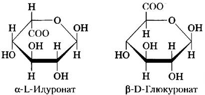
One of the two monosaccharides in the repeating disaccharide is always either N-acetylglucosamine or N-acetylgalactosamine; the second is typically a uronic acid—most commonly D-glucuronic or L-iduronic acid. Some glycosaminoglycans also contain esterified sulfate groups. The combination of sulfate groups and carboxylate groups on the uronic acid residues confers an extremely high density of negative charges on glycosaminoglycan molecules. To minimize electrostatic repulsion between adjacent charged groups, these molecules adopt an extended, highly expanded conformation in solution, forming a stiffened helix with negatively charged carboxylate groups protruding outward (as shown for heparin in Fig. 7-22). This extended geometry also serves to space out the negatively charged sulfate groups as far apart as possible. The specific spatial distribution of sulfated and unsulfated sugar residues allows glycosaminoglycans to bind specific protein ligands via electrostatic interactions. Glycosaminoglycans associate with extracellular proteins to form Proteoglycans (Section 7.3).
Fig. 7-22. Repeating units in the structure of several extracellular matrix glycosaminoglycans. Glycosaminoglycans are copolymers composed of alternating residues of uronic acids and amino sugars (with the exception of keratan sulfate), which may be sulfated at various hydroxyl positions (except in hyaluronic acid). Ionized carboxyl and sulfate groups (highlighted in red in the projection formulas) impart a characteristic high negative charge to the polymer molecules. Pharmaceutical-grade heparin consists predominantly of iduronic acid (IdoA) and small amounts of glucuronic acid (GlcA; not shown); heparin molecules are typically heavily sulfated and exhibit considerable size heterogeneity. A spatial molecular model of a heparin segment in its solution conformation (based on NMR spectroscopy data; PDB ID 1HPN). Carbon atoms of iduronic acid are shown in blue, and those of glucosamine in green. Oxygen and sulfur atoms are colored red and yellow, respectively, as is conventional. Hydrogen atoms are omitted for clarity. Heparan sulfate (not shown) resembles heparin but contains a higher proportion of GlcA and fewer, less regularly distributed sulfate groups.

The glycosaminoglycan hyaluronan (or hyaluronic acid, which exists as the hyaluronate anion at physiological pH) is composed of alternating D-glucuronic acid and N-acetylglucosamine units (Fig. 7-22). Hyaluronates can consist of up to 50,000 primary disaccharide units, yielding molecular weights exceeding 1 million Da. These substances form clear, highly viscous solutions that serve as lubricants in synovial fluid of joints and give the vertebrate vitreous humor of the eye its jelly-like consistency (derived from the Greek hyalos, meaning Glass; hyaluronan solutions are crystal-clear or translucent). Furthermore, hyaluronates are crucial Components of the extracellular matrix in Cartilage and tendons, imparting tensile strength and elasticity through their interactions with other matrix macromolecules. Certain pathogenic bacteria secrete the enzyme hyaluronidase, which degrades the glycosaminoglycan bonds of hyaluronate, thereby rendering tissues more susceptible to bacterial invasion. In many organisms, a similar enzyme found in sperm hydrolyzes the outer glycosaminoglycan protective coat of egg cells, enabling sperm penetration during Fertilization.
Other glycosaminoglycans differ from hyaluronates in three respects: first, their molecular sizes are much smaller; second, they are covalently linked to specific proteins, forming proteoglycans; and third, one or both of their monomers differ from those of hyaluronate. Chondroitin sulfate (from the Greek chondros, meaning cartilage) confers tensile strength upon cartilage, tendons, ligaments, and the walls of the aorta. Dermatan sulfate (from the Greek derma, meaning Skin) contributes to the elasticity of skin, Blood Vessels, and Heart Valves. In this polymer, many of the glucuronate (GlcA) residues present in chondroitin are replaced by its 5-epimer, L-iduronate (IdoA).
Keratan sulfates (from the Greek keras, meaning horn) lack uronic acid, and the number of sulfated groups in them varies. They are found in the cornea, cartilage, bones, and various cornified structures formed from dead cells, such as horns, Hair, hooves, Nails, and claws. Heparan sulfate (from the Greek hepar, meaning liver) is synthesized by all animal cells and contains sulfated and nonsulfated sugars in varying proportions. The sulfated Regions of the chain allow heparin to interact with a variety of proteins, including growth factors and ECM components, as well as enzymes and factors present in Blood Plasma. Heparin is a fractionated form of heparan sulfate extracted primarily from mast cells (a type of leukocyte). It is widely used clinically as an anticoagulant due to its ability to bind to the proteinase inhibitor antithrombin. This binding induces antithrombin to bind and inhibit Thrombin, a protease playing a crucial role in Blood Coagulation. The interaction between heparin and antithrombin is driven by electrostatic forces; the heparin molecule carries a high negative charge density, exceeding that of any other known biological molecule (Fig. 7-23). Purified heparin is added to blood samples in clinical assays and to donated blood to prevent clotting. Table 7-2 summarizes the composition, properties, biological roles, and distribution of the polysaccharides described in Section 7.2.
Figure 7-23. Interaction between a glycosaminoglycan and a protein. The structure shown is derived from the co-crystal of fibroblast growth factor (FGF1), its cell-surface receptor, and a short glycosaminoglycan (heparin) fragment (PDB ID 1E00). The protein surfaces are rendered in solid colors reflecting electrostatic potential: red indicates a predominantly negative charge, and blue indicates a predominantly positive charge. Heparin is depicted in a ball-and-stick model; its negatively charged groups (-SO3- and -COO-) interact with the positively charged (blue) region of the protein molecule. Although heparin was used in this experiment, heparan sulfate is the physiological Ligand that interacts with FGF in vivo.

Table 7-2. Structures and Functions of Selected Polysaccharides
Polymer |
Type* |
Repeating unit** |
Size (number of monosaccharide units) |
Biological function |
Starch Amylose Amylopectin |
Homo- Homo- |
(α1—>4)Glc, linear |
50–5,000 Up to 106 |
Energy storage in plant cells |
Glycogen |
Homo- |
(α1—>4)Glc with (α1—>6)Glc branches every 8–12 residues |
Up to 50,000 |
Energy storage in bacterial and animal cells |
Cellulose |
Homo- |
(β1—>4)Glc |
Up to 15,000 |
Structural: imparts rigidity and tensile strength to plant cell walls |
Chitin |
Homo- |
(β1—>4)GlcNAc |
Very large |
Structural: provides mechanical support in the exoskeletons of insects, arachnids, and crustaceans |
Dextran |
Homo- |
(α1—>6)Glc with (α1—>3) branches |
Variable |
Structural: bacterial Cell Adhesion |
Peptidoglycan |
Hetero-; peptide-linked |
Mur2Ac(β1—>4)GlcNAc |
Very large |
Structural: provides rigidity and strength to bacterial cell walls |
Agarose |
Hetero- |
D-Gal(β1—>4)3,6-anhydro-L-Gal(α1—>3) |
~1,000 |
Structural: component of algal cell walls |
Hyaluronan (glycosaminoglycan) |
Hetero-; acidic |
GlcUA(β1—>3)GlcNAc(β1—>4) |
Up to 100,000 |
Structural: extracellular matrix of skin and Connective Tissues, vertebrate joint lubrication |
* Each polymer is classified as a homopolysaccharide (homo-) or heteropolysaccharide (hetero-).
** The abbreviated names of the repeating units in peptidoglycan, agarose, and hyaluronan indicate that their polymers are built from disaccharide subunits. For instance, in peptidoglycan, the GlcNAc residue of one disaccharide is linked via a (β1—>4) bond to the first residue of the adjacent disaccharide.
Summary of Section 7.2 Polysaccharides
■ Polysaccharides (glycans) serve as energy storage reserves and as Structural components of cell walls and the extracellular matrix.
■ The homopolysaccharides starch and glycogen function as energy storage forms in plant, animal, and bacterial cells. They consist of D-glucose residues joined by (α1—>4) linkages and feature branched architectures.
■ The homopolysaccharides cellulose, chitin, and dextran play structural roles. Cellulose, composed of D-glucose units linked by (β1—>4) bonds, confers strength and rigidity to plant cell walls. Chitin, consisting of N-acetylglucosamine residues connected via (β1—>4) linkages, provides the structural foundation for arthropod exoskeletons. Dextran forms protective capsules around certain bacterial cells.
■ Polysaccharides adopt well-defined three-dimensional structures. Because the chair conformation of the pyranose ring is quite rigid, the overall polymer conformation is determined by rotation angles about the glycosidic bonds linking the sugar residues. Starch and glycogen form helical structures stabilized by extensive hydrogen bonding. Cellulose and chitin form extended, straight chains that interact with adjacent strands via hydrogen bonds.
■ The mechanical strength of bacterial and algal cell walls stems from heteropolysaccharides: peptidoglycan in bacteria and agarose in red algae. The repeating disaccharide unit of peptidoglycan has the structure Mur2Ac(β1—>4)GlcNAc, whereas that of agarose is composed of D-Gal(β1—>4)3,6-anhydro-L-Gal.
■ Glycosaminoglycans are extracellular heteropolysaccharides in which one of the two monosaccharide units is a uronic acid and the other is an N-acetylated amino sugar. Certain hydroxyl groups and amino groups of the constituent sugar residues in these polymers are sulfated, imparting a high negative charge density that forces the chains into extended, hydrated conformations. These polymers (hyaluronan, chondroitin sulfate, dermatan sulfate, keratan sulfate, and heparin) provide viscosity, adhesiveness, and structural resilience to the extracellular matrix.
Last update: 06/08/2026
Editorial and Educational Adaptation: This material has been compiled based on the primary/original source text. The project team performed an editorial review, corrected technical inaccuracies, structured sections, and adapted the content for an educational format.
What was processed:
- elimination of formatting defects (OCR errors, structural breaks, corrupted characters);
- editorial organization of content;
- standardization of terminology in accordance with academic sources;
- verification of factual statements against the original source text.
All mentions of the author, publication year, and origin of the primary text have been preserved in accordance with the source.