LEHNINGER PRINCIPLES OF BIOCHEMISTRY - VOL. 1. THE FOUNDATIONS OF BIOCHEMISTRY: STRUCTURE AND CATALYSIS - 2011
PART I. STRUCTURE AND CATALYSIS
Class="center">I would have more optimism about a bright future for mankind if it spent less time proving that it can outwit Nature and more time tasting her sweetness and holding her in respect.
E. B. White, The Trees of Eden, 1977
7. CARBOHYDRATES AND GLYCOBIOLOGY
CARBOHYDRATES are the most abundant Biomolecules on Earth. Each year, Photosynthesis converts over 100 billion metric tons of CO2 and H2O into Cellulose and other plant products. Carbohydrates (sugars and starches) serve as the primary dietary staple almost everywhere; The oxidation of carbohydrates is the principal energy-yielding pathway in most nonphotosynthetic Cells. Insoluble high-molecular-weight carbohydrates provide structural and protective elements in The Cell walls of Bacteria and plants, and in the Connective Tissues of animals. Other complex carbohydrates act as lubricants for joints and participate in Cell Recognition and adhesion. More intricate, polymeric carbohydrates covalently joined to Proteins or Lipids function as signaling molecules that determine the intracellular localization or metabolic fate of these hybrid molecules, known as Glycoconjugates. This chapter introduces The main classes of carbohydrates and glycoconjugates, along with Examples of their diverse Structural and functional roles.
Carbohydrates are polyhydroxy aldehydes or ketones, or substances that yield these compounds on Hydrolysis. For many, though not all, carbohydrates, the empirical formula is (CH2O)n; some also contain nitrogen, phosphorus, or sulfur.
Carbohydrates are divided into three major size classes: Monosaccharides, Oligosaccharides, and Polysaccharides (the word saccharide derives from the Greek sarkharon, meaning "sugar"). Monosaccharides, or simple sugars, consist of a single polyhydroxy aldehyde or ketone unit. The most abundant monosaccharide in nature is the six-carbon sugar D-glucose, sometimes referred to as dextrose. Monosaccharides with four or more carbon atoms typically occur as cyclic structures.
Oligosaccharides consist of short chains of monosaccharide units, or residues, joined by glycosidic bonds. The most abundant are Disaccharides, which consist of two monosaccharide units. A typical example is sucrose (cane sugar), which is composed of the six-carbon sugars D-glucose and D-fructose. The names of all mono- and disaccharides end in the suffix "-ose." Most oligosaccharides with three or more units do not exist as free molecules in cells; instead, they are covalently attached to other molecules, such as proteins or lipids, to form glycoconjugates.
Polysaccharides are sugar polymers containing 20 or more monosaccharide units; some have hundreds or thousands of units. Certain polysaccharides, such as cellulose, are linear chains, whereas others, such as Glycogen, are branched. Both glycogen and cellulose are polymers of D-glucose, but they differ in the type of glycosidic linkage, which gives them strikingly different properties and biological Functions.
7.1. Monosaccharides and Disaccharides
The simplest carbohydrates are the monosaccharides. Chemically, they are aldehydes or ketones with two or more hydroxyl groups; the six-carbon monosaccharides glucose and fructose each have five hydroxyl groups. Because many of the carbon atoms bearing hydroxyl groups are chiral centers, a great variety of sugar stereoisomers are found in nature. We begin with a description of monosaccharide families whose carbon backbones contain from three to seven carbon atoms, discussing their Structure, stereoisomeric forms, and conventions for representing their three-dimensional structures. We then examine several Chemical Reactions Involving the carbonyl groups of monosaccharides. One such reaction is the intramolecular addition of a hydroxyl group to the carbonyl carbon, which yields the cyclic five- and six-membered ring structures characteristic of sugars in aqueous solution and creates a new chiral center, further enriching the stereochemistry of this class of compounds. Consequently, we will review in detail the nomenclature used to unambiguously designate THE POSITION OF each carbon atom within a ring, as well as the Methods for depicting these structures graphically. This Background will be useful when we discuss monosaccharide METABOLISM in Part II. In addition, we will introduce several important Monosaccharide Derivatives that will be encountered later.
The Monosaccharides Are of Two Families: Aldoses and Ketoses
Monosaccharides are colorless, crystalline solids that are freely soluble in Water but insoluble in nonpolar Solvents. Most monosaccharides have a sweet taste. The carbon Skeleton of typical monosaccharides is an unbranched chain in which all carbon atoms are linked by single bonds. In the open-chain form, one of the carbon atoms is double-bonded to an oxygen atom to form a carbonyl group; each of the other carbon atoms bears a hydroxyl group. If the carbonyl group is at the end of the carbon chain (an aldehyde group), the monosaccharide is an aldose; if the carbonyl group is at any other position (a ketone group), the monosaccharide is a ketose. The simplest monosaccharides are the two three-carbon sugars: the aldotriose glyceraldehyde and the ketotriose dihydroxyacetone (Fig. 7-1a).
Figure 7-1. Examples of monosaccharides. (a) Two trioses: an aldose and a ketose. The carbonyl carbon in each compound is highlighted in color. (b) Two commonly occurring hexoses. (c) Pentoses, which are Components of nucleic acids. D-Ribose is a constituent of Ribonucleic Acids (RNA), and 2-deoxy-D-ribose is a component of Deoxyribonucleic Acids (DNA).

Monosaccharides with four, five, six, and seven carbon atoms are called tetroses, pentoses, hexoses, and heptoses, respectively. Each can exist as an aldose or a ketose, yielding aldotetroses and ketotetroses, aldopentoses and ketopentoses, aldohexoses and ketohexoses, and so on. Hexoses, which include the aldohexose D-glucose and the ketohexose D-fructose (Fig. 7-1b), are the most abundant monosaccharides in nature. The aldopentoses D-ribose and 2-deoxy-D-ribose (Fig. 7-1c) are essential components of NUCLEOTIDES and Nucleic Acids (Chapter 8).
Monosaccharides Contain Asymmetric Carbon Atoms
All monosaccharides except dihydroxyacetone contain one or more asymmetric (chiral) carbon atoms and thus occur in optically active isomeric forms (pp. 36–38). The simplest aldose, glyceraldehyde, has a single chiral center (the middle carbon atom) and therefore exists as two optical isomers, or enantiomers (Fig. 7-2).
Figure 7-2. Three ways to represent the two stereoisomers of glyceraldehyde. The stereoisomers are nonsuperimposable mirror images of each other. Ball-and-stick models reflect the actual molecular geometry. By convention, in Fischer projection formulas, horizontal bonds project out of the plane of the paper toward the reader, and vertical bonds project behind the plane of the paper away from the reader. Recall (Fig. 1-17) that in perspective formulas, bonds pointing toward the reader are drawn as solid wedges, and those pointing away are drawn as dashed wedges.
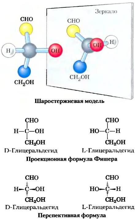
Key Conventions. The dextrorotatory form is designated the D isomer, and the levorotatory form is the L isomer. As with other biomolecules containing chiral centers, the absolute configuration of sugars is determined by X-ray crystallography. To represent the three-dimensional structures of sugars in two dimensions, chemists most commonly use Fischer projection formulas (Fig. 7-2). In a Fischer projection, horizontal bonds point toward the reader out of the plane of the paper, whereas vertical bonds point away from the reader behind the plane of the paper.
In general, a molecule with $n$ chiral centers can have $2^n$ stereoisomers. Glyceraldehyde has $2^1 = 2$ isomers; an aldohexose with four chiral centers has $2^4 = 16$ stereoisomers. Monosaccharide stereoisomers of a given chain length can be divided into two groups according to their configuration at the chiral center most distant from the carbonyl carbon. Those stereoisomers whose chiral carbon configuration matches that of D-glyceraldehyde are designated D isomers, and those with the configuration of L-glyceraldehyde are L isomers. In a Fischer projection, if the hydroxyl group on the reference carbon is on the right, the sugar is a D isomer; if it is on the left, it is an L isomer. Of the 16 possible aldohexoses, 8 are L isomers and 8 are D isomers. Most hexoses found in Living organisms are D isomers. Figure 7-3 illustrates the structures of the D stereoisomers of all aldoses and ketoses containing three to six carbon atoms. Carbon atoms in sugar molecules are numbered consecutively from the end of the chain nearest the carbonyl group. Each of the eight D-aldohexoses, which differ in the stereochemistry at C-2, C-3, and C-4, has its own unique name: D-glucose, D-galactose, D-mannose, and so forth (Fig. 7-3a). The names of four- and five-carbon ketoses are derived by adding the suffix "-ulose" to the name of the corresponding aldose; for example, D-ribulose is the ketopentose corresponding to the aldopentose D-ribose. Ketohexoses are named similarly in some cases, or have traditional names: fructose comes from the Latin fructus ("fruit"), as fruits are rich in this sugar, and sorbose comes from Sorbus, the genus of the rowan tree, whose berries yield a related sugar alcohol, sorbitol. Two sugars that differ only in the configuration around one specific carbon atom are called epimers. For example, D-glucose and D-mannose are epimers that differ only in the stereochemistry at C-2, whereas D-glucose and D-galactose differ in the stereochemistry at C-4 (Fig. 7-4).
Figure 7-3. Aldoses and ketoses. Fischer projections show the structures of (a) D-aldoses and (b) D-ketoses containing from three to six carbon atoms. Chiral carbon atoms are highlighted in red. In all D isomers, the chiral carbon most distant from the carbonyl carbon has the same configuration as the chiral carbon in D-glyceraldehyde. The names of the most biologically prevalent sugars are enclosed in boxes.
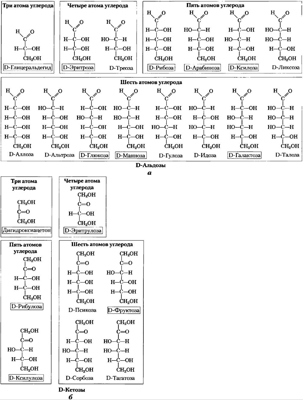
Fig. 7-4. Epimers. D-glucose and two of its epimers are shown using projection formulas. Each epimer differs from D-glucose in the configuration of a single chiral center (highlighted in color).

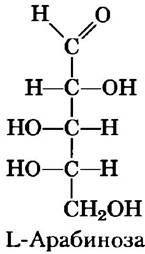
Some sugars occur in nature in the L form. Examples include L-arabinose and the L-isomers of certain sugar derivatives found in glycoconjugates (Section 7-3).
Common monosaccharides have cyclic structures
For simplicity, we have so far depicted all aldoses and ketoses as linear molecules (Figs. 7-3, 7-4). In reality, however, in aqueous solution, aldotetroses and all monosaccharides with a greater number of carbon atoms in the chain predominantly exist as cyclic (ring) molecules, in which the carbonyl group forms a covalent bond with the oxygen of a hydroxyl group on the same molecule. The formation of such ring structures is a special case of the reaction between alcohols and aldehydes or ketones, leading to the formation of hemiacetals and hemiketals (Fig. 7-5), which contain an additional asymmetric carbon atom and therefore can exist in two stereoisomeric forms. For example, D-glucose exists in solution as an intramolecular hemiacetal in which the free hydroxyl group at the C-5 carbon has reacted with the aldehyde group at the C-1 carbon, making the C-1 carbon asymmetric; the two resulting Stereoisomers are designated by the letters α and β (Fig. 7-6). Such six-membered rings are called pyranoses because they resemble the six-membered pyran ring (Fig. 7-7). According to systematic nomenclature, the two ring structures formed by D-glucose are called α-D-glucopyranose and β-D-glucopyranose.
Fig. 7-5. Formation of hemiacetals and hemiketals. Aldehydes and ketones can react with alcohols in a 1:1 ratio to form hemiacetals and hemiketals, respectively, containing an additional chiral center. Addition of a second alcohol molecule leads to the formation of acetals and ketals. If the second alcohol group is part of another sugar molecule, the resulting bond is called a glycosidic bond.

Aldohexoses can also exist in another cyclic form (a five-membered ring). Because of their similarity to the five-membered furan ring, such structures are called furanoses. However, six-membered aldopyranose rings are much more stable than aldofuranose rings and therefore predominate in solutions of aldohexoses. Pyranose rings can be formed only by aldoses whose molecules contain more than five carbon atoms.
Isomeric forms of monosaccharides that differ only in the configuration of the hemiacetal or hemiketal carbon atom are called anomers, and the hemiacetal (carbonyl) carbon atom is referred to as the anomeric carbon. In solution, the α- and β-anomeric forms interconvert through The process of mutarotation (Fig. 7-6). Thus, solutions of α-D-glucose and β-D-glucose eventually form a uniform equilibrium mixture with identical optical properties. This mixture consists of approximately one-third α-D-glucose and two-thirds β-D-glucose; in addition, very small amounts of glucose are present in the linear form and as a five-membered ring (glucofuranose).
Fig. 7-6. Formation of the two cyclic forms of D-glucose. The reaction between the aldehyde group at C-1 and the hydroxyl group at C-5 leads to the formation of a hemiacetal having two stereoisomers (α- and β-anomers) that differ only in the stereochemical Properties of the C-1 carbon. The interconversion of α- and β-anomers is called mutarotation.

Ketohexoses also exist as α- and β-anomers. In these compounds, the hydroxyl group at C-5 (or C-6) reacts with the keto group at C-2, leading to the formation of a furanose (or pyranose) ring containing a hemiketal bond (Fig. 7-5). D-Fructose readily forms furanose rings (Fig. 7-7); the most common anomeric form of this sugar is β-D-fructofuranose.
Fig. 7-7. Pyranoses and furanoses. The pyranose forms of D-glucose and furanose forms of D-fructose are depicted using Haworth projection formulas. The side of the ring closer to the reader is shown with a thick line. Hydroxyl groups located below the plane of the ring in these formulas correspond to those positioned on the right in Fischer projection formulas (cf. Fig. 7-6). The formulas of pyran and furan are shown at the bottom for comparison.

To depict the cyclic forms of monosaccharides, Haworth projection formulas such as those shown in Fig. 7-7 are commonly used. It should be borne in mind that the six-membered pyranose ring does not lie in a single plane, as the Haworth projection implies, but rather assumes a chair conformation (Fig. 7-8). Recall (from Chapter 1) that Conformations refer to the different shapes that a molecule can assume without breaking covalent bonds, whereas configurations refer to molecular states whose interconversion requires the breaking of covalent bonds. For example, the interconversion between the α- and β-configurations takes place with the Cleavage of the ring oxygen bond. As we will see later, the biological Properties and functions of many polysaccharides are largely determined by the conformational features of their constituent monosaccharide units.
Fig. 7-8. Conformational formulas of a pyranose. (a) Two possible chair conformations of the pyranose ring. Substituents at the carbon atoms in the ring can be either axial (ax), oriented parallel to a vertical axis passing through the ring, or equatorial (eq), i.e., positioned approximately perpendicular to the axis. The two conformations shown in the figure cannot readily interconvert without breaking the ring. However, under molecular strain (as shown by atomic force Microscopy studies, see Box 11-1), an energy input of approximately 46 kJ/mol of sugar can drive the interconversion of the two forms. In general, substituents in the equatorial position experience less steric hindrance from neighboring groups; therefore, when bulky substituents are present in the molecule, the conformer with these substituents in the equatorial position is favored. Another possible conformation—the boat conformation (not shown here)—is observed only in molecules with very bulky substituents. (b) α-D-Glucopyranose in its most energetically favorable chair conformation.

Living organisms contain a variety of hexose derivatives
In addition to simple hexoses such as glucose, galactose, and mannose, there are A number of derivatives in which either a hydroxyl group of the parent compound is replaced by another group, or a carbon atom is oxidized to form a carboxyl group (Fig. 7-9). In glucosamine, galactosamine, and mannosamine, the hydroxyl at the C-2 position of the parent sugar is replaced by an amino group. The amino group is almost invariably condensed with acetic acid, as in N-acetylglucosamine, for example. This compound is a constituent of many structural polymers, including those that make up the Introduction/37.html">Bacterial Cell wall. The latter contains yet another glucosamine derivative—N-acetylmuramic acid. In this compound, the oxygen at the C-3 position of N-acetylglucosamine is linked by an ether bond to lactic acid (a three-carbon carboxylic acid). Replacement of the hydroxyl group by hydrogen at the C-6 position in L-galactose or L-mannose yields L-fucose or L-rhamnose, respectively. L-Fucose is found in complex oligosaccharide components of Glycoproteins and Glycolipids, whereas L-rhamnose is a constituent of plant polysaccharides.
Fig. 7-9. Hexose derivatives of major biological importance. In aminosugars, one of the -OH groups of the hexose is replaced by -NH3. Deoxy sugars arise when an -OH is replaced by -H; these deoxy sugars occur in nature as L-isomers. Acidic sugars contain a carboxyl group, conferring a negative charge at neutral pH. D-Glucono-δ-lactone is formed by the creation of an ester bond between the carboxyl group and the hydroxyl group at the C-5 carbon (δ-carbon) in D-gluconate.

Oxidation of the carbonyl (aldehyde) carbon of glucose to a carboxyl group yields gluconic acid; oxidation of other aldoses yields the corresponding aldonic acids. Oxidation of the carbon at the opposite end of the molecule (the C-6 carbon of glucose, galactose, or mannose) leads to the formation of the corresponding uronic acids—glucuronic, galacturonic, or mannuronic acid. Both aldonic and uronic acids form stable intramolecular esters known as lactones (Fig. 7-9, bottom left). In addition to these acidic hexose derivatives, mention should be made of a nine-carbon sugar, N-acetylneuraminic acid (one of the sialic acids, often referred to simply as sialic acid). N-Acetylneuraminic acid is a derivative of N-acetylmannosamine and is a component of many animal cell glycoproteins and glycolipids. At pH 7.0, the carboxyl groups of acidic sugar derivatives are ionized; therefore, by analogy with carboxylates, these compounds are named glucuronate, galacturonate, and so forth.
Intermediates in carbohydrate synthesis and metabolism are very frequently not free sugars, but rather their phosphorylated derivatives. The Condensation reaction between phosphoric acid and one of the hydroxyl groups of a sugar yields a phosphoric acid ester, such as glucose-6-phosphate (Fig. 7-9). Sugar phosphates are quite stable at neutral pH values and carry a negative charge. One consequence of phosphorylation is the retention of sugars within the cell; most cells lack transport systems for crossing The Plasma Membrane with phosphorylated sugars. Furthermore, phosphorylation activates sugars for subsequent chemical transformations. Certain phosphorylated sugar derivatives are vital components of nucleotides (discussed in the next chapter).
Monosaccharides are reducing agents
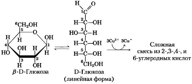
■ Monosaccharides are oxidized by relatively mild oxidizing agents, such as Fe3+ or Cu2+ ions (Fig. 7-10). In this process, the carbonyl group is oxidized to a carboxyl group. Glucose and other sugars capable of reducing Fe3+ or Cu2+ ions are referred to as reducing sugars. They form endiols, which are converted into aldonic acids and subsequently into a complex mixture of 2-, 3-, 4-, and 6-carbon acids. This property forms The basis of several Methods for the Quantitative determination of reducing sugars. By measuring The amount of oxidizing agent reduced by a sugar solution, the sugar concentration can be determined. For many years, such tests were used to detect and quantify glucose in Blood and urine for the Diagnosis of Diabetes Mellitus (Box 7-1). ■
Figure 7-10. Sugars as reducing agents. Fehling's reaction is based on the oxidation of the anomeric carbon atom in a glucose molecule or other sugars. Under alkaline conditions, Cu+ copper ions form a red copper oxide precipitate. In the hemiacetal form (cyclic form), the C-1 atom of glucose is not oxidized by Cu2+ ions. However, an equilibrium exists between the linear and cyclic forms, so the reaction ultimately goes to completion. The interaction with Cu2+ ions is complex, leads to the formation of a mixture of products, and requires 3 mol of Cu2+ per 1 mol of glucose.
Disaccharides contain a glycosidic bond
Disaccharides (maltose, lactose, sucrose) consist of two monosaccharides linked together by a covalent O-glycosidic bond, which is formed by the reaction between the hydroxyl group of one sugar molecule and the anomeric carbon atom of another molecule (Fig. 7-11). In this process, an acetal is formed from a hemiacetal (e.g., glucopyranose) and an alcohol (the hydroxyl group of the second sugar molecule) (Fig. 7-5). Glycosidic bonds are readily hydrolyzed in acidic media, but are resistant to bases. Upon boiling in dilute acid, disaccharides are hydrolyzed, releasing their constituent monosaccharide components. An N-glycosidic bond connects the anomeric carbon atom of a sugar to a nitrogen atom in glycoproteins (Fig. 7-29) and nucleotides (Fig. 8-1).
Box 7-1. MEDICINE. Blood Glucose Measurement in the Diagnosis and Management of Diabetes
Glucose is the primary energy source for the Brain. A compromised glucose supply to the brain can have life-threatening consequences: lethargy, coma, irreversible brain damage, and death (Fig. 23-25). Animals possess a sophisticated hormonal mechanism that maintains blood glucose levels high enough (about 5 mM) to meet the brain's demands, yet not excessively high, as excess blood glucose can also lead to severe physiological consequences.
Individuals suffering from Insulin-dependent diabetes mellitus produce insufficient amounts of the hormone insulin, which normally serves to lower blood glucose concentrations. If left untreated, the blood glucose concentration in patients can exceed normal levels severalfold. Elevated blood glucose is one of the causes of such severe complications of progressive diabetes as renal failure, cardiovascular disease, blindness, and impaired wound healing. Therefore, a primary goal in treating diabetes is to ensure an adequate supply of insulin in the body (via injections) to maintain a normal Blood Glucose Level. To determine the optimal diet, physical activity, and medication regimen, it is necessary to measure blood glucose concentrations several times a day and adjust the administered insulin dose accordingly.
Blood glucose concentration can be determined using simple methods for quantifying reducing sugars, such as Fehling's test, which has been used for many years as a diagnostic screening test for suspected diabetes (Fig. 7-10). Modern analysis requires only a drop of blood placed on a glucose oxidase test strip (Fig. 1); a simple photometer detects the color development resulting from the reaction of a dye with H2O2, which is released during the enzymatic reaction, and determines the blood glucose concentration.
Figure 1. The reaction catalyzed by glucose oxidase is used to determine blood glucose concentration. A second enzyme, peroxidase, catalyzes the reaction of H2O2 with a colorless substrate, leading to the formation of a colored product, the concentration of which is measured using a spectrophotometer.

Blood glucose levels fluctuate after meals and intense physical exertion, so a single measurement does not necessarily reflect the average glucose concentration over a day or several days, and dangerous deviations above the norm may go unnoticed. The average glucose concentration can be determined by examining Hemoglobin, the oxygen-carrying protein found in erythrocytes (p. 233). The function of this carrier protein in The erythrocyte membrane is to equilibrate glucose concentrations inside the cells and in Blood Plasma, making hemoglobin constantly accessible for glucose binding regardless of its blood concentration. A nonenzymatic reaction occurs between glucose and the primary amino group of hemoglobin (the amino group of the N-terminal valine residue or the ε-amino group of a Lysine residue; see Fig. 2). The rate of this reaction is proportional to the glucose concentration, allowing it to be used to estimate the average blood glucose concentration over several weeks. The level of glycated hemoglobin (GHb) at any given time reflects the average blood glucose concentration for a period corresponding to the erythrocyte lifespan (about 120 days), although the measurement over the preceding two weeks is the most critical for determining GHb levels.
The degree of hemoglobin glycation (glycation, unlike glycosylation, refers to the nonenzymatic transfer of glucose to a protein molecule) is measured in clinical practice by extracting hemoglobin from a small blood sample and electrophoretically separating GHb from unmodified hemoglobin (Separation is based on charge differences resulting from amino acid modification). Normally, GHb accounts for about 5% of total hemoglobin (corresponding to a blood glucose concentration of 120 mg / 100 mL). In untreated diabetic patients, this value can reach 13%, reflecting an elevation in blood glucose to a dangerous level of 300 mg / 100 mL. One of the criteria for properly selecting an individualized insulin administration program (injection time, frequency, and dosage) is maintaining GHb at around 7%.
During hemoglobin glycation, the initial reaction step (formation of a Schiff base) is followed by a series of rearrangements involving oxidation and dehydration, resulting in a heterogeneous mixture of advanced glycation end products (AGEs). These products can exit erythrocytes and cross-link with proteins, interfering with normal protein function (Fig. 2). The accumulation of relatively large amounts of AGEs in diabetic patients can lead to impaired Kidney, retinal, and cardiovascular function. Research is underway to find pharmacological agents capable of counteracting this destructive process in the body.
Figure 2. Nonenzymatic reaction of glucose with the primary amino group in hemoglobin: 1) formation of a Schiff base; 2) Amadori rearrangement yielding a stable product; 3) cyclization of the ketoamine to form GHb; 4) subsequent reactions yield advanced glycation end products (AGEs), such as E-N-carboxymethyllysine and methylglyoxal; 5) these substances can interfere with the function of other proteins by cross-linking with them, causing severe pathological Changes in the body.

The anomeric carbon atom is oxidized by Fe3+ or Cu2+ ions (the reaction used to identify reducing sugars) only when the molecule is in the linear form, which is in equilibrium with the cyclic form. If the anomeric carbon is involved in a glycosidic bond, the sugar residue cannot convert into the linear form and becomes nonreducing. In Disaccharides and Polysaccharides in general, the end of the carbohydrate chain featuring a free anomeric carbon atom (not bound in a glycosidic bond) is called the reducing end.
The disaccharide maltose (Fig. 7-11) consists of two D-glucose residues joined by a glycosidic bond between the C-1 atom (the anomeric atom) of one glucose residue and the C-4 atom of the other residue. Because this disaccharide retains a free anomeric carbon (C-1 in the right-hand glucose residue in Fig. 7-11), maltose is a reducing sugar. The anomeric carbon atom in the glycosidic bond has the α-configuration. The second glucose residue with the free anomeric carbon atom can exist in either the α- or β-form.
Figure 7-11. Formation of maltose. The disaccharide is formed from two monosaccharides (in this case, two D-glucose molecules), where the -OH group (alcoholic) of one glucose molecule (right) reacts with the intramolecular hemiacetal of another molecule (left) with the release of an H2O molecule and the formation of an O-glycosidic bond. The reverse reaction (hydrolysis) represents the attack of the glycosidic bond by a water molecule. The maltose molecule retains reducing properties because one anomeric C-1 atom does not participate in the formation of the glycosidic bond. Mutarotation leads to the interconversion of the α- and β-forms of the hemiacetal (sometimes depicted with a wavy line, as shown here); thus, this maltose residue can exist in both α- and β-forms.
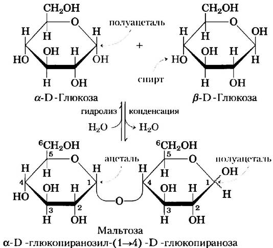
Nomenclature Conventions. Certain rules govern the naming of reducing disaccharides such as maltose, and especially more complex oligosaccharides. By convention, the name refers to a compound in which the nonreducing end of the molecule is placed on the left, and the compound is named as follows: 1) Specify the configuration (α or β) of the anomeric carbon linking the first monosaccharide residue (on the left) to the second. 2) Name the nonreducing residue; for five- and six-membered ring structures, use the roots "furano" or "pyrano," respectively. 3) Enclose in parentheses the numbers of the two carbon atoms connected by the glycosidic bond, joining the figures with an arrow; for example, the notation (1->4) indicates that C-1 of the first sugar residue is linked to C-4 of the second. 4) Name the second residue. If a third residue is present, the second glycosidic bond is described using the same rules. To abbreviate the names of complex polysaccharides, three-letter designations of monosaccharide units are used, as shown in Table 7-1. According to the rules for designating oligosaccharides, maltose is correctly named α-D-glucopyranosyl-(1->4)-D-glucopyranose. Because most sugars discussed in this book are D-sugars, and the predominant form of hexoses is pyranose, we usually abbreviate the formal names of such compounds by indicating only the configuration of the anomeric carbon and the numbers of the atoms connected by the glycosidic bond. Using this nomenclature, maltose is written as Glc(α1->4)Glc. ■
Table 7-1.

Note: hexoses are conventionally depicted as circles, N-acetylhexosamines as squares, and hexosamines as bisected circles. All sugars with a "gluco" configuration are blue, those with a "galacto" configuration are yellow, and those with a "manno" configuration are green. Other substituents are added to the name as needed: sulfate (S), phosphate (P), O-acetyl (OAc), or O-methyl (OMe).
The disaccharide lactose (Fig. 7-12), which yields D-galactose and D-glucose upon hydrolysis, occurs naturally only in milk. The anomeric carbon of the glucose residue is accessible for oxidation, making lactose a reducing disaccharide. This compound can be abbreviated as Gal(β1->4)Glc. Sucrose (table sugar) is a disaccharide composed of glucose and fructose. Sucrose is synthesized in plant cells, but not in animal cells. Unlike maltose and lactose, sucrose lacks a free anomeric carbon; the anomeric carbon atoms of both monosaccharide residues participate in the formation of the glycosidic bond (Fig. 7-12). Consequently, sucrose is a nonreducing sugar. Nonreducing sugars are designated as Glycosides, where the anomeric carbon atoms are linked by a glycosidic bond. In abbreviated names, the numbers and configurations of the anomeric atoms are connected by a bidirectional arrow. For example, sucrose can be abbreviated as either Glc(α1⇌2β)Fru or Fru(β2⇌1α)Glc. Sucrose is a major intermediate product of photosynthesis; in many plants, sugar transport from leaves to other tissues is carried out precisely in the form of sucrose. Trehalose, Glc(α1⇌1α)Glc (Fig. 7-12), is a disaccharide composed of D-glucose residues and, similar to sucrose, lacks reducing activity. Trehalose is a major component of insect circulating fluid (hemolymph), where it serves as a reserve energy source. Fungi also contain trehalose, from which this sugar is obtained commercially.
Fig. 7-12. Some commonly occurring disaccharides. Here, as in Fig. 7-11, molecules are depicted using Haworth projection formulas. For each disaccharide, the trivial, full systematic, and abbreviated names are given. According to the formal nomenclature for sucrose, glucose is the parent glycoside; it is usually depicted, as shown here, with the glucose residue on the left.

Summary of Section 7.1 Monosaccharides and Disaccharides
■ Sugars (or saccharides) are compounds containing an aldehyde or ketone group and two or more hydroxyl groups.
■ Monosaccharides typically contain multiple chiral carbon atoms and therefore exist in several stereoisomeric forms, which can be represented on a plane using Fischer projection formulas. Epimers are sugars that differ only in the configuration at a single carbon atom.
■ Monosaccharides frequently form intramolecular hemiacetals or hemiketals, in which the aldehyde or ketone group reacts with a hydroxyl group of the same molecule, resulting in a cyclic structure. Such structures are depicted using Haworth projection formulas. The carbon atom originally belonging to the aldehyde or ketone group (the anomeric carbon atom) can exist in one of two configurations ($\alpha$ or $\beta$), which interconvert during mutarotation. In the linear form of a monosaccharide, which is in equilibrium with the cyclic form, the anomeric carbon atom is readily oxidized.
■ The hydroxyl group of one monosaccharide can react with the anomeric carbon atom of a second monosaccharide to form an acetal. In such a disaccharide, the glycosidic bond protects the anomeric carbon from oxidation.
■ Oligosaccharides are oligomers composed of a small number of monosaccharide units linked by glycosidic bonds. One end of the chain (the reducing end) retains an anomeric carbon that is not involved in a glycosidic bond.
■ The nomenclature adopted for designating di- and oligosaccharides specifies The sequence of monosaccharide units, the configuration of each anomeric carbon, and the numbers of the carbon atoms participating in the formation of glycosidic bonds.
Last update: 06/08/2026
Editorial and Educational Adaptation: This material has been compiled based on the primary/original source text. The project team performed an editorial review, corrected technical inaccuracies, structured sections, and adapted the content for an educational format.
What was processed:
- elimination of formatting defects (OCR errors, structural breaks, corrupted characters);
- editorial organization of content;
- standardization of terminology in accordance with academic sources;
- verification of factual statements against the original source text.
All mentions of the author, publication year, and origin of the primary text have been preserved in accordance with the source.